. 1998 Aug 1;12(15):2293-304.
doi: 10.1101/gad.12.15.2293.

Switch from translation to RNA replication in a positive-stranded RNA virus

Affiliations

Switch from translation to RNA replication in a positive-stranded RNA virus

A V Gamarnik et al. Genes Dev. .

Abstract

In positive-stranded viruses, the genomic RNA serves as a template for both translation and RNA replication. Using poliovirus as a model, we examined the interaction between these two processes. We show that the RNA polymerase is unable to replicate RNA templates undergoing translation. We discovered that an RNA structure at the 5' end of the viral genome, next to the internal ribosomal entry site, carries signals that control both viral translation and RNA synthesis. The interaction of this RNA structure with the cellular factor PCBP up-regulates viral translation, while the binding of the viral protein 3CD represses translation and promotes negative-strand RNA synthesis. We propose that the interaction of 3CD with this RNA structure controls whether the genomic RNA is used for translation or RNA replication.


Figures
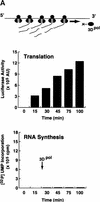
Figure 1
Figure 1
Poliovirus translation inhibits 3Dpol RNA elongation activity. (A) A poliovirus replicon (Polio–Luc, Fig. 2A) was in vitro-translated in a translation system supplemented with purified poliovirus 3D polymerase (3Dpol). Translation (top) was determined by the amount of luciferase activity produced and was expressed as arbitrary units (AU). RNA synthesis (bottom) was measured by [32P]UMP incorporation into an acid-insoluble fraction. (B) The same experiment described in A was performed in the presence of cycloheximide (100 μg/μl).
Figure 1
Figure 1
Poliovirus translation inhibits 3Dpol RNA elongation activity. (A) A poliovirus replicon (Polio–Luc, Fig. 2A) was in vitro-translated in a translation system supplemented with purified poliovirus 3D polymerase (3Dpol). Translation (top) was determined by the amount of luciferase activity produced and was expressed as arbitrary units (AU). RNA synthesis (bottom) was measured by [32P]UMP incorporation into an acid-insoluble fraction. (B) The same experiment described in A was performed in the presence of cycloheximide (100 μg/μl).
Figure 2
Figure 2
Poliovirus-infected cell extracts contain an activity that specifically inhibits poliovirus translation. (A) Schematic representation of the chimeric poliovirus luciferase RNA (Polio–Luc) and capped luciferase RNA (Cap–Luc). In Polio–Luc, the coding region of the poliovirus capsid proteins was replaced by the luciferase reporter gene, and a cleavage site for 2Apro has been introduced between luciferase and 2Apro (represented by the arrow). The Cap–Luc RNA consists of the luciferase gene flanked by the 5‘ and 3‘ uncoding regions of the β-globin mRNA. (B) Microinjection of infected S100 HeLa cell extract into Xenopus oocytes specifically inhibits poliovirus cap-independent translation. Polio–Luc or Cap–Luc RNA was injected into oocytes together with uninfected (open bars) or poliovirus-infected S10 or S100 HeLa cell fractions (solid bars) as indicated in each case. Luciferase activity was determined in oocytes after 3 hr of incubation at 22°C and expressed in arbitrary units (AU). (C) Elution profile of the translation inhibitory activity after ion-exchange chromatography. Infected S100 HeLa cell extract was loaded onto a HiTrap SP column (Pharmacia) and eluted with a KCl gradient, as indicated at right. The translation inhibitory activity was determined by coinjection of 20 nl of each fraction (1–14) together with 5 nl of HeLa S10 (to provide the cellular factor essential for poliovirus translation in oocytes, PTF) and 20 ng of Polio–Luc RNA (▪) or Cap–Luc RNA (formula image) into oocytes. Luciferase activity was determined in oocyte extracts after 3 hr of incubation at 22°C and expressed in AU. (D) Viral proteins 3Dpol, 3CD, and P3 copurified with the viral translation inhibitory activity. Western blot analysis of fractions 1–14 eluted from the HiTrap SP column is shown. Two microliters of each fraction was resolved in a 10% SDS–polyacrylamide gel, transferred to nitrocelluose membrane, and probed with specific anti-3CD antibodies. The electrophoretic mobility of P3, 3CD, and 3D is indicated at left.
Figure 2
Figure 2
Poliovirus-infected cell extracts contain an activity that specifically inhibits poliovirus translation. (A) Schematic representation of the chimeric poliovirus luciferase RNA (Polio–Luc) and capped luciferase RNA (Cap–Luc). In Polio–Luc, the coding region of the poliovirus capsid proteins was replaced by the luciferase reporter gene, and a cleavage site for 2Apro has been introduced between luciferase and 2Apro (represented by the arrow). The Cap–Luc RNA consists of the luciferase gene flanked by the 5‘ and 3‘ uncoding regions of the β-globin mRNA. (B) Microinjection of infected S100 HeLa cell extract into Xenopus oocytes specifically inhibits poliovirus cap-independent translation. Polio–Luc or Cap–Luc RNA was injected into oocytes together with uninfected (open bars) or poliovirus-infected S10 or S100 HeLa cell fractions (solid bars) as indicated in each case. Luciferase activity was determined in oocytes after 3 hr of incubation at 22°C and expressed in arbitrary units (AU). (C) Elution profile of the translation inhibitory activity after ion-exchange chromatography. Infected S100 HeLa cell extract was loaded onto a HiTrap SP column (Pharmacia) and eluted with a KCl gradient, as indicated at right. The translation inhibitory activity was determined by coinjection of 20 nl of each fraction (1–14) together with 5 nl of HeLa S10 (to provide the cellular factor essential for poliovirus translation in oocytes, PTF) and 20 ng of Polio–Luc RNA (▪) or Cap–Luc RNA (formula image) into oocytes. Luciferase activity was determined in oocyte extracts after 3 hr of incubation at 22°C and expressed in AU. (D) Viral proteins 3Dpol, 3CD, and P3 copurified with the viral translation inhibitory activity. Western blot analysis of fractions 1–14 eluted from the HiTrap SP column is shown. Two microliters of each fraction was resolved in a 10% SDS–polyacrylamide gel, transferred to nitrocelluose membrane, and probed with specific anti-3CD antibodies. The electrophoretic mobility of P3, 3CD, and 3D is indicated at left.
Figure 2
Figure 2
Poliovirus-infected cell extracts contain an activity that specifically inhibits poliovirus translation. (A) Schematic representation of the chimeric poliovirus luciferase RNA (Polio–Luc) and capped luciferase RNA (Cap–Luc). In Polio–Luc, the coding region of the poliovirus capsid proteins was replaced by the luciferase reporter gene, and a cleavage site for 2Apro has been introduced between luciferase and 2Apro (represented by the arrow). The Cap–Luc RNA consists of the luciferase gene flanked by the 5‘ and 3‘ uncoding regions of the β-globin mRNA. (B) Microinjection of infected S100 HeLa cell extract into Xenopus oocytes specifically inhibits poliovirus cap-independent translation. Polio–Luc or Cap–Luc RNA was injected into oocytes together with uninfected (open bars) or poliovirus-infected S10 or S100 HeLa cell fractions (solid bars) as indicated in each case. Luciferase activity was determined in oocytes after 3 hr of incubation at 22°C and expressed in arbitrary units (AU). (C) Elution profile of the translation inhibitory activity after ion-exchange chromatography. Infected S100 HeLa cell extract was loaded onto a HiTrap SP column (Pharmacia) and eluted with a KCl gradient, as indicated at right. The translation inhibitory activity was determined by coinjection of 20 nl of each fraction (1–14) together with 5 nl of HeLa S10 (to provide the cellular factor essential for poliovirus translation in oocytes, PTF) and 20 ng of Polio–Luc RNA (▪) or Cap–Luc RNA (formula image) into oocytes. Luciferase activity was determined in oocyte extracts after 3 hr of incubation at 22°C and expressed in AU. (D) Viral proteins 3Dpol, 3CD, and P3 copurified with the viral translation inhibitory activity. Western blot analysis of fractions 1–14 eluted from the HiTrap SP column is shown. Two microliters of each fraction was resolved in a 10% SDS–polyacrylamide gel, transferred to nitrocelluose membrane, and probed with specific anti-3CD antibodies. The electrophoretic mobility of P3, 3CD, and 3D is indicated at left.
Figure 2
Figure 2
Poliovirus-infected cell extracts contain an activity that specifically inhibits poliovirus translation. (A) Schematic representation of the chimeric poliovirus luciferase RNA (Polio–Luc) and capped luciferase RNA (Cap–Luc). In Polio–Luc, the coding region of the poliovirus capsid proteins was replaced by the luciferase reporter gene, and a cleavage site for 2Apro has been introduced between luciferase and 2Apro (represented by the arrow). The Cap–Luc RNA consists of the luciferase gene flanked by the 5‘ and 3‘ uncoding regions of the β-globin mRNA. (B) Microinjection of infected S100 HeLa cell extract into Xenopus oocytes specifically inhibits poliovirus cap-independent translation. Polio–Luc or Cap–Luc RNA was injected into oocytes together with uninfected (open bars) or poliovirus-infected S10 or S100 HeLa cell fractions (solid bars) as indicated in each case. Luciferase activity was determined in oocytes after 3 hr of incubation at 22°C and expressed in arbitrary units (AU). (C) Elution profile of the translation inhibitory activity after ion-exchange chromatography. Infected S100 HeLa cell extract was loaded onto a HiTrap SP column (Pharmacia) and eluted with a KCl gradient, as indicated at right. The translation inhibitory activity was determined by coinjection of 20 nl of each fraction (1–14) together with 5 nl of HeLa S10 (to provide the cellular factor essential for poliovirus translation in oocytes, PTF) and 20 ng of Polio–Luc RNA (▪) or Cap–Luc RNA (formula image) into oocytes. Luciferase activity was determined in oocyte extracts after 3 hr of incubation at 22°C and expressed in AU. (D) Viral proteins 3Dpol, 3CD, and P3 copurified with the viral translation inhibitory activity. Western blot analysis of fractions 1–14 eluted from the HiTrap SP column is shown. Two microliters of each fraction was resolved in a 10% SDS–polyacrylamide gel, transferred to nitrocelluose membrane, and probed with specific anti-3CD antibodies. The electrophoretic mobility of P3, 3CD, and 3D is indicated at left.
Figure 3
Figure 3
The polymerase–protease precursor, 3CD, represses viral translation. (A) Depletion of 3CD from infected cell extracts correlates with loss of translation inhibition. The viral protein 3CD was depleted from a partially purified infected HeLa fraction by affinity chromatography by use of an immobilized cloverleaf RNA (see Materials and Methods). (Left) Two microliters of 3CD-depleted extract (lane 2) and 2 μl of a nondepleted control (lane 1) were subjected to Western blot analysis as described for Fig. 2C. (Right) Luciferase activity was determined in oocyte extracts 3 hr after coinjection of Polio–Luc RNA with buffer, nondepleted control, or 3CD-depleted fractions as indicated in the bottom. (B) Overexpression of mutated 3CD (Gln-182 → Asn) in Xenopus oocytes inhibits poliovirus translation. (Top) Schematic diagram of the microinjection protocol. Oocytes were injected with 4 ng of a capped RNA encoding for 3CD, 3C, 3D, or an unrelated RNA encoding GFP, and incubated at 17°C for 15 hr. Then, oocytes were microinjected a second time with 40 ng of Polio–Luc or Cap–Luc RNA. Luciferase expression in oocytes was measured by enzymatic activity 6 hr after the second microinjection. Translation of Polio–Luc and Cap–Luc RNA was determined in oocytes that were preinjected with the mRNAs or with buffer control (−) as indicated at the bottom. Luciferase activity was expressed in arbitrary units (AU).
Figure 3
Figure 3
The polymerase–protease precursor, 3CD, represses viral translation. (A) Depletion of 3CD from infected cell extracts correlates with loss of translation inhibition. The viral protein 3CD was depleted from a partially purified infected HeLa fraction by affinity chromatography by use of an immobilized cloverleaf RNA (see Materials and Methods). (Left) Two microliters of 3CD-depleted extract (lane 2) and 2 μl of a nondepleted control (lane 1) were subjected to Western blot analysis as described for Fig. 2C. (Right) Luciferase activity was determined in oocyte extracts 3 hr after coinjection of Polio–Luc RNA with buffer, nondepleted control, or 3CD-depleted fractions as indicated in the bottom. (B) Overexpression of mutated 3CD (Gln-182 → Asn) in Xenopus oocytes inhibits poliovirus translation. (Top) Schematic diagram of the microinjection protocol. Oocytes were injected with 4 ng of a capped RNA encoding for 3CD, 3C, 3D, or an unrelated RNA encoding GFP, and incubated at 17°C for 15 hr. Then, oocytes were microinjected a second time with 40 ng of Polio–Luc or Cap–Luc RNA. Luciferase expression in oocytes was measured by enzymatic activity 6 hr after the second microinjection. Translation of Polio–Luc and Cap–Luc RNA was determined in oocytes that were preinjected with the mRNAs or with buffer control (−) as indicated at the bottom. Luciferase activity was expressed in arbitrary units (AU).
Figure 4
Figure 4
The cloverleaf structure formed at the 5′ end of the viral genome controls viral translation. (A) Schematic representation of the ribonucleoprotein complex formed around the cloverleaf RNA. The predicted cloverleaf structure is composed of stem–loop B (nucleotides 10–34), stem–loop C (nucleotides 35–45), and stem–loop D (nucleotides 51–78). Viral factor 3CD and cellular protein PCBP are shown interacting with their specific target sequences. The locations of the mutations introduced into the cloverleaf structure of the Polio–Luc RNAs are indicated by arrows: LB.14 (nucleotides 23–26, CCCA, were deleted in loop B); SB.212 (nucleotides 14–16, GGG, and nucleotides 28–30, CCC, were replaced with AAA and UUU, respectively, which maintain the stem B structure); and LD.73 (nucleotides GUAC were inserted in position 70 of loop D). (B) Luciferase activity produced by Polio–Luc constructs carrying wild-type or mutated cloverleaf structures. In vitro-transcribed Polio–Luc RNAs were either transfected into HeLa cells (top) or microinjected into Xenopus oocytes (bottom). The RNAs are indicated as WT (wild-type), ΔCL (cloverleaf-deleted), LB.14 (loop B muted), SB.212 (stem B mutated), and LD.73 (loop D mutant). Luciferase activity was measured in HeLa cell extracts 2 hr after electroporation and in oocyte extracts 10 hr after injection, and expressed in arbitrary units (AU). (C) Microinjection of decoy cloverleaf RNAs into Xenopus oocytes interferes with poliovirus translation. Wild-type Polio-Luc RNA was coinjected with buffer (−), 30 ng of wild-type cloverleaf (WT), or 30 ng of mutant cloverleaf decoys (LB, nucleotides C23 to A26 deleted, or LD, nucleotides GUAC inserted in position 70). Luciferase activity was determined in oocyte extracts 10 hr after injection.
Figure 4
Figure 4
The cloverleaf structure formed at the 5′ end of the viral genome controls viral translation. (A) Schematic representation of the ribonucleoprotein complex formed around the cloverleaf RNA. The predicted cloverleaf structure is composed of stem–loop B (nucleotides 10–34), stem–loop C (nucleotides 35–45), and stem–loop D (nucleotides 51–78). Viral factor 3CD and cellular protein PCBP are shown interacting with their specific target sequences. The locations of the mutations introduced into the cloverleaf structure of the Polio–Luc RNAs are indicated by arrows: LB.14 (nucleotides 23–26, CCCA, were deleted in loop B); SB.212 (nucleotides 14–16, GGG, and nucleotides 28–30, CCC, were replaced with AAA and UUU, respectively, which maintain the stem B structure); and LD.73 (nucleotides GUAC were inserted in position 70 of loop D). (B) Luciferase activity produced by Polio–Luc constructs carrying wild-type or mutated cloverleaf structures. In vitro-transcribed Polio–Luc RNAs were either transfected into HeLa cells (top) or microinjected into Xenopus oocytes (bottom). The RNAs are indicated as WT (wild-type), ΔCL (cloverleaf-deleted), LB.14 (loop B muted), SB.212 (stem B mutated), and LD.73 (loop D mutant). Luciferase activity was measured in HeLa cell extracts 2 hr after electroporation and in oocyte extracts 10 hr after injection, and expressed in arbitrary units (AU). (C) Microinjection of decoy cloverleaf RNAs into Xenopus oocytes interferes with poliovirus translation. Wild-type Polio-Luc RNA was coinjected with buffer (−), 30 ng of wild-type cloverleaf (WT), or 30 ng of mutant cloverleaf decoys (LB, nucleotides C23 to A26 deleted, or LD, nucleotides GUAC inserted in position 70). Luciferase activity was determined in oocyte extracts 10 hr after injection.
Figure 5
Figure 5
Synthesis of viral negative-strand RNA in Xenopus oocytes. (A) Oocytes were microinjected with in vitro-transcribed 32P-labeled poliovirus RNA together with HeLa S10 extracts (Gamarnik and Andino 1996), and the conversion of the input RNA into a double-stranded form was analyzed as a function of the time in 1% native agarose gels electrophoresis, as indicated on the top. For comparison, 32P-labeled poliovirus RNA was synthesized in crude replication complexes (CRC) obtained from poliovirus-infected HeLa cells (lane 1). Single stranded viral RNA (ssRNA) and the double stranded replicative form (RF), are indicated at left. (B) Northern blot analysis confirms that the oocytes produce negative-strand RNA. Oocyte extracts obtained from 200 oocytes at 20 hr after microinjection of unlabeled viral RNA were used to detect newly synthesized negative-strand RNA (lane 2). DNase- and RNase-treated samples were extracted with phenol–chloroform, ethanol precipitated, and resolved under denaturing conditions through 1% agarose gel electrophoresis. Then, RNA was transferred to a nylon filter, and hybridized with a specific probe complementary to the viral negative strand. As a control, infected HeLa extracts treated in similar conditions was analyzed (lane 1). (C) The replicative form synthesized in oocytes contains a covalently linked Vpg molecule. Oocytes injected with 32P-labeled poliovirus RNA were lysed at 0 and 20 hr post-injection (as described in Materials and Methods), immunoprecipitated with anti-Vpg antibodies (α-Vpg) or preimmune sera (Preimm.), and analyzed in 1% native agarose gel (lanes 1–3). Total RNA from oocytes injected with 32P-labeled poliovirus RNA obtained 20 hr after incubation (lane 4) and 32P-labeled RNA synthesized in crude replication complexes obtained from infected HeLa cells (lanes 5) are shown.
Figure 5
Figure 5
Synthesis of viral negative-strand RNA in Xenopus oocytes. (A) Oocytes were microinjected with in vitro-transcribed 32P-labeled poliovirus RNA together with HeLa S10 extracts (Gamarnik and Andino 1996), and the conversion of the input RNA into a double-stranded form was analyzed as a function of the time in 1% native agarose gels electrophoresis, as indicated on the top. For comparison, 32P-labeled poliovirus RNA was synthesized in crude replication complexes (CRC) obtained from poliovirus-infected HeLa cells (lane 1). Single stranded viral RNA (ssRNA) and the double stranded replicative form (RF), are indicated at left. (B) Northern blot analysis confirms that the oocytes produce negative-strand RNA. Oocyte extracts obtained from 200 oocytes at 20 hr after microinjection of unlabeled viral RNA were used to detect newly synthesized negative-strand RNA (lane 2). DNase- and RNase-treated samples were extracted with phenol–chloroform, ethanol precipitated, and resolved under denaturing conditions through 1% agarose gel electrophoresis. Then, RNA was transferred to a nylon filter, and hybridized with a specific probe complementary to the viral negative strand. As a control, infected HeLa extracts treated in similar conditions was analyzed (lane 1). (C) The replicative form synthesized in oocytes contains a covalently linked Vpg molecule. Oocytes injected with 32P-labeled poliovirus RNA were lysed at 0 and 20 hr post-injection (as described in Materials and Methods), immunoprecipitated with anti-Vpg antibodies (α-Vpg) or preimmune sera (Preimm.), and analyzed in 1% native agarose gel (lanes 1–3). Total RNA from oocytes injected with 32P-labeled poliovirus RNA obtained 20 hr after incubation (lane 4) and 32P-labeled RNA synthesized in crude replication complexes obtained from infected HeLa cells (lanes 5) are shown.
Figure 6
Figure 6
The interaction between 3CD and the cloverleaf RNA is required for negative strand RNA synthesis. (A) Schematic representation of wild-type poliovirus genome (Polio-WT), a mutant in the cloverleaf structure in which two Us in positions 60 and 68 were replaced by Cs to disrupt stem–loop D (Polio-315), and a mutant in 3CD-coding sequence in which the Asp-85 was replaced by Glu, which abrogates RNA binding (Polio-181). (B) Disrupting 3CD–cloverleaf interaction increases viral translation. Oocytes were microinjected with Polio–Luc WT (black bars), Polio–Luc 315 (white bars), or Polio-Luc 181 (gray bars) RNAs and incubated at 22°C. Cytoplasmic extracts were obtained, and luciferase activity was measured at the indicated times (0.5, 2, 4, and 8 hr). (C) Disruption of 3CD–cloverleaf interaction abolishes negative-strand RNA synthesis. Oocytes were microinjected with 32P-labeled Polio–WT (lanes 1–5), mutant Polio-315 (lanes 6–10), or mutant Polio-181 (lanes 11–15) and incubated at 30°C. Total RNA was extracted at 0 , 2, 4, 8, and 12 hr, and analyzed on 1% agarose gels. For comparison, 32P-labeled poliovirus RNA was synthesized in crude replication complexes (CRC) obtained from poliovirus-infected HeLa cells (lane 16). Double-stranded (ds) replicative form and single-stranded (ss) poliovirus RNAs are indicated.

Similar articles

Cited by

References

    1. Alexander L, Lu HH, Wimmer E. Polioviruses containing picornavirus type 1 and/or type 2 internal ribosomal entry site elements: Genetic hybrids and the expression of a foreign gene. Proc Natl Acad Sci. 1994;91:1406–1410. - PMC - PubMed
    1. Andino R, Rieckhof GE, Baltimore D. A functional ribonucleoprotein complex forms around the 5′ end of poliovirus RNA. Cell. 1990a;63:369–380. - PubMed
    1. Andino R, Rieckhof GE, Trono D, Baltimore D. Substitutions in the protease (3Cpro) gene of poliovirus can suppress a mutation in the 5′ noncoding region. J Virol. 1990b;64:607–612. - PMC - PubMed
    1. Andino R, Rieckhof GE, Achacoso PL, Baltimore D. Poliovirus RNA synthesis utilizes an RNP complex formed around the 5′-end of viral RNA. EMBO J. 1993;12:3587–3598. - PMC - PubMed
    1. Barton DJ, Flanegan JB. Coupled translation and replication of poliovirus RNA in vitro: Synthesis of functional 3D polymerase and infectious virus. J Virol. 1993;67:822–831. - PMC - PubMed

Publication types

MeSH terms

LinkOut - more resources