

Differential regulation of the interleukin-12 receptor during the innate immune response to Leishmania major
- PMID: 9673267
- PMCID: PMC108425
- DOI: 10.1128/IAI.66.8.3818-3824.1998
Differential regulation of the interleukin-12 receptor during the innate immune response to Leishmania major
Abstract
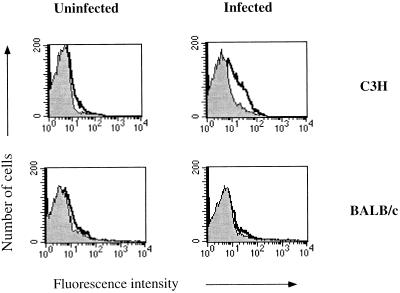
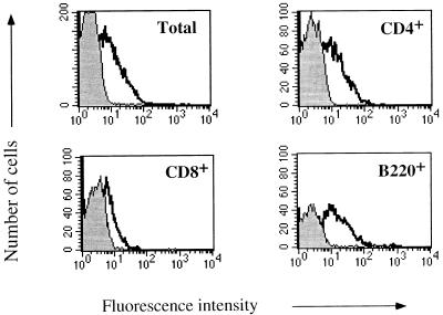
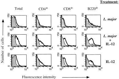
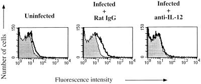
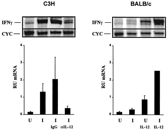
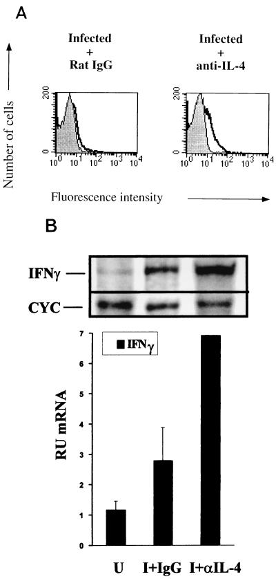
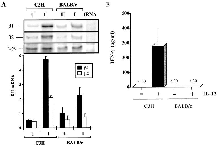
Previous studies have shown the central role of interleukin 12 (IL-12) in the development of resistance to Leishmania major infection in C3H mice. We now show that during the innate immune response the lymph node cells of L. major-infected C3H mice upregulate the IL-12 receptor on CD4(+), CD8(+), and B220(+) cells. An increase in the ability of the lymph node cells to bind IL-12 correlates with 9.3- and 4.6-fold increases in the mRNA expression levels of the IL-12Rbeta1 and -beta2 subunits, respectively. In contrast, BALB/c mice, which are susceptible to L. major infection, have no increase in the ability of the lymph node cells to bind IL-12 and correspondingly smaller increases in the mRNA expression levels of the IL-12Rbeta1 and -beta2 subunits of 2- and 1.5-fold, respectively. Neutralizing IL-4 and the administration of exogenous IL-12 upregulate IL-12R expression in BALB/c mice, while the neutralization of IL-12 in C3H mice blocks increased IL-12 receptor expression. These experiments reveal an important role for the regulation of the IL-12 receptor during the innate immune response after infection of mice with a pathogen.
Figures
Similar articles
-
In BALB/c mice, IL-4 production during the initial phase of infection with Leishmania major is necessary and sufficient to instruct Th2 cell development resulting in progressive disease.J Immunol. 2000 May 1;164(9):4819-25. doi: 10.4049/jimmunol.164.9.4819. J Immunol. 2000. PMID: 10779790
-
IL-4-independent inhibition of IL-12 responsiveness during Leishmania amazonensis infection.J Immunol. 2000 Jul 1;165(1):364-72. doi: 10.4049/jimmunol.165.1.364. J Immunol. 2000. PMID: 10861073
-
The IL-4 rapidly produced in BALB/c mice after infection with Leishmania major down-regulates IL-12 receptor beta 2-chain expression on CD4+ T cells resulting in a state of unresponsiveness to IL-12.J Immunol. 1998 Dec 1;161(11):6156-63. J Immunol. 1998. PMID: 9834101
-
Anti-leishmania effector functions of CD4+ Th1 cells and early events instructing Th2 cell development and susceptibility to Leishmania major in BALB/c mice.Adv Exp Med Biol. 1998;452:53-60. doi: 10.1007/978-1-4615-5355-7_7. Adv Exp Med Biol. 1998. PMID: 9889959 Review.
-
The use of the murine model of infection with Leishmania major to reveal the antagonistic effects that IL-4 can exert on T helper cell development and demonstrate that these opposite effects depend upon the nature of the cells targeted for IL-4 signaling.Pathol Biol (Paris). 2003 Mar;51(2):71-3. doi: 10.1016/s0369-8114(03)00101-9. Pathol Biol (Paris). 2003. PMID: 12801805 Review.
Cited by
-
Cure of progressive murine leishmaniasis: interleukin 4 dominance is abolished by transient CD4(+) T cell depletion and T helper cell type 1-selective cytokine therapy.J Exp Med. 1999 Jun 21;189(12):1895-906. doi: 10.1084/jem.189.12.1895. J Exp Med. 1999. PMID: 10377185 Free PMC article.
-
Beryllium, an adjuvant that promotes gamma interferon production.Infect Immun. 2000 Jul;68(7):4032-9. doi: 10.1128/IAI.68.7.4032-4039.2000. Infect Immun. 2000. PMID: 10858219 Free PMC article.
-
Interleukin-12 as an adjuvant for induction of protective antibody responses.Cytokine. 2010 Oct-Nov;52(1-2):102-7. doi: 10.1016/j.cyto.2010.06.011. Epub 2010 Jul 22. Cytokine. 2010. PMID: 20650650 Free PMC article. Review.
-
Development and regulation of cell-mediated immunity in experimental leishmaniasis.Immunol Res. 2003;27(2-3):489-98. doi: 10.1385/IR:27:2-3:489. Immunol Res. 2003. PMID: 12857992 Review.
-
Effects of CXCL10 on dendritic cell and CD4+ T-cell functions during Leishmania amazonensis infection.Infect Immun. 2008 Jan;76(1):161-9. doi: 10.1128/IAI.00825-07. Epub 2007 Nov 12. Infect Immun. 2008. PMID: 17998308 Free PMC article.
References
-
- Afonso L C C, Scharton T M, Vieira L Q, Wysocka M, Trinchieri G, Scott P. The adjuvant effect of interleukin-12 in a vaccine against Leishmania major. Science. 1994;263:235–237. - PubMed
-
- Chizzonite R, Truitt T, Desai B B, Nunes P, Podlaski F J, Stern A S, Gately M K. IL-12 receptor. I. Characterization of the receptor on phytohemagglutinin-activated human lymphoblasts. J Immunol. 1992;148:3117–3124. - PubMed
-
- Chua A O, Wilkinson V L, Presky D H, Gubler U. Cloning and characterization of a mouse IL-12 receptor-β component. J Immunol. 1995;155:4286–4294. - PubMed
-
- Desai B B, Quinn P M, Wolitzky A G, Mongini P K, Chizzonite R, Gately M K. IL-12 receptor. II. Distribution and regulation of receptor expression. J Immunol. 1992;148:3125–3132. - PubMed
-
- Galbiati F, Rogge L, Guery J, Smiroldo S, Adorini L. Regulation of the IL-12 receptor β2 subunit by soluble antigen and IL-12 in vivo. Eur J Immunol. 1998;28:209–220. - PubMed
Publication types
MeSH terms
Substances
Grants and funding
LinkOut - more resources
Full Text Sources
Research Materials
