

Contribution of virus-specific CD8+ cytotoxic T cells to virus clearance or pathologic manifestations of influenza virus infection in a T cell receptor transgenic mouse model
- PMID: 9670035
- PMCID: PMC2212460
- DOI: 10.1084/jem.188.2.223
Contribution of virus-specific CD8+ cytotoxic T cells to virus clearance or pathologic manifestations of influenza virus infection in a T cell receptor transgenic mouse model
Abstract
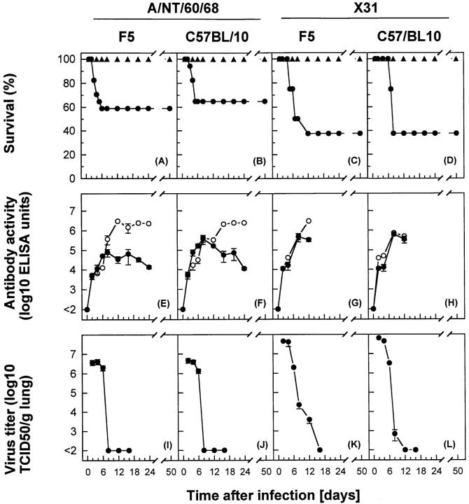
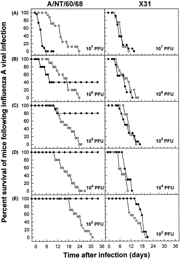
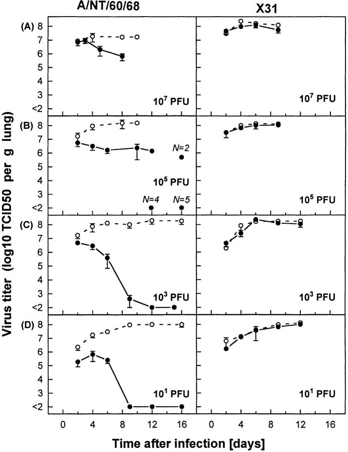
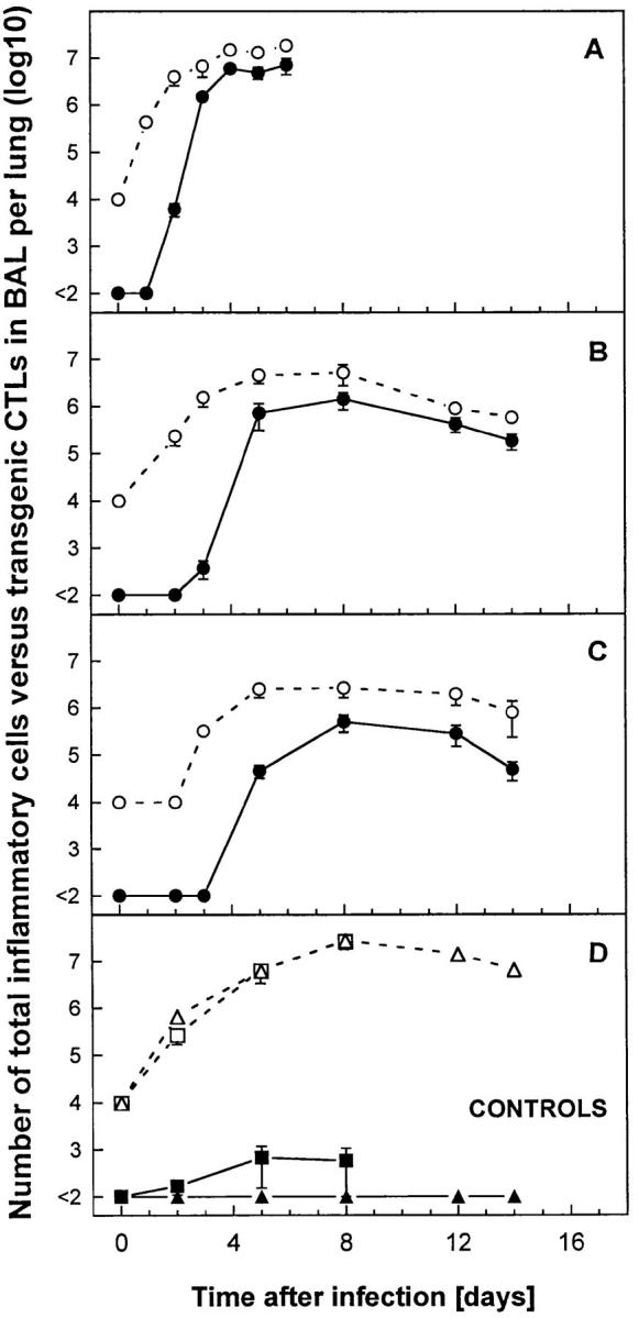
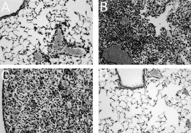
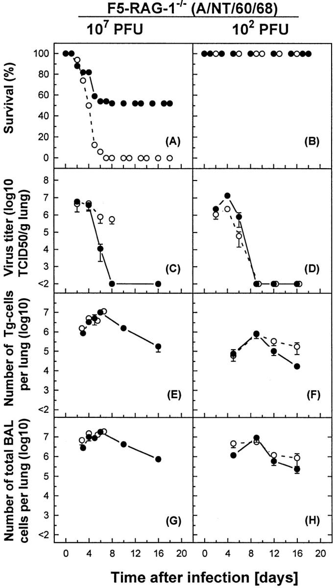
The ability of influenza virus to evade immune surveillance by neutralizing antibodies (Abs) directed against its variable surface antigens provides a challenge to the development of effective vaccines. CD8+ cytotoxic T lymphocytes (CTLs) restricted by class I major histocompatibility complex molecules are important in establishing immunity to influenza virus because they recognize internal viral proteins which are conserved between multiple viral strains. In contrast, protective Abs are strain-specific. However, the precise role of effector CD8+ CTLs in protection from influenza virus infection, critical for understanding disease pathogenesis, has not been well defined. In transgenic mice with a very high frequency of antiinfluenza CTL precursors, but without protective Abs, CD8+ CTLs conferred protection against low dose viral challenge, but exacerbated viral pathology and caused mortality at high viral dose. The data suggest a dual role for CD8+ CTLs against influenza, which may present a challenge to the development of effective CTL vaccines. Effector mechanisms used by CD8+ CTLs in orchestrating clearance of virus and recovery from experimental influenza infection, or potentiation of lethal pathology, are discussed.
Figures
Similar articles
-
A Role of Influenza Virus Exposure History in Determining Pandemic Susceptibility and CD8+ T Cell Responses.J Virol. 2016 Jul 11;90(15):6936-6947. doi: 10.1128/JVI.00349-16. Print 2016 Aug 1. J Virol. 2016. PMID: 27226365 Free PMC article.
-
Cellular mechanisms involved in protection against influenza virus infection in transgenic mice expressing a TCR receptor specific for class II hemagglutinin peptide in CD4+ and CD8+ T cells.J Immunol. 1998 May 1;160(9):4500-7. J Immunol. 1998. PMID: 9574556
-
Deficiency of the NOD-Like Receptor NLRC5 Results in Decreased CD8+ T Cell Function and Impaired Viral Clearance.J Virol. 2017 Aug 10;91(17):e00377-17. doi: 10.1128/JVI.00377-17. Print 2017 Sep 1. J Virol. 2017. PMID: 28615208 Free PMC article.
-
Defense mechanisms against influenza virus infection in the respiratory tract mucosa.Jpn J Infect Dis. 2004 Dec;57(6):236-47. Jpn J Infect Dis. 2004. PMID: 15623947 Review.
-
Virus-specific T cells as correlate of (cross-)protective immunity against influenza.Vaccine. 2015 Jan 15;33(4):500-6. doi: 10.1016/j.vaccine.2014.11.054. Epub 2014 Dec 9. Vaccine. 2015. PMID: 25498210 Review.
Cited by
-
Partial depletion of natural CD4⁺CD25⁺ regulatory T cells with anti-CD25 antibody does not alter the course of acute influenza A virus infection.PLoS One. 2011;6(11):e27849. doi: 10.1371/journal.pone.0027849. Epub 2011 Nov 18. PLoS One. 2011. PMID: 22125630 Free PMC article.
-
Harnessing alveolar macrophages for sustained mucosal T-cell recall confers long-term protection to mice against lethal influenza challenge without clinical disease.Mucosal Immunol. 2014 Jan;7(1):89-100. doi: 10.1038/mi.2013.27. Epub 2013 May 29. Mucosal Immunol. 2014. PMID: 23715172
-
Cell-mediated protection in influenza infection.Emerg Infect Dis. 2006 Jan;12(1):48-54. doi: 10.3201/eid1201.051237. Emerg Infect Dis. 2006. PMID: 16494717 Free PMC article. Review.
-
Detrimental contribution of the Toll-like receptor (TLR)3 to influenza A virus-induced acute pneumonia.PLoS Pathog. 2006 Jun;2(6):e53. doi: 10.1371/journal.ppat.0020053. Epub 2006 Jun 9. PLoS Pathog. 2006. PMID: 16789835 Free PMC article.
-
Aryl hydrocarbon receptor activation reduces dendritic cell function during influenza virus infection.Toxicol Sci. 2010 Aug;116(2):514-22. doi: 10.1093/toxsci/kfq153. Epub 2010 May 23. Toxicol Sci. 2010. PMID: 20498003 Free PMC article.
References
-
- Palese P, Young JF. Variation of influenza A, B, and C viruses. Science. 1982;215:1468–1474. - PubMed
-
- Webster, R.G., W.G. Laver, and G.M. Air. 1983. Antigenic variation among type A influenza viruses. In Genetics of Influenza Viruses. P. Palese and D.W. Kingsbury, editors. Springer-Verlag, Vienna/New York. 127–168.
-
- Gorman OT, Bean WJ, Webster RG. Evolutionary processes in influenza viruses: divergence, rapid evolution, and stasis. Curr Top Microbiol Immunol. 1992;176:75–97. - PubMed
-
- Yewdell JW, Webster RG, Gerhard W. Antigen variation in three distinct determinants of an influenza A haemagglutinin molecule. Nature. 1979;279:246–248. - PubMed
-
- Townsend ARM, Bodmer H. Antigen recognition by class I-restricted T lymphocytes. Annu Rev Immunol. 1989;7:601–624. - PubMed
Publication types
MeSH terms
Substances
LinkOut - more resources
Full Text Sources
Other Literature Sources
Molecular Biology Databases
Research Materials
