. 1998 Aug 1;510 ( Pt 3)(Pt 3):721-34.
doi: 10.1111/j.1469-7793.1998.721bj.x.

Kainate induces an intracellular Na+-activated current in cultured embryonic rat hippocampal neurones

Affiliations

Kainate induces an intracellular Na+-activated current in cultured embryonic rat hippocampal neurones

Q Y Liu et al. J Physiol. .

Abstract

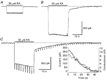
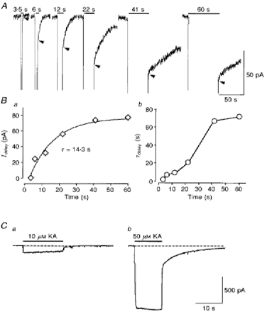
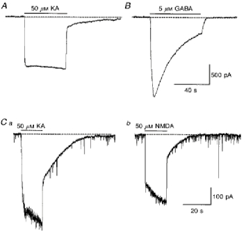
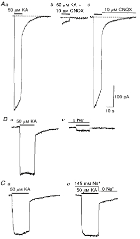
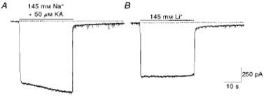
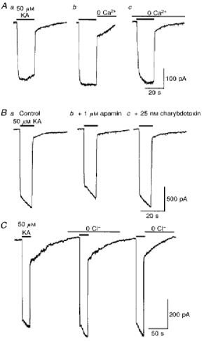
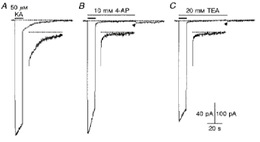
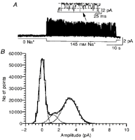
1. In embryonic rat hippocampal neurones cultured for < 3 days, kainate induced an inward current at negative potentials that recovered to baseline levels immediately upon termination of agonist application. However, in neurones cultured for longer, the kainate-induced current was often followed by a long-lasting inward current that slowly recovered to baseline levels. The amplitude of the delayed current (Idelay) triggered by kainate was positively related both to the duration of application at constant agonist concentration and to concentration at constant application duration. 2. Idelay could last for several minutes and was accompanied by a conductance increase, which closely paralleled current amplitude. Depression of the kainate-induced current response at receptor level with CNQX or at ionic level with Na+-free solution eliminated Idelay. However, when applied during Idelay neither CNQX nor Na+-free solution had any effect on Idelay. Li+ effected the same response as Na+ in mediating kainate-induced Idelay. 3. GABA-activated Cl- current, which was associated with the same amount of inwardly directed charge flow at the same potential as that induced by kainate, did not trigger a long-lasting delayed current. 4. Idelay depended on the existence of extracellular K+ and its amplitude increased with the increase in K+ concentration. Neither applying Cl-- or Ca2+-free solutions nor increasing intracellular Ca2+ buffering speed and capacity altered Idelay. Exposure to the specific KCa channel blockers apamin and charybdotoxin also failed to alter Idelay. However, Idelay could be blocked by Cs+, Ba2+ and high concentrations of 4-aminopyridine (4-AP) and TEA. 5. Inside-out excised patch-clamp recordings revealed that low density or highly clustered Na+-activated K+ channels were expressed in the cell bodies of cultured embryonic rat hippocampal neurones. These could be the elementary channels underlying Idelay.


Figures

Figure 1
Figure 1. Kainate induced an inwardly directed slowly decaying current (Idelay) in embryonic rat hippocampal neurones cultured for more than 1 week
A neurone cultured for 3 days did not exhibit any residual trace of current following cessation of the kainate application (A) while another neurone grown in culture for 10 days exhibited significantly more kainate-elicited current, which was immediately followed by an inwardly directed, slowly decaying current (Idelay, B). The tail-like current was accompanied by an increase in membrane conductance as revealed by the current response to intermittent 10 mV hyperpolarizing commands in another neurone cultured for 12 days (C). Inset in C is a plot of Idelay amplitude (□) and the corresponding increase in membrane conductance (^) after termination of the kainate application. Membrane potential was clamped at -80 mV. KA, kainate. Here and in subsequent figures the dashed horizontal line shows the baseline holding current.
Figure 2
Figure 2. The amplitude and duration of Idelay depended on the duration of kainate application and kainate concentration
A, the baseline before and immediately after kainate application is amplified to illustrate the duration-dependent appearance of Idelay. While 3 s application of kainate (not shown) did not induce Idelay, applications lasting longer than 6 s triggered Idelay with amplitude and duration increasing as the application duration increased. Arrowheads indicate the peak amplitudes of Idelay. Ba, plot of Idelay amplitude against duration of the kainate application, which can be best fitted with an exponential function (τ, 14.3 s). b, the time constants of Idelaydelay) were related to the duration of kainate application in a bimodal manner. C, in another neurone, the current evoked by 10 μm kainate (a) decayed to baseline level immediately after the termination of application but the current induced by 50 μm kainate (b) was followed by Idelay.
Figure 3
Figure 3. The delayed current was not caused by non-specific inwardly directed charge movement
A, inward charge movement evoked by 50 μm kainate was followed by Idelay. B, in the same cell, a comparable accumulation of inward charge movement (6.3 × 10⁻⁹ C compared with 6.2 × 10⁻⁹ C for kainate) evoked by 5 μm GABA was not followed by any detectable Idelay. In another neurone, currents induced by both kainate (50 μm, Ca) and NMDA (50 μm, Cb) were followed by Idelay. (Brief downward transients are synaptic-like GABAergic currents.)
Figure 4
Figure 4. Idelay depended on Na+ influx
A, under control conditions, application of 50 μm kainate to a neurone cultured for 14 days (a) induced a sizable inward current which was followed by Idelay; when kainate was applied with CNQX (10 μm, b) the majority of kainate-evoked current was blocked and Idelay disappeared; however, when CNQX was applied after termination of the kainate application (c) it had no effect on Idelay. B, in another neurone, suppressing the majority of kainate current (a) by replacing extracellular Na+ with non-permeable N-methyl-D-glucamine (b) prevented induction of Idelay. Ca and b, application of Na+-free saline after termination of the kainate application had little, if any effect on Idelay.
Figure 5
Figure 5. Li+ effected the same response as Na+ in kainate-induced Idelay
A, in control conditions with 145 mm extracellular Na+, application of 50 μm kainate evoked an inward current followed by Idelay in a neurone cultured for 10 days. B, in the presence of 145 mm Li+ (and 0 Na+), kainate evoked a slightly lower amplitude current response while Idelay remained comparable in amplitude and duration to that recorded under control (145 mm Na+) conditions.
Figure 6
Figure 6. The induction and maintenance of Idelay did not depend on intracellular Ca2+ or extracellular Ca2+ and Cl
A, Ca2+-free solution applied after termination of the kainate application (b) or throughout the recording period (c) had no obvious effect on Idelay when compared with that recorded under control conditions (a). B, compared with control (a) Ca2+-activated K+ (KCa) channel blockers apamin (b) and charybdotoxin (c) had no effects on Idelay, indicating that small- and large-conductance KCa channels are not involved in the generation of Idelay. C, the amplitude and appearance of Idelay recorded in Cl-free solution applied immediately after termination of the kainate application or during the recording period were comparable to that recorded under control conditions.
Figure 7
Figure 7. Idelay depended on extracellular K+
A, in the presence of 5.4 mm potassium, kainate induced a sizable Idelay (a), which became outward when kainate was applied in Ko+-free solution (b). Upon returning to K+-containing solution, an inwardly directed, slowly decaying Idelay appeared (b). B, in another neurone, the amplitude of Idelay was severalfold larger in 15 mm K+ than in 5.4 mm K+ solution (a). If kainate was applied in the presence of 5 mm Cs+, no observable Idelay could be detected (b). However, removal of Cs+ from the solution immediately revealed a delayed current. Brief applications of a 15 mm K+ solution induced a large inwardly directed, slowly decaying current shortly after removal of Cs+ but a noticeably smaller non-decaying inward current later. The non-decaying current probably reflects the contribution of the inward-rectifier K+ current.
Figure 8
Figure 8. Current-voltage relations of Idelay
In neurones showing clear Idelay, 120 mV ramp protocols (1 s, -80 to +40 mV) were applied 1 s after the termination of kainate application. The baseline currents obtained before kainate application were subtracted and the putative Idelay plotted against membrane potentials. The chord conductance was about 130 pS. A negative slope appeared at positive membrane potentials. Data were obtained from 7 cells and were plotted as means ±s.e.m.
Figure 9
Figure 9. Extracellular Cs+ blocked kainate-induced Idelay
A, Cs+ (5 mm) added to the extracellular solution blocked Idelay which is present under control conditions (a), when applied immediately after kainate (c) or during the whole kainate application-termination period (b). B, in another neurone, 50 μm kainate induced a delayed current response in the absence of 5 mm Cs+ (a) but not in the presence of Cs+ (b). Upon removal of Cs+, an inwardly directed, slowly decaying Idelay appears immediately (b). Another inorganic K+ channel blocker, Ba2+ (5 mm), also completely blocks Idelay when applied immediately after the termination of kainate application (C).
Figure 10
Figure 10. Blocking of Idelay by TEA and 4-AP
Kainate (50 μm) induced a sizable Idelay under control conditions (A) but not in the presence of 10 mm 4-AP (B) or 20 mm TEA (C) (vertical calibration, 100 pA). Note that an inwardly directed, slowly decaying Idelay appears upon removal of 4-AP and TEA (arrowheads). Insets show Idelay at higher gains (vertical calibration, 40 pA).
Figure 11
Figure 11. Na+-activated K+ single-channel currents in cultured embryonic rat hippocampal neurones
A, Na+ (145 mm) reversibly induced elementary-sized, all-or-none transitions in current in an inside-out membrane patch excised from the soma of a neurone cultured for 14 days. At higher time resolution, two preferred current levels are evident (inset). B, all-points histogram shows two preferred current levels of 1.3 and 3.3 pA. The potential across the membrane patch was held at 0 mV, the equilibrium potential for Cl under these recording conditions. The continuous line in the inset indicates the closed state of the channel and the dotted lines open states. Upward deflection represents inward (to the cytoplasmic side of the membrane) flow of currents.
Figure 12
Figure 12. Induction of Idelay by high frequency activation of Na + current
Membrane potential was clamped at -80 mV. A train of 12 ms voltage clamp steps to 0 mV was applied for 30 s at 20 Hz under control conditions (A) and in the presence of 10 mm Cs+ (B). Insets show Idelay at higher gain (left calibration).

Similar articles

Cited by

References

    1. Bader CR, Bernheim L, Bertrand D. Sodium activated potassium current in cultured avian neurons. Nature. 1985;317:540–542. - PubMed
    1. Choi DW. Glutamate neurotoxicity and diseases of the nervous system. Neuron. 1988;1:623–634. - PubMed
    1. Collingridge GL, Lester RAS. Excitatory amino acid receptors in the vertebrate central nervous system. Pharmacological Reviews. 1989;40:145–195. - PubMed
    1. Colquhoun D, Jonas P, Sakmann B. Action of brief pulses of glutamate on AMPA/kainate receptors in patches from different neurones of rat hippocampal slices. The Journal of Physiology. 1992;458:261–287. - PMC - PubMed
    1. Dale N. A large, sustained Na+- and voltage-dependent K+ current in spinal neurons of the frog embryo. The Journal of Physiology. 1993;462:349–372. - PMC - PubMed

MeSH terms

LinkOut - more resources