

doi: 10.1128/JVI.72.8.6932-6936.1998.
Apoptosis in feline panleukopenia virus-infected lymphocytes
Affiliations
- PMID: 9658149
- PMCID: PMC109909
- DOI: 10.1128/JVI.72.8.6932-6936.1998
Item in Clipboard
Apoptosis in feline panleukopenia virus-infected lymphocytes
J Virol.
1998 Aug.
Abstract
Feline panleukopenia virus (FPLV) was shown to induce apoptosis to feline lymphoid cells and to reduce the expression of interleukin-2 receptor alpha on the cells. FPLV-induced apoptosis might be a key element in the pathophysiology of atrophy of lymphoid tissues associated with feline panleukopenia caused by FPLV.
Figures
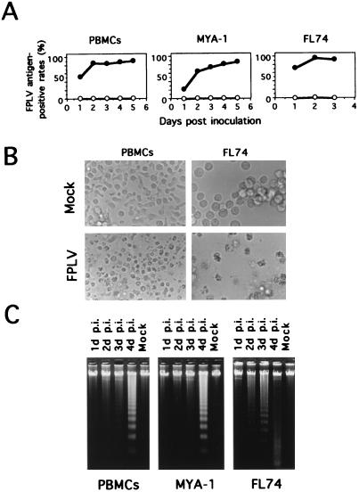
Susceptibilities of feline cells to FPLV. (A) Growth curves of strain TU1 in feline lymphoid cells. Feline cells were inoculated with TU1 and collected at the indicated times. FPLV antigen-positive rates in the cells were measured with an anti-FPLV VP2 MAb. Symbols: ○, mock-inoculated cells; •, TU1-infected cells. (B) CPEs observed in FPLV-inoculated feline PBMCs and FL74 cells. FPLV-inoculated or mock-inoculated feline PBMCs and FL74 cells were harvested at 2 days and 1 day p.i., respectively. (C) Electrophoresis of total cellular DNA. Cellular DNA was extracted from cultures of inoculated or mock-inoculated PBMCs and MYA-1 and FL74 cells. Lanes 1, FPLV-inoculated cells at 1 day p.i.; lanes 2, 2 days p.i.; lanes 3, 3 days p.i.; lanes 4, 4 days p.i.; Mock, mock-inoculated cells at 4 days p.i. The extracted DNAs were analyzed by 1.7% agarose gel electrophoresis.
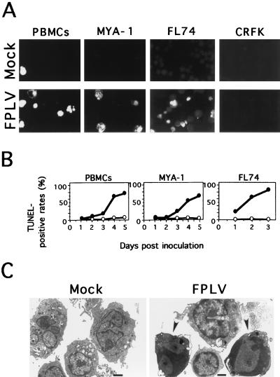
Morphologies of FPLV-infected lymphocytes. (A) Detection of DNA strand breaks in FPLV-inoculated cells by the TUNEL assay. FPLV-inoculated or mock-inoculated PBMCs and MYA-1, FL74, and CRFK cells were harvested at 2, 2, 1, and 8 days p.i., respectively (B) Flow cytometric analysis of TUNEL-positive rates in the inoculated cells. The inoculated cells were collected at the indicated times and analyzed by FACScan. Symbols: ○, mock-inoculated cells; •, TU1-inoculated cells. (C) Electron microscopic analysis of infected PBMCs. FPLV-inoculated or mock-inoculated PBMCs were harvested at 2 days p.i. Arrowheads indicate cells showing chromatin condensation along the inner aspect of the nuclear membrane. Bars, 1 μm.
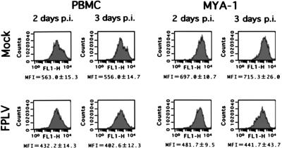
Effects of FPLV infection on feline IL-2Rα of inoculated feline lymphoid cells. Flow cytometric analysis was performed with FPLV-inoculated feline PBMCs and MYA-1 cells at 2 or 3 days p.i. Three independent experiments were performed, and the averages and standard deviations of mean fluorescence intensities (MFI) are presented. The results of flow cytometric analyses are representative of one of the three independent experiments.
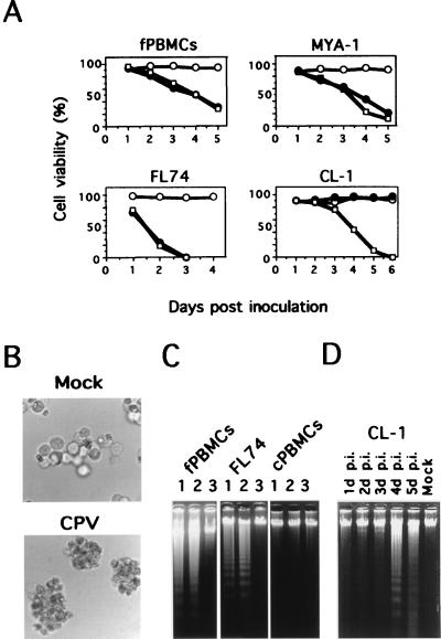
Susceptibilities of feline and canine cells to CPV. (A) Cell viabilities of FPLV- or CPV-inoculated feline and canine lymphoid cells. Feline and canine cells were inoculated with strain TU1 or strain Cp49 and collected at the indicated times. Symbols: ○, mock-inoculated cells; •, TU1-inoculated cells; □, Cp49-inoculated cells. (B) CPEs observed in CPV-inoculated CL-1 cells. CPV-inoculated or mock-inoculated CL-1 cells were harvested at 5 days p.i. (C) Electrophoresis of total cellular DNA. Cellular DNA was extracted from cultures of FPLV-inoculated (lanes 1), CPV-inoculated (lanes 2), or mock-inoculated (lanes 3) feline PBMCs (fPBMCs), FL74 cells, and canine PBMCs (cPBMCs). The inoculated fPBMCs, FL74 cells and cPBMCs were harvested at 4, 2, and 4 days p.i., respectively. The extracted DNAs were analyzed by 1.7% agarose gel electrophoresis. (D) Electrophoresis of total cellular DNA of the CPV-inoculated CL-1 cells. Cellular DNA was extracted from cultures of inoculated or mock-inoculated CL-1 cells. Lane 1, CPV-inoculated cells at 1 day p.i.; lane 2, 2 days p.i.; lane 3, 3 days p.i.; lane 4, 4 days p.i.; lane 5, 5 days p.i.; Mock, mock-inoculated cells at 4 days p.i. The extracted DNAs were analyzed by 1.7% agarose gel electrophoresis.
Similar articles
-
Panleukopenia-like syndrome of FeLV caused by co-infection with FeLV and feline panleukopenia virus.Vet Immunol Immunopathol. 1995 May;46(1-2):21-33. doi: 10.1016/0165-2427(94)07003-p. Vet Immunol Immunopathol. 1995. PMID: 7618258
-
Characterisation of cross-reactivity of virus neutralising antibodies induced by feline panleukopenia virus and canine parvoviruses.Res Vet Sci. 2001 Dec;71(3):219-22. doi: 10.1053/rvsc.2001.0492. Res Vet Sci. 2001. PMID: 11798298
-
Regional adaptations and parallel mutations in Feline panleukopenia virus strains from China revealed by nearly-full length genome analysis.PLoS One. 2020 Jan 16;15(1):e0227705. doi: 10.1371/journal.pone.0227705. eCollection 2020. PLoS One. 2020. PMID: 31945103 Free PMC article.
-
Feline parvovirus infection and associated diseases.Vet J. 2014 Aug;201(2):150-5. doi: 10.1016/j.tvjl.2014.05.027. Epub 2014 May 22. Vet J. 2014. PMID: 24923754 Review.
-
Pathogenesis of feline panleukopenia virus and canine parvovirus.Baillieres Clin Haematol. 1995 Mar;8(1):57-71. doi: 10.1016/s0950-3536(05)80232-x. Baillieres Clin Haematol. 1995. PMID: 7663051 Free PMC article. Review.
Cited by
-
Human Parvovirus Infection of Human Airway Epithelia Induces Pyroptotic Cell Death by Inhibiting Apoptosis.J Virol. 2017 Nov 30;91(24):e01533-17. doi: 10.1128/JVI.01533-17. Print 2017 Dec 15. J Virol. 2017. PMID: 29021400 Free PMC article.
-
Nuclear envelope disruption involving host caspases plays a role in the parvovirus replication cycle.J Virol. 2011 May;85(10):4863-74. doi: 10.1128/JVI.01999-10. Epub 2011 Mar 2. J Virol. 2011. PMID: 21367902 Free PMC article.
-
True versus false parasite interactions: a robust method to take risk factors into account and its application to feline viruses.PLoS One. 2012;7(1):e29618. doi: 10.1371/journal.pone.0029618. Epub 2012 Jan 3. PLoS One. 2012. PMID: 22235312 Free PMC article.
-
Feline host range of canine parvovirus: recent emergence of new antigenic types in cats.Emerg Infect Dis. 2002 Apr;8(4):341-6. doi: 10.3201/eid0804.010228. Emerg Infect Dis. 2002. PMID: 11971764 Free PMC article.
-
Apoptosis in murine norovirus-infected RAW264.7 cells is associated with downregulation of survivin.J Virol. 2009 Apr;83(8):3647-56. doi: 10.1128/JVI.02028-08. Epub 2009 Feb 11. J Virol. 2009. PMID: 19211757 Free PMC article.
References
-
- Ackley C D, Cooper M D. Characterization of a feline T-cell-specific monoclonal antibody reactive with a CD5-like molecule. Am J Vet Res. 1992;53:466–471. - PubMed
-
- Azetaka M, Hirasawa T, Konishi S, Ogata M. Studies on canine parvovirus isolation, experimental infection and serologic survey. Jpn J Vet Sci. 1981;43:243–255. - PubMed
-
- Crandell R A, Fabricant C G, Nelson-Rees W A. Development, characterization and viral susceptibility of a feline (Felis catus) renal cell line (CRFK) In Vitro (Rockville) 1973;9:176–185. - PubMed
-
- Goto H, Hirano T, Uchida E, Watanabe K, Shinagawa M, Ichijo S, Shimizu K. Comparative studies of physicochemical and biological properties between canine parvovirus and feline panleukopenia virus. Jpn J Vet Sci. 1984;46:519–526. - PubMed
Publication types
MeSH terms
LinkOut - more resources
Full Text Sources
