. 1998 Aug;72(8):6581-91.
doi: 10.1128/JVI.72.8.6581-6591.1998.

The disruption of ND10 during herpes simplex virus infection correlates with the Vmw110- and proteasome-dependent loss of several PML isoforms

Affiliations

The disruption of ND10 during herpes simplex virus infection correlates with the Vmw110- and proteasome-dependent loss of several PML isoforms

R D Everett et al. J Virol. 1998 Aug.

Abstract

The small nuclear structures known as ND10 or PML nuclear bodies have been implicated in a variety of cellular processes including response to stress and interferons, oncogenesis, and viral infection, but little is known about their biochemical properties. Recently, a ubiquitin-specific protease enzyme (named HAUSP) and a ubiquitin-homology family protein (PIC1) have been found associated with ND10. HAUSP binds strongly to Vmw110, a herpesvirus regulatory protein which has the ability to disrupt ND10, while PIC1 was identified as a protein which interacts with PML, the prototype ND10 protein. We have investigated the role of ubiquitin-related pathways in the mechanism of ND10 disruption by Vmw110 and the effect of virus infection on PML stability. The results show that the disruption of ND10 during virus infection correlates with the loss of several PML isoforms and this process is dependent on active proteasomes. The PML isoforms that are most sensitive to virus infection correspond closely to those which have recently been identified as being covalently conjugated to PIC1. In addition, a large number of PIC1-protein conjugates can be detected following transfection of a PIC1 expression plasmid, and many of these are also eliminated in a Vmw110-dependent manner during virus infection. These observations provide a biochemical mechanism to explain the observed effects of Vmw110 on ND10 and suggest a simple yet powerful mechanism by which Vmw110 might function during virus infection.


Figures
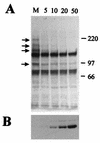
FIG. 1
FIG. 1
HSV-1 infection stimulates the loss of several high-molecular-weight PML isoforms. (A) HEp-2 cells were infected with HSV-1 at multiplicities of 5, 10, 20, and 50 PFU per cell as indicated and harvested 4 h later. The samples were analyzed by Western blotting using anti-PML MAb 5E10 in comparison with an uninfected control (M). Positions of molecular weight markers are indicated on the right in kilodaltons, and PML isoforms most sensitive to elimination are indicated by arrows. (B) The same filter was reprobed with MAb Z1F11 to compare the efficiency of virus infection by detection of the early viral protein UL42.
FIG. 2
FIG. 2
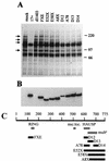
Vmw110 is absolutely required for the virally induced loss of high-molecular-weight PML isoforms. (A) HEp-2 cells were infected with viruses as indicated (as described in the text) at a multiplicity of 20 PFU per cell and harvested 4 h later. The samples were analyzed by western blotting using anti-PML MAb 5E10 in comparison with an uninfected control (mock). Positions of molecular weight markers are indicated on the right in kilodaltons, and PML isoforms most sensitive to elimination are indicated by arrows. (B) The same filter was reprobed with MAb 11060 to compare the efficiency of the virus infections by detection of Vmw110. (C) A schematic representation of the viruses used in this experiment. The 775-codon open reading frame of Vmw110 is depicted, with the locations of the RING finger, nuclear localization signal (nuc.loc.), HAUSP binding region, and sequences implicated in multimerization (multn) indicated below. The solid bars indicate the extents of the deleted sequences in the mutant viruses, as labeled.
FIG. 3
FIG. 3
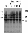
Inhibitors of proteasome activity eliminate the virally induced loss of the high-molecular-weight PML isoforms. HEp-2 cells were infected (I) or left uninfected (U) with HSV-1 strain 17+ at a multiplicity of 20 PFU per cell in the absence of drug or the presence of lactacystin lactone (lact.; 5 μM) or MG132 (5 μM) as indicated. The cells were harvested 4 h later, and the samples were analyzed by Western blotting using anti-PML MAb 5E10. Positions of molecular weight markers are indicated on the right in kilodaltons, and PML isoforms most sensitive to elimination are indicated by arrows.
FIG. 4
FIG. 4
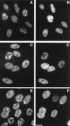
Proteasome inhibitors inhibit the disruption of ND10 during HSV-1
FIG. 5
FIG. 5
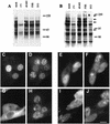
Correlation of ND10 disruption and PML isoform loss in different cell lines. BHK cells (A) or HFL cells (B) were infected with the indicated viruses at multiplicities of 20 PFU per cell, and total cell proteins were harvested 4 h later. The proteins were separated by SDS-polyacrylamide gel electrophoresis and PML was detected by Western blotting using MAb 5E10. Positions of molecular weight markers are indicated in kilodaltons, and the PML isoforms in HFL cells equivalent to those in HEp-2 cells indicated in Fig. 1 and 2 are marked by arrows in panel B. A similar but not identical pattern of isoform bands is present in BHK cells (A). In parallel, BHK cells (C, D, G, and H) or HFL cells (E, F, I, and J) were infected with wild-type virus (C to F) or mutant D12 (G to J) and stained for Vmw110 and PML. The paired panels (for example, C and D) show Vmw110 staining on the left and PML staining on the right. The bar indicates 5 μm.
FIG. 6
FIG. 6
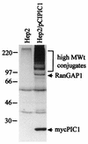
High-level expression of an epitope-tagged form of PIC1 and formation of PIC1-conjugated proteins. HEp-2 cells were electroporated with plasmid pCIPIC1 and harvested 40 h later. The samples were analyzed by Western blotting using MAb 9E10 in comparison with control untreated cells. Positions of molecular weight markers, the monomeric Myc-tagged PIC1, a band likely to represent PIC1 conjugated to RanGAP1, and a multitude of other high-molecular-weight PIC1 conjugate proteins are indicated.
FIG. 7
FIG. 7
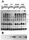
HSV-1 infection leads to the loss of several high-molecular-weight PIC1-conjugated proteins. HEp-2 cells were electroporated with plasmid pCIPIC1 and the following day either left uninfected (mock) or infected at a multiplicity of 20 PFU per cell with wild-type virus (17+), Vmw110 deletion mutant dl1403, or Vmw110 RING finger mutant FXE. Samples were taken at 4, 8, and 22 h after infection, loaded from left to right in each set of three lanes, and analyzed by Western blotting using the anti-Myc tag MAb 9E10 (A) and anti-UL42 MAb Z1F11 (B) to control for infection. The leftmost lane contains a sample from untreated HEp-2 cells. Positions of the 220-, 97-, and 66-kDa molecular weight markers are shown to the right in panel A.
FIG. 8
FIG. 8
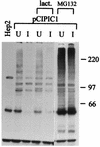
Proteasome inhibitor MG132 inhibits the virus-induced loss of high-molecular-weight PIC1 conjugates. HEp-2 cells were electroporated with plasmid pCIPIC1 and the following day either left uninfected (U) or infected at a multiplicity of 50 PFU per cell with wild-type virus (I) in the presence of 10 μM lactacystin lactone (lact.) or 5 μM MG132 as marked. Samples were taken 22 h after infection and analyzed by Western blotting using MAb 9E10. The positions of the molecular weight markers are shown in kilodaltons on the right.
FIG. 9
FIG. 9
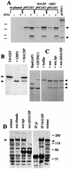
Evidence that HAUSP does not cleave PIC1 conjugates. (A) Bacterial strains were established and enzyme activity was induced as described in Materials and Methods section. The far right-hand lane contains proteins expressed by pCIPIC1 in HEp-2 cells (Fig. 6). The arrows indicate the substrate band (upper arrow) and the Myc-tagged PIC1 protein with the precursor C terminus (i.e., 4 residues longer than the predicted specific cleavage product) (lower arrow). Induction of HAUSP and UBP2 activities has no effect on the amount of the PIC1-GST fusion protein substrate, and there is no evidence for a specific cleavage product. A dominant breakdown product appears as readily in the control as in the HAUSP- and UBP2-expressing cells. Positions of the 46-, 30-, 21-, and 14-kDa molecular weight markers are indicated on the left. (B) Cleavage of Ub-GST by HAUSP and UBP2. An experiment analogous to that in panel A was conducted except that the substrate was Ub-GST expressed by plasmid pRB307. Only the IPTG-induced lanes are shown. The arrows on the left indicate the Ub-GST substrate and the GST product. Cleavage is complete in bacteria carrying UBP2. The partial cleavage in bacteria carrying HAUSP is probably due to the toxicity of this protein in bacteria, leading to instability of the expression plasmid. (C) Conjugation of GSP-PIC1 to RanGAP1 and assay of isopeptidase activity on the purified product. The left-most lane shows 35S-labeled RanGAP1 synthesized in reticulocyte lysates, which appears as a 65-kDa unmodified protein and an 88-kDa endogenous PIC1-conjugated form (49). In the presence of excess GST-PIC1, RanGAP1 is preferentially conjugated with GST-PIC1 to produce a 120-kDa protein. The right-hand four lanes show the 120-kDa substrate (purified on glutathione beads) incubated with buffer, untreated egg extract (ext.), mock-depleted egg extract, or HAUSP-depleted egg extract, as described in Materials and Methods. The arrows on the right indicate the 120-kDa GST-PIC1-RanGAP1 substrate, the 88-kDa RanGAP1-endogenous PIC1 conjugate (which is formed by reconjugation of released RanGAP1 with endogenous PIC1), and the 65-kDa free form of RanGAP1. (D) Depletion of Xenopus HAUSP from egg extracts. The left-hand four lanes show proteins detected by Western blotting using anti-HAUSP r201 serum in untreated egg extracts, in extracts treated with Sepharose beads, and in extracts treated with beads charged with preimmune (pi) r201 antibodies and immune r201 antibodies, respectively. The extracts analyzed in the latter two lanes were used for the incubations in the rightmost two lanes in panel C. On the right, the proteins in the immunoprecipitate (IP) pellets obtained with preimmune and immune r201 sera are shown. The arrows indicate the position of the approximately 130-kDa Xenopus HAUSP homolog.

Similar articles

Cited by

References

    1. Ahn J-H, Hayward G S. The major immediate-early proteins IE1 and IE2 of human cytomegalovirus colocalize with and disrupt PML-associated nuclear bodies at very early times in infected permissive cells. J Virol. 1997;71:4599–4613. - PMC - PubMed
    1. Ascoli C A, Maul G G. Identification of a novel nuclear domain. J Cell Biol. 1991;112:785–795. - PMC - PubMed
    1. Boddy M N, Howe K, Etkin L D, Solomon E, Freemont P S. PIC1, a novel ubiquitin-like protein which interacts with the PML component of a multiprotein complex that is disrupted in acute promyelocytic leukaemia. Oncogene. 1996;13:971–982. - PubMed
    1. Boddy M N, Duprez E, Borden K L B, Freemont P S. Surface residue mutations of the PML RING finger domain alter the formation of nuclear-matrix-associated PML bodies. J Cell Sci. 1997;110:2197–2205. - PubMed
    1. Cai W, Schaffer P A. A cellular function can enhance gene expression and plating efficiency of a mutant defective in the gene for ICP0, a transactivating protein of herpes simplex virus type 1. J Virol. 1991;65:4078–4090. - PMC - PubMed

Publication types

MeSH terms

Substances