

Persistent inhibition of cell respiration by nitric oxide: crucial role of S-nitrosylation of mitochondrial complex I and protective action of glutathione
- PMID: 9636201
- PMCID: PMC22706
- DOI: 10.1073/pnas.95.13.7631
Persistent inhibition of cell respiration by nitric oxide: crucial role of S-nitrosylation of mitochondrial complex I and protective action of glutathione
Abstract
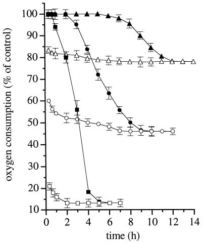
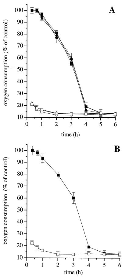
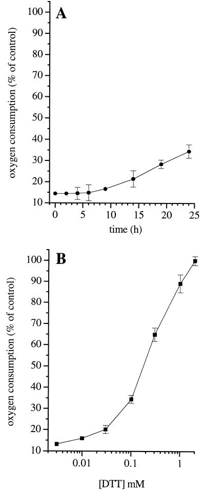
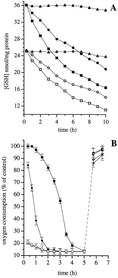
Both reversible and irreversible inhibition of mitochondrial respiration have been reported following the generation of nitric oxide (NO) by cells. Using J774 cells, we have studied the effect of long-term exposure to NO on different enzymes of the respiratory chain. Our results show that, although NO inhibits complex IV in a way that is always reversible, prolonged exposure to NO results in a gradual and persistent inhibition of complex I that is concomitant with a reduction in the intracellular concentration of reduced glutathione. This inhibition appears to result from S-nitrosylation of critical thiols in the enzyme complex because it can be immediately reversed by exposing the cells to high intensity light or by replenishment of intracellular reduced glutathione. Furthermore, decreasing the concentration of reduced glutathione accelerates the process of persistent inhibition. Our results suggest that, although NO may regulate cell respiration physiologically by its action on complex IV, long-term exposure to NO leads to persistent inhibition of complex I and potentially to cell pathology.
Figures
Similar articles
-
Nitric oxide and cell respiration: physiology and pathology.Verh K Acad Geneeskd Belg. 2000;62(3):171-9; discussion 179-81. Verh K Acad Geneeskd Belg. 2000. PMID: 10905118 Review.
-
Oxidative stress and S-nitrosylation of proteins in cells.Br J Pharmacol. 2000 Mar;129(5):953-60. doi: 10.1038/sj.bjp.0703147. Br J Pharmacol. 2000. PMID: 10696095 Free PMC article.
-
Increased sensitivity of mitochondrial respiration to inhibition by nitric oxide in cardiac hypertrophy.J Mol Cell Cardiol. 2001 Jan;33(1):69-82. doi: 10.1006/jmcc.2000.1276. J Mol Cell Cardiol. 2001. PMID: 11133224
-
Continuous exposure to high concentrations of nitric oxide leads to persistent inhibition of oxygen consumption by J774 cells as well as extraction of oxygen by the extracellular medium.Biochem J. 2000 Mar 1;346 Pt 2(Pt 2):407-12. Biochem J. 2000. PMID: 10677360 Free PMC article.
-
Inhibition of mitochondrial respiratory complex I by nitric oxide, peroxynitrite and S-nitrosothiols.Biochim Biophys Acta. 2004 Jul 23;1658(1-2):44-9. doi: 10.1016/j.bbabio.2004.03.016. Biochim Biophys Acta. 2004. PMID: 15282173 Review.
Cited by
-
Imaging Axonal Degeneration and Repair in Preclinical Animal Models of Multiple Sclerosis.Front Immunol. 2016 May 19;7:189. doi: 10.3389/fimmu.2016.00189. eCollection 2016. Front Immunol. 2016. PMID: 27242796 Free PMC article. Review.
-
Suppression of oxidative stress by grape seed supplementation in rats.Nutr Res Pract. 2012 Feb;6(1):3-8. doi: 10.4162/nrp.2012.6.1.3. Epub 2012 Feb 29. Nutr Res Pract. 2012. PMID: 22413034 Free PMC article.
-
Targeting eNOS and beyond: emerging heterogeneity of the role of endothelial Rho proteins in stroke protection.Expert Rev Neurother. 2009 Aug;9(8):1171-86. doi: 10.1586/ern.09.70. Expert Rev Neurother. 2009. PMID: 19673606 Free PMC article. Review.
-
Role of nitric oxide in the pathophysiology of heart failure.Heart Fail Rev. 2003 Jan;8(1):35-46. doi: 10.1023/a:1022142904202. Heart Fail Rev. 2003. PMID: 12652158 Review.
-
The in vitro manipulation of carbohydrate metabolism: a new strategy for deciphering the cellular defence mechanisms against nitric oxide attack.Biochem J. 1999 Dec 15;344 Pt 3(Pt 3):643-8. doi: 10.1042/0264-6021:3440643. Biochem J. 1999. PMID: 10585850 Free PMC article.
References
-
- Malmström B G. Q Rev Biophys. 1973;6:389–431. - PubMed
-
- Bolaños J P, Almeida A, Stewart V, Peuchen S, Land J M, Clark J B, Heales S J R. J Neurochem. 1997;68:2227–2241. - PubMed
-
- Palmer R M, Ashton D S, Moncada S. Nature (London) 1988;333:664–666. - PubMed
-
- Brown G C, Cooper C E. FEBS Lett. 1994;356:295–298. - PubMed
-
- Cleeter M W J, Cooper J M, Darley-Usmar V M, Moncada S, Schapira A H V. FEBS Lett. 1994;345:50–54. - PubMed
Publication types
MeSH terms
Substances
Grants and funding
LinkOut - more resources
Full Text Sources
Other Literature Sources
