. 1998 Jul;18(7):4131-40.
doi: 10.1128/MCB.18.7.4131.

Tyrosine 1101 of Tie2 is the major site of association of p85 and is required for activation of phosphatidylinositol 3-kinase and Akt

Affiliations

Tyrosine 1101 of Tie2 is the major site of association of p85 and is required for activation of phosphatidylinositol 3-kinase and Akt

C D Kontos et al. Mol Cell Biol. 1998 Jul.

Abstract

Tie2 is an endothelium-specific receptor tyrosine kinase that is required for both normal embryonic vascular development and tumor angiogenesis and is thought to play a role in vascular maintenance. However, the signaling pathways responsible for the function of Tie2 remain unknown. In this report, we demonstrate that the p85 subunit of phosphatidylinositol 3-kinase (PI3-kinase) associates with Tie2 and that this association confers functional lipid kinase activity. Mutation of tyrosine 1101 of Tie2 abrogated p85 association both in vitro and in vivo in yeast. Tie2 was found to activate PI3-kinase in vivo as demonstrated by direct measurement of increases in cellular phosphatidylinositol 3-phosphate and phosphatidylinositol 3, 4-bisphosphate, by plasma membrane translocation of a green fluorescent protein-Akt pleckstrin homology domain fusion protein, and by downstream activation of the Akt kinase. Activation of PI3-kinase was abrogated in these assays by mutation of Y1101 to phenylalanine, consistent with a requirement for this residue for p85 association with Tie2. These results suggest that activation of PI3-kinase and Akt may in part account for Tie2's role in both embryonic vascular development and pathologic angiogenesis, and they are consistent with a role for Tie2 in endothelial cell survival.


Figures
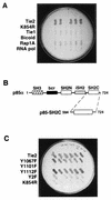
FIG. 1
FIG. 1
Identification of p85 as a specific Tie2 interactor by the yeast two-hybrid system. (A) Specific, autophosphorylation-dependent interaction of a human fetal heart library-encoded protein with Tie2. A plasmid encoding a 1.3-kb Tie2 interactor, from a fetal heart cDNA library, was transformed into yeast expressing the original lexA-Tie2 kinase bait, lexA-Tie2-K854R, lexA-Tie1 kinase, or one of three unrelated lexA bait proteins (bicoid protein, the small G protein Rap1A, and subunit RPB7 of S. cerevisiae RNA polymerase II). Six colonies from each transformation (horizontal rows of colonies) were picked and replica plated as described in Materials and Methods and tested for galactose-dependent activation of LEU2 on medium lacking leucine. (B) Schematic representation of structural domains within the full-length p85α and p85-SH2C. cDNA sequencing demonstrated that the 1.3-kb interactor cDNA encoded the entire C-terminal SH2 domain of p85α (p85-SH2C) (first and last amino acid residues of each protein are shown). bcr, breakpoint cluster region; iSH2, inter-SH2 domain. (C) Mutational analysis of the p85-SH2C–Tie2 interaction. The plasmid encoding p85-SH2C was transformed into yeast expressing either wild-type or mutant lexA-Tie2 kinase baits. Six colonies from each transformation were replica plated as described for panel A and tested for galactose-dependent LEU2 transcription. Y2F, Y1101F plus Y1112F.
FIG. 2
FIG. 2
Endothelial p85 associates preferentially with Y1101 of the Tie2 kinase in vitro. Similar amounts of recombinant GST-Tie2 kinase fusion proteins, with or without specific mutations, were immobilized on glutathione-Sepharose, subjected to in vitro kinase reactions, and incubated with endothelial cell lysates. Kinase-associated proteins were separated by SDS–8 to 16% PAGE and analyzed by Western blotting with antibodies against Tie2, phosphotyrosine (PTyr), p85, and Grb2. An aliquot of crude endothelial cell lysate (lysate) was used as a comparison for p85 and Grb2, and glutathione-Sepharose alone, without recombinant kinase, was incubated with endothelial cell lysates as a negative control (beads).
FIG. 3
FIG. 3
Inhibition of p85-Tie2 kinase association with a synthetic phosphopeptide mimicking tyrosine 1101. In vitro association assays were performed as described for Fig. 2 except that endothelial cell lysates were preincubated with increasing concentrations of synthetic phosphopeptides derived from sequences flanking Y1101 (pY1101, left panels) or Y1112 (pY1112, right panels). GST-Tie2-associated p85 and Grb2 were separated by SDS–8 to 16% PAGE and analyzed by Western blotting as described for Fig. 2. Blots were probed with anti-Tie2 to demonstrate equal loading of GST-Tie2 in each lane (upper panels).
FIG. 4
FIG. 4
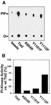
Tie2-associated p85 confers association of PI3 kinase activity in vitro. (A) Representative thin-layer chromatogram demonstrating GST-Tie2 kinase-associated PI3-kinase activity. Autophosphorylated wild-type or mutant GST-kinase fusion proteins were immobilized on glutathione-Sepharose and incubated with endothelial cell lysates as described for Fig. 2 except that, after the final wash, protein complexes were incubated with PI and [γ-32P]ATP in kinase buffer. Radiolabeled PI3P was separated by thin-layer chromatography and visualized by autoradiography. As a control, glutathione-Sepharose without recombinant kinase was incubated with endothelial cell lysates (beads). The positions of the origin (O) and PI3P (PIP) are shown. (B) Quantitation of Tie2-associated PI3-kinase activity. PI3-kinase activity associated with wild-type or mutant GST-Tie2 from the chromatogram in panel A was quantitated with a Molecular Dynamics PhosphorImager. Activity was corrected for that associated with beads alone and expressed as a percentage of activity associated with the wild-type (wt) kinase.
FIG. 5
FIG. 5
Chimeric fms-Tie2 receptors are functionally active in NIH 3T3 cells. NIH 3T3 fibroblasts stably expressing fms-Tie2 (fTie2) chimeric receptors, with or without specific mutations, were stimulated with CSF-1 (500 ng/ml) or left unstimulated, all in the presence of 1 mM sodium orthovanadate. Lysates were immunoprecipitated with an anti-c-fms MAb, separated by SDS–6% PAGE, and analyzed by Western blotting with anti-Tie2 and antiphosphotyrosine (anti-PTyr) antibodies to demonstrate CSF-1-dependent receptor autophosphorylation.
FIG. 6
FIG. 6
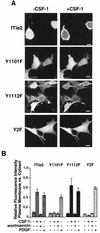
fTie2 activation induces plasma membrane translocation of GFP-PH in vivo. (A) Representative images of wild-type or mutant fTie2 cells expressing GFP-PH, before and after stimulation with CSF-1. NIH 3T3 fibroblasts expressing fTie2 chimeric receptors, with or without specific mutations, were plated on coverslips, electroporated with RNA encoding GFP-PH, briefly serum starved, and then stimulated with CSF-1 (500 ng/ml) and vanadate (1 mM). Cells were analyzed by confocal laser scanning fluorescence microscopy before and after stimulation. Bar, 10 μm. (B) Quantitation of plasma membrane translocation of GFP-PH induced by either fTie2 or PDGFR stimulation. The relative fluorescence intensity of plasma membrane versus that of cytosol was assessed as described in Materials and Methods before and after stimulation with CSF-1 (500 ng/ml) or PDGF-BB (40 ng/ml), both in the presence of 1 mM sodium orthovanadate. Each cell line was also assessed after stimulation with CSF-1 and vanadate following pretreatment with 50 nM wortmannin. The relative fluorescence intensity in each experiment is expressed as the mean ± SEM from 10 to 20 cells.
FIG. 7
FIG. 7
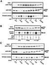
Stimulation of fTie2 in vivo induces phosphorylation and activation of Akt. (A) Phosphorylation and activation of Akt requires an intact Y1101 residue on Tie2. Untransfected NIH 3T3 cells or those expressing fTie2 chimeric receptors were left unstimulated or stimulated with CSF-1 (500 ng/ml), all in the presence of 1 mM sodium orthovanadate. Aliquots of total cell lysates were separated by SDS–8% PAGE and blotted with anti-Tie2 PAb to demonstrate similar receptor expression levels (top) and anti-Akt PAb to evaluate Akt expression and demonstrate gel mobility shift secondary to phosphorylation (middle). Anti-Akt immunoprecipitates from the same cell lysates were used in kinase assays with the substrate histone H2B and [γ-32P]ATP. Proteins were separated by SDS–15% PAGE and evaluated by autoradiography to assess Akt activation after CSF-1 stimulation (bottom). (B) Tie2-mediated phosphorylation and activation of Akt is PI3-kinase dependent. Fibroblasts expressing wild-type fTie2 or Y1112F were either unstimulated, stimulated with CSF-1, or stimulated with CSF-1 following 15 min of pretreatment with 100 nM wortmannin, all in the presence of 1 mM vanadate. Untransfected 3T3 cells were treated similarly with PDGF-BB (30 ng/ml) as a positive control. Cell lysates were used to assess Akt phosphorylation by Western blotting and in kinase assays to evaluate Akt activation as described for panel A. (C) Wild-type- and mutant fTie2-expressing cells exhibit intact Akt signaling pathways. Untransfected 3T3 cells and cells expressing wild-type or mutant fTie2 receptors were evaluated as described for panel A, except that stimulation of all cells was with PDGF (30 ng/ml) to demonstrate intact signaling pathways upstream of Akt. The positions of Akt, phosphorylated Akt (pAkt), and histone H2B are shown.
FIG. 8
FIG. 8
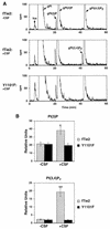
fTie2 activation of PI3-kinase and increases in cellular PI3P and PI 3,4-bisphosphate require an intact Y1101 residue on Tie2. (A) Representative chromatograms from wild-type fTie2- and Y1101F-expressing cells. Cells labeled for 72 h with 5 μCi of [3H]myo-inositol per ml were serum starved for 2 h and then were either left unstimulated or stimulated with CSF-1, all in the presence of 1 mM vanadate. Cellular lipids were extracted and deacylated, and then the resultant gPIPs were resolved by HPLC as described in Materials and Methods. Positions of the different gPIPs are indicated. cpm, counts per minute; Ins, free inositol. (B) Quantitative analysis of d-3-phosphoinositide production by wild-type fTie2 and Y1101F. Estimated baseline counts per minute on each chromatogram were subtracted from total counts under each peak and then expressed relative to the total cellular gPI in each experiment. Each bar represents the mean ± SEM from three separate experiments. ∗, P < 0.05 versus fTie2 minus CSF; P < 0.02 versus Y1101F plus CSF. ∗∗, P = 0.02 versus fTie2 minus CSF; P < 0.03 versus Y1101F plus CSF.
FIG. 9
FIG. 9
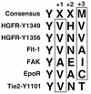
Comparison of amino acid residues flanking tyrosine 1101 of Tie2 with consensus and nonconsensus p85 binding motifs. Two nonconsensus p85 binding motifs on the hepatocyte growth factor receptor (HGFR) as well as those on the endothelium-specific vascular endothelial growth factor receptor Flt-1, focal adhesion kinase (FAK), and the erythropoietin receptor (EpoR) are shown in comparison to residues flanking Y1101 of Tie2 and the consensus motif determined from phosphopeptide binding studies (58). There is a strong preference for hydrophobic or neutral residues in the Y+1 and Y+3 positions (boxed residues). Y1101 of Tie2 differs by the presence of a threonine in the Y+3 position. Amino acid residues are designated according to the single-letter code: A, alanine; C, cysteine; E, glutamate; H, histidine; I, isoleucine; M, methionine; N, glutamine; V, valine; Y, tyrosine; X, any amino acid residue.

Similar articles

Cited by

References

    1. Ahmed N N, Grimes H L, Bellacosa A, Chan T O, Tsichlis P N. Transduction of interleukin-2 antiapoptotic and proliferative signals via Akt protein kinase. Proc Natl Acad Sci USA. 1997;94:3627–3632. - PMC - PubMed
    1. Antonetti D A, Algenstaedt P, Kahn C R. Insulin receptor substrate 1 binds two novel splice variants of the regulatory subunit of phosphatidylinositol 3-kinase in muscle and brain. Mol Cell Biol. 1996;16:2195–2203. - PMC - PubMed
    1. Bae Y S, Cantley L G, Chen C-S, Kim S-R, Kwon K-S, Rhee S G. Activation of phospholipase C-γ by phosphatidylinositol 3,4,5-triphosphate. J Biol Chem. 1998;273:4465–4469. - PubMed
    1. Carpenter C L, Cantley L C. Phosphoinositide kinases. Curr Opin Cell Biol. 1996;8:153–158. - PubMed
    1. Chen H-C, Appeddu P A, Isoda H, Guan J-L. Phosphorylation of tyrosine 397 in focal adhesion kinase is required for binding phosphatidylinositol 3-kinase. J Biol Chem. 1996;271:26329–26334. - PubMed

Publication types

MeSH terms

Substances

LinkOut - more resources