. 1998 Jun 15;141(6):1291-300.
doi: 10.1083/jcb.141.6.1291.

Different cis-regulatory DNA elements mediate developmental stage- and tissue-specific expression of the human COL2A1 gene in transgenic mice

Affiliations

Different cis-regulatory DNA elements mediate developmental stage- and tissue-specific expression of the human COL2A1 gene in transgenic mice

K K Leung et al. J Cell Biol. .

Abstract

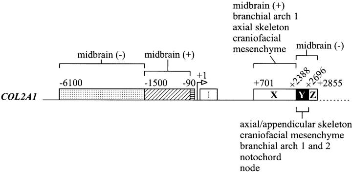
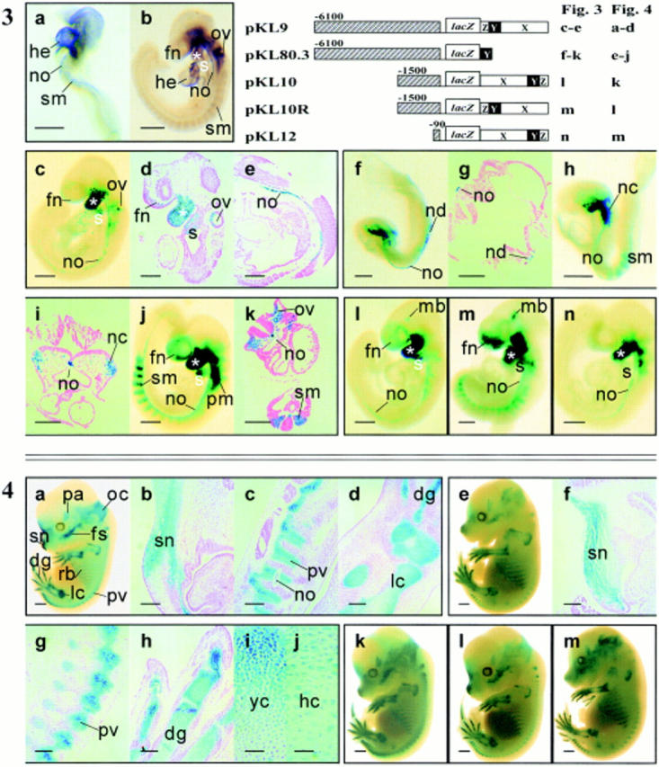
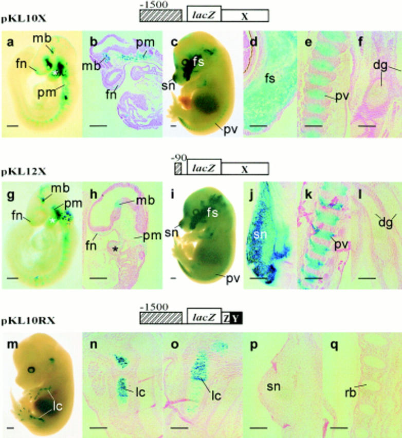
Expression of the type II collagen gene (human COL2A1, mouse Col2a1) heralds the differentiation of chondrocytes. It is also expressed in progenitor cells of some nonchondrogenic tissues during embryogenesis. DNA sequences in the 5' flanking region and intron 1 are known to control tissue-specific expression in vitro, but the regulation of COL2A1 expression in vivo is not clearly understood. We have tested the regulatory activity of DNA sequences from COL2A1 on the expression of a lacZ reporter gene in transgenic mice. We have found that type II collagen characteristic expression of the transgene requires the enhancer activity of a 309-bp fragment (+2, 388 to +2,696) in intron 1 in conjunction with 6.1-kb 5' sequences. Different regulatory elements were found in the 1.6-kb region (+701 to +2,387) of intron 1 which only needs 90-bp 5' sequences for tissue-specific expression in different components of the developing cartilaginous skeleton. Distinct positive and negative regulatory elements act together to control tissue-specific transgene expression in the developing midbrain neuroepithelium. Positive elements affecting expression in the midbrain were found in the region from -90 to -1,500 and from +701 to +2,387, whereas negatively acting elements were detected in the regions from -1,500 to -6,100 and +2,388 to +2,855.


Figures

Figure 1
Figure 1
Schematic representation of constructs used to generate transgenic mice in this study. Each construct contains a lacZ cassette flanked on the 5′ side by upstream sequences from the following genes: COL2A1 (hatched box); human β-globin gene promoter (cross-striped box); mouse Col10a1 5′ flanking DNA (stippled box) and on the 3′ side by COL2A1 first intron sequences (black or white boxes) or the SV-40 enhancer (vertically striped box). Numbers indicate the positions of the nucleotide in the sequence relative to the start site of transcription in the COL2A1 gene. For ease of reference, the intron 1 sequences analyzed have been subdivided into three regions, designated X, Y, and Z (refer to Materials and Methods). ATG, translation start site; pA, polyadenylation signal.
Figure 6
Figure 6
A summary of the organization of promoter and enhancer elements of the human COL2A1 gene as revealed by in vivo reporter activity in transgenic mouse embryos. The 1.6-kbp intron 1 (+701 to +2,387) sequence (region X) contains the enhancer elements that are required, in conjunction with a minimal 90-bp fragment of the 5′ flanking region of the COL2A1 gene, to direct prechondrogenic and chondrogenic and some nonchondrogenic expression of the reporter. Expression of the reporter is found in the midbrain, first branchial arch, craniofacial mesenchyme, and axial skeleton. Region Y of intron 1 (+2,388 to +2,696) in conjunction with 6.1-kbp 5′ flanking sequence confers similar prechondrogenic and chondrogenic enhancer activity. With this combination of regulatory elements, the transgene is expressed widely in the chondrogenic tissues (axial and appendicular skeleton and craniofacial mesenchyme), branchial arch 1 and 2, notochord, and node. Midbrain-positive elements may be present in the sequences from −90 to −1,500 and from +701 to +2,387 (region X). On the other hand, midbrain-negative elements may be present in the sequences from −1,500 to −6,100 and +2,388 to +2,855 (region Y+Z). Our study also shows that chondrogenic expression of the reporter can be achieved only by using homologous 5′ flanking and intron 1 sequences of the COL2A1 gene and substituting either one with heterologous regulatory elements results in nonspecific reporter expression.
Figure 2
Figure 2
A comparison of the expression of the human COL2A1 (a and c, 9.5-dpc; e and g, 14.5-dpc embryo) and mouse Col2a1 (b and d, 9.5-dpc; f and h, 14.5-dpc embryo) genes in transgenic (a–f) and nontransgenic (g and h) embryos. The transgenic embryos are heterozygous for a transgenic locus containing a 9.1-kbp 5′ and a 1.7-kbp 3′ flanking sequences and the COL2A1 gene (cosHcol2 construct). In the transgenic embryos, transcripts of the cosHcol2 transgene are found in the first branchial arch (asterisk), frontonasal (fn) and cranial paraxial mesenchyme (cm) (a), the otic vesicle (ov) and prechondrogenic trunk mesenchyme (tm) (c), the prevertebrae (pv) and the chondrocranial cartilage (cc) (e). These tissues in the transgenic mice also express the endogenous mouse Col2a1 (b, d, and f). The cosHcol2 transgene is not expressed in the heart tissues (he) that express the endogenous Col2a1 gene (compare c with d). In the nontransgenic embryo, no COL2A1 mRNA is detected in the chondrogenic mesenchyme (g) and expression of the endogenous Col2a1 transcript is shown in (h). Bar, 200 μm.
Figure 3
Figure 3
The delineation of minimal COL2A1 sequences required for expression of the lacZ reporter in prechondrogenic tissues. Col2a1 is expressed in the heart (he), notochord (no), and somites (sm) of (a) 8.5- and (b) 9.5-dpc embryos. The tissue-specific pattern of Col2a1 expression is emulated by that of the COL2A1-lacZ transgene with a 6.1-kbp 5′ flanking and regions X, Y, and Z of intron 1 in the pKL9 transgenic embryo (c–e). The transgene is expressed in the frontonasal mesenchyme (fn), otic vesicle (ov), first (asterisk) and second (s) branchial arches (c and d) and notochord (no) (c and e). Reduction of the first intron to region Y in pKL80.3 embryos still allows the full expression of the reporter gene in prechondrogenic tissues both in 8.5- (f–i) and 9.5-dpc (j and k) embryos and mimics the expression of Col2a1 gene (a, 8.5 dpc; b, 9.5 dpc). Transgene expression is found in the node (nd) and notochord (no) of the three-somite stage embryo but not in the embryonic heart (f and g). In the eight-somite stage pKL80.3 embryo, transgene expression is found in the notochord (no), neural crest cells (nc) (h and i), and somites (sm) (h). At 9.5 dpc, pKL80.3 is expressed in a pattern (j and k) similar to that of the endogenous Col2a1 (b). In addition, it is found to express in the paraxial mesenchyme (pm) (j). Reduction of the 5′ sequence of pKL9 to 1.5 kbp (l, pKL10; m, pKL10R) and 90 bp (n, pKL12) allows the full expression of the reporter gene, suggesting that the −1 to−90 fragment of the COL2A1 gene acts like a minimal promoter in conjunction with regions X, Y, and Z of intron 1. The intron 1 sequence functions as an enhancer element and gives the same reporter gene expression when placed in either orientation (compare l with m). It is interesting to find transgene expression in the midbrain (mb) of pKL10 and pKL10R (l and m). Bars: (a–c, h, j, and l–n) 500 μm; (d and e) 200 μm; (g, i, and k) 100 μm.Figure 4. Delineation of the minimal COL2A1 sequences needed to direct expression of the transgene in chondrogenic tissues. pKL9 embryo at 13.5 dpc (a) shows characteristic chondrogenic expression in the snout (sn), digits (dg), limb (lc), ribs (rb), prevertebrae (pv), facial skeleton (fs) and parietal (pa), and occipital (oc) mesenchyme. Histological sections of pKL9 13.5-dpc embryos show reporter gene expression in the condensation of the nasal mesenchyme (sn) (b), prevertebrae (pv), notochord (no) (c), and mesenchymal condensation of the zygopods (lc), and digits (dg) of the limb (d). Deletion of the intron 1 to region Y in pKL80.3 does not affect expression of the transgene in chondrogenic tissues (e–j), indicating that region Y of intron 1, in conjunction with 6.1-kbp 5′ flanking sequence, contains sufficient cis-acting tissue-specific elements to direct chondrogenic expression. Histological sections of pKL80.3 14.5-dpc embryos show similar lacZ expression in chondrogenic tissues (f–h) as in pKL9 embryos (b–d). Transgene expression in the hypertrophic chondrocytes (hc) (j) is weaker than that in young chondrocytes (yc) (i), which mimics the endogenous Col2a1 expression pattern. Reduction of the 5′ flanking sequences to 1.5 kbp in pKL10 (k) and pKL10R (l) and 90 bp in pKL12 (m) also has no effect, indicating that only 90-bp 5′ flanking sequence is enough to give chondrogenic expression in conjunction with regions X, Y, and Z of intron 1. Bars: (a, e, and k–m) 500 μm; (b–d and f–h) 200 μm; (i and j) 50 μm.
Figure 5
Figure 5
Demonstration of separable regulatory modules in the first intron. Deletion of regions Y and Z from pKL10 and pKL12 causes restricted and patchy expression of the reporter in the paraxial mesoderm (pm), the first branchial arch mesenchyme (asterisk), and the mesenchyme in the midbrain (mb) and frontonasal (fn) regions of the 9.5-dpc pKL10X (a and b) and pKL12X (g and h) embryos. lacZ expression is found in the snout (sn), facial skeleton (fs), and the prevertebrae (pv) but not in the cartilage of the digits (dg) of the 14.5-dpc pKL10X transgenic fetus (c–f) and that of the 15.5-dpc pKL12X transgenic fetus (i–l). Deletion of region X from pKL10R (refer to Fig. 1, pKL10RX) leads to the loss of expression in most tissues except the digits and limbs (lc) (m–o). No expression in the snout (sn) (p) or ribs (rb) (q) is found. Bars: (a, c, g, i, and m) 500 μm; (b, d–f, h, j–l, and n–q) 200 μm.

Similar articles

Cited by

References

    1. Bell DM, Leung KK, Wheatley SC, Ng LJ, Zhou S, Ling KW, Sham MH, Koopman P, Tam PP, Cheah KS. SOX9 directly regulates the type-II collagen gene. Nat Genet. 1997;16:174–178. - PubMed
    1. Braghetta P, Fabbro C, Piccolo S, Marvulli D, Bonaldo P, Volpin D, Bressan GM. Distinct regions control transcriptional activation of the alpha 1(VI) collagen promoter in different tissues of transgenic mice. J Cell Biol. 1996;135:1163–1177. - PMC - PubMed
    1. Cheah KS, Lau ET, Au PK, Tam PP. Expression of the mouse alpha 1(II) collagen gene is not restricted to cartilage during development. Development (Camb) 1991;111:945–953. - PubMed
    1. Cheah KS, Levy A, Trainor PA, Wai AW, Kuffner T, So CL, Leung KK, Lovell RH, Badge, Tam PP. Human COL2A1-directed SV40 T antigen expression in transgenic and chimeric mice results in abnormal skeletal development. J Cell Biol. 1995;128:223–237. - PMC - PubMed
    1. Darnell JE. Variety in the level of gene control in eukaryotic cells. Nature. 1982;297:365–371. - PubMed

Publication types