. 1998 Jul;72(7):5589-98.
doi: 10.1128/JVI.72.7.5589-5598.1998.

Sequences just upstream of the simian immunodeficiency virus core enhancer allow efficient replication in the absence of NF-kappaB and Sp1 binding elements

Affiliations

Sequences just upstream of the simian immunodeficiency virus core enhancer allow efficient replication in the absence of NF-kappaB and Sp1 binding elements

S Pöhlmann et al. J Virol. 1998 Jul.

Abstract

Large deletions of the upstream U3 sequences in the long terminal repeats (LTRs) of human immunodeficiency virus and simian immunodeficiency virus (SIV) accumulate in vivo in the absence of an intact nef gene. In the SIV U3 region, about 65 bp just upstream of the single NF-kappaB binding site always remained intact, and some evidence for a novel enhancer element in this region exists. We analyzed the transcriptional and replicative capacities of SIVmac239 mutants containing deletions or mutations in these upstream U3 sequences and/or the NF-kappaB and Sp1 binding sites. Even in the absence of 400 bp of upstream U3 sequences, the NF-kappaB site and all four Sp1 binding sites, the SIV promoter maintained about 15% of the wild-type LTR activity and was fully responsive to Tat activation in transient reporter assays. The effects of these deletions on virus production after transfection of COS-1 cells with full-length proviral constructs were much greater. Deletion of the upstream U3 sequences had no significant influence on viral replication when either the single NF-kappaB site or the Sp1 binding sites were intact. In contrast, the 26 bp of sequence located immediately upstream of the NF-kappaB site was essential for efficient replication when all core enhancer elements were deleted. A purine-rich site in this region binds specifically to the transcription factor Elf-1, a member of the ets proto-oncogene-encoded family. Our results indicate a high degree of functional redundancy in the SIVmac U3 region. Furthermore, we defined a novel regulatory element located immediately upstream of the NF-kappaB binding site that allows efficient viral replication in the absence of the entire core enhancer region.


Figures
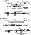
FIG. 1
FIG. 1
Locations of deletions in the nef-unique and U3 regions observed in HIV-1 and SIV infection. (A) Deletions observed in a long-term nonprogressor of HIV-1 infection at early and late time points (23). (B) Additional deletions observed in the U3 region in macaques infected with nef-deleted SIVmac239 (22). Nucleotide numbers refer to the HIV-1 NL4-3 (27) and SIVmac239 (32) sequences. Below, a schematic presentation of sequences close to the 3′ end that were maintained is given. The shaded boxes labeled with triangles indicate the deletions observed in the nef-unique and U3 regions in a long-term nonprogressor of HIV-1 infection (23) and rhesus macaques experimentally infected with nef-deleted SIVmac239 (22). As indicated, sequence elements of well-documented functional relevance, e.g., the polypurine tract, the core enhancer, and the TATA box, were preserved. Abbreviations: INT, U3 sequences required for integration; PPT, polypurine tract; USF, upstream stimulatory factor; Ets, Ets family transcription factor; SF, simian factor (40).
FIG. 2
FIG. 2
Mutations in the SIVmac239 U3 region. The SIVmac239 sequence has been published by Regier and Desrosiers (32). The NF-κB and Sp1 binding sites, the stop codon of nef, two PuB sites, a region with homology to the USF binding site, the peri-κB binding site (2), and the SF1 to SF3 sites (40) are indicated. Dots indicate deletions; dashes indicate identities in sequence. The deleted areas are shaded for clarity.
FIG. 3
FIG. 3
Activities of mutated SIVmac239 promoters with (+) or without (−) addition of a Tat expression plasmid. COS-1 cells were transfected with the indicated LTR-luciferase plasmids alone or cotransfected with an SIVmac Tat expression plasmid. For better comparison between independent experiments, stock DNA preparations from the wild-type LTR-luciferase and SIVmac Tat expression plasmids were always included in duplicate as positive controls (100% activity). Promoter activities relative to the 239wt promoter together with values for standard errors are shown above the bars. The results were obtained from four to six independent experiments. Background values for the luciferase plasmid without SIV LTR sequences were 0.16% ± 0.06% in the presence and 0.1% ± 0.04% in the absence of Tat. Background values were deducted from the measurements. Numbers at the bottom indicate the ratios of luciferase activities observed in the presence and in the absence of Tat. The ΔUSK mutant contains a 334-bp deletion in the US region, which is also present in ΔNU, and SstII and XhoI sites just upstream of the US65 region.
FIG. 4
FIG. 4
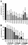
Production of p27 core antigen by COS-1 cells transfected with proviral constructs containing mutations in both LTRs. (A) p27 production measured after transfection with clones containing deletions in the US65 region, mutations in the SF binding sites, or a premature in-frame TAA stop signal at codon 93 of nef (nef*) (19). (B) Virus production obtained after transfection with proviral constructs bearing upstream U3 deletions in conjunction with NF-κB and/or Sp1 deletions. For clarity, panel B is shown in a logarithmic scale. Stock preparations of 239wt DNA were always transfected in parallel, and virus production is shown as a percentage of the wild-type value. Exact values with standard deviations are indicated above the bars. Average p27 production obtained with wild-type virus was about 50 ng of p27 per ml. The results are mean values obtained from four independent experiments.
FIG. 5
FIG. 5
Replication of SIVmac239 US and SF mutants in rPBMC and CEMx174 cells. Virus containing 2 ng of p27 derived from transfected COS-1 cells was used for infection. Similar results were obtained in three independent experiments. RT, reverse transcriptase; P.S.L., photo-stimulated luminescence.
FIG. 6
FIG. 6
Replication of SIVmac239 LTR variants containing deletions in the US region in conjunction with NF-κB or Sp1 site deletions. Virus containing 2 ng of p27 derived from transfected CEMx174 cells was used for infection. The results were confirmed in four independent experiments. For abbreviations, see the legend to Fig. 5.
FIG. 7
FIG. 7
Replication of SIVmac239 LTR variants containing deletions in the US region in conjunction with a deletion of all NF-κB and Sp1 sites. Virus containing 2 ng of p27 derived from transfected CEMx174 cells was used for infection of rPBMC (A), CEMx174 (B), and MmHF7062A (C) cells. For abbreviations, see the legend to Fig. 5.
FIG. 8
FIG. 8
Replication of SIVmac239 LTR variants containing point mutations in the SF1 to SF3 binding sites in conjunction with a deletion of all NF-κB and Sp1 sites. Virus containing 2 ng of p27 derived from transfected CEMx174 cells was used for infection of rPBMC (A), CEMx174 (B), and MmHF7062A (C) cells. In about one-third of the experiments we observed a less efficient replication of nef-defective SIVmac239 even in PHA-stimulated rPBMC. Replication of the ΔNF/Sp/NU variant was always comparable to that of the nef* variant, indicating that the slightly reduced and delayed replication kinetics are a Nef and not an LTR effect. For abbreviations, see the legend to Fig. 5.
FIG. 9
FIG. 9
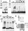
Binding of Elf-1 to the SIVmac239 PuB sites. (A) Elf-1 and Ets-1 bind to the HIV-2 PuB2 element. In vitro-transcribed and -translated Ets-1 (lane 4) and Elf-1 (lane 5), and as a control reticulocyte lysate (lane 3), were incubated with a radiolabeled probe corresponding to the HIV-2 PuB2 site (see Materials and Methods). The Elf-1 extract was incubated with an antibody directed against Elf-1 (Elf-Ab; lane 6). (B) Elf-1 binding to SIV PuB sites. In vitro-transcribed and -translated Elf-1 and a control reticulocyte lysate were incubated with the radiolabeled MSV and SIV probes in the presence or absence of Elf-1 antibody as indicated. The specific probes are described in Materials and Methods. (C) Elf-1 binds to the SIV PuB2 site in MmHF7062A cells. Extracts from MmHF7062A cells were incubated with the SIV PuB2 probe in the presence or absence of Elf-specific antibody as indicated. Asterisks indicate PuB2- and MSV-specific complexes. Arrows indicate supershift by Elf-1 antibody. (D) The Elf-1 antibody is highly specific. In vitro-transcribed and -translated Elf-1 (lane 1), a control reticulocyte lysate (lane 2), in vitro-transcribed and -translated Ets-1 (lane 3), and nuclear extracts from MmHF7062A cells were separated on a sodium dodecyl sulfate–12.5% polyacrylamide gel, immunoblotted, and probed with Elf-1 antibody as described in Materials and Methods. (E) Purine-rich sequences in the probes used for EMSA. The conserved central GGA motif is boxed; the consensus binding site for members of the Ets family of transcription factors (39) is indicated at the bottom. Results of the EMSA are shown at the right [++, strong binding; +, binding; (+), weak binding; −, no binding].
FIG. 10
FIG. 10
Biological effects of some mutations in the SIVmac239 U3 region investigated in this study. 1Promoter activity in the presence of Tat in COS-1 cells. 2p27 antigen production in COS-1 cells relative to the nef-defective SIVmac239 nef* variant. 3−, no replication detected; (+) maximal levels of virus production detected were <10% of the parental 239wt clone; +, maximal levels of virus production detected were 10 to 50% of the original clone; ++, virus production comparable to that of 239wt; d, delayed growth kinetics; dd, strongly delayed growth kinetics. The arrows indicate the positions of points mutations in the SF123 mutant.

Similar articles

Cited by

References

    1. Bassuk A G, Anandappa R T, Leiden J M. Physical interactions between Ets and NF-κB/NFAT proteins play an important role in their cooperative activation of the human immunodeficiency virus enhancer in T cells. J Virol. 1997;71:3563–3573. - PMC - PubMed
    1. Clark N M, Hannibal M C, Markovitz D M. The peri-κB site mediates human immunodeficiency virus type 2 enhancer activation in monocytes but not in T cells. J Virol. 1995;69:4854–4862. - PMC - PubMed
    1. Cullen B R. Use of eukaryotic expression technology in the functional analysis of cloned genes. Methods Enzymol. 1987;152:684–703. - PubMed
    1. Deacon N J, Tsykin A, Solomon A, Smith K, Ludford-Menting M, Hooker D J, McPhee D A, Greenway A L, Sonza S, Learmont J, Sullivan J S, Cunningham A, Dwyer D, Dowton D, Mills J. Genomic structure of an attenuated quasi species of HIV-1 from a blood transfusion donor and recipients. Science. 1995;270:988–991. - PubMed
    1. Dewhurst S, Embretson J E, Anderson D C, Mullins J I, Fultz P N. Sequence analysis and acute pathogenicity of molecularly cloned SIVSMM-PBj14. Nature. 1990;345:636–640. - PubMed

Publication types

LinkOut - more resources