. 1998 Jun 1;12(11):1652-64.
doi: 10.1101/gad.12.11.1652.

Localization-dependent translation requires a functional interaction between the 5' and 3' ends of oskar mRNA

Affiliations

Localization-dependent translation requires a functional interaction between the 5' and 3' ends of oskar mRNA

N Gunkel et al. Genes Dev. .

Abstract

The precise restriction of proteins to specific domains within a cell plays an important role in early development and differentiation. An efficient way to localize and concentrate proteins is by localization of mRNA in a translationally repressed state, followed by activation of translation when the mRNA reaches its destination. A central issue is how localized mRNAs are derepressed. In this study we demonstrate that, when oskar mRNA reaches the posterior pole of the Drosophila oocyte, its translation is derepressed by an active process that requires a specific element in the 5' region of the mRNA. We demonstrate that this novel type of element is a translational derepressor element, whose functional interaction with the previously identified repressor region in the oskar 3' UTR is required for activation of oskar mRNA translation at the posterior pole. The derepressor element only functions at the posterior pole, suggesting that a locally restricted interaction between trans-acting factors and the derepressor element may be the link between mRNA localization and translational activation. We also show specific interaction of two proteins with the oskar mRNA 5' region; one of these also recognizes the 3' repressor element. We discuss the possible involvement of these factors as well as known genes in the process of localization-dependent translation.


Figures

Figure 1
Figure 1
Identification of a translation control element between the first and second start codons of osk mRNA. The cartoon shows the alternative usage of the two start codons, m1 and m2, in wild-type osk, which gives rise to a long and a short isoform of Oskar (Markussen et al. 1995). Full rescue activity of the wild-type construct was used as a standard and is indicated (+). Failure to rescue is also indicated (−). (oskM1L) Mutation of m1 into a leucine (CTG) turns the sequence upstream of m2 into a 5′ UTR. This transcript encodes short Oskar, which shows full rescue activity with respect to abdomen formation and fertility (Markussen et al. 1995). (oskM1LΔ1) Deletion of 249 nucleotides from the 5′ UTR of oskM1L abolishes osk activity; the two independent lines tested, oskM1LΔ1–2 and oskM1LΔ1–9 produced 0% and 2% hatchers, respectively, and none of the hatched females were fertile (males were not tested).
Figure 2
Figure 2
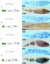
The 5′ end of osk mRNA contains sequences required to alleviate translational repression at the posterior pole. Functional analysis of the 5′ end of osk mRNA in chimeric reporter transgenes by analysis of RNA distribution and reporter gene activity. In the schematic representation of the transcripts, sequences derived from the osk 5′ end are indicated in green, the osk 3′ UTR is presented as a line, and the TAA proximal translation repressor element is indicated as a black box (region AB, Kim-Ha et al. 1995). Panels on the right shows in situ transcript distribution (RNA) and translation profile for the lacZ reporter as β-galactosidase staining (β-Gal). (A) m1414lacwt RNA, containing 414 nucleotides downstream of m1, is localized and regulated translationally like wild-type osk mRNA. (B) m1117lacwt RNA, consisting of the osk 15-nucleotide 5′ UTR and 117 nucleotides downstream of m1 fused in-frame to lacZ reading frame, and followed by the wild-type osk 3′ UTR, is localized and repressed like wild-type osk RNA, but fails to be translated at the posterior pole. (C) m1414lacLS5 contains a 5-nucleotide substitution (see Materials and Methods) in the second Bruno-binding consensus sequence (Kim-Ha et al. 1995) in an otherwise wild-type reporter transcript. This mutation causes moderate premature translation, detectable as light blue β-galactosidase staining in nurse cells. (D) m1117lacLS5 is identical to m1117lacwt, with the exception of the 5-nucleotide substitution (LS5) in the BRE. This RNA is translated efficiently in spite of the absence of the 5′ element. The LS5 mutation was identified in a linker-scanning mutagenesis series spanning what we find to be the essential part of the proximal BRE. Premature translation of LS5-containing transcripts is detected by blue β-galactosidase staining in the nurse cells and young oocytes. Mutation of additional Bruno-binding consensus sequences results in stronger staining of nurse cells and early oocytes (data not shown). Interestingly, premature translation in m1414lacLS5 is always less pronounced than in m1117lacLS5. All transgenes were assayed in the w1118 background and support similar steady-state levels of RNA (see Fig. 7, lanes 4–7).
Figure 3
Figure 3
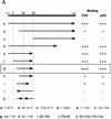
Mapping specific protein/RNA interactions in the m1–m2 fragment of osk mRNA that is essential for its translation. (A) The 5′ end of osk mRNA, including the first and second start codons, m1 and m2, respectively, is diagrammed schematically. A partial restriction map of the region, showing sites used for subcloning is shown at the top. (E) EcoRI; (Bs) BstEII; (Bg) BglII. Transcripts used as probes for the UV cross-linking assay are identified by lowercase letters. The coordinates of the subfragments are indicated at the bottom. Position 1 is the first nucleotide of m1, position 417 is the last nucleotide of m2. The entire m1–m2 region (a) or subfragments of the region, created with available restriction sites or by PCR were labeled radioactively and incubated with oocyte extract. Proteins cross-linked to labeled RNA were separated by SDS-PAGE and visualized by autoradiography. The signal intensity obtained with the full-length m1–m2 probe was used as a standard and is indicated by (+++). Significantly reduced but detectable binding, and complete loss of binding are indicated (+) and (−), respectively. (B) UV cross-linking assay revealing the specific interaction of p50 and p68 with RNA fragment g. RNA probes (bottom) were incubated with oocyte extract and cross-linked by method 1 (lanes 1–4 and 9,10) or method 2 (lanes 5–8). (Lane 10) p68 complex with RNA fragment g was incubated with anti-Bruno antiserum and subjected to immunoprecipitation. The minimal region recognized (fragment g) was determined as shown in A. Probes used in each lane are indicated below. Arrows pointing rightward indicate RNA in sense orientation; arrows pointing leftward indicate RNA in antisense orientation.
Figure 3
Figure 3
Mapping specific protein/RNA interactions in the m1–m2 fragment of osk mRNA that is essential for its translation. (A) The 5′ end of osk mRNA, including the first and second start codons, m1 and m2, respectively, is diagrammed schematically. A partial restriction map of the region, showing sites used for subcloning is shown at the top. (E) EcoRI; (Bs) BstEII; (Bg) BglII. Transcripts used as probes for the UV cross-linking assay are identified by lowercase letters. The coordinates of the subfragments are indicated at the bottom. Position 1 is the first nucleotide of m1, position 417 is the last nucleotide of m2. The entire m1–m2 region (a) or subfragments of the region, created with available restriction sites or by PCR were labeled radioactively and incubated with oocyte extract. Proteins cross-linked to labeled RNA were separated by SDS-PAGE and visualized by autoradiography. The signal intensity obtained with the full-length m1–m2 probe was used as a standard and is indicated by (+++). Significantly reduced but detectable binding, and complete loss of binding are indicated (+) and (−), respectively. (B) UV cross-linking assay revealing the specific interaction of p50 and p68 with RNA fragment g. RNA probes (bottom) were incubated with oocyte extract and cross-linked by method 1 (lanes 1–4 and 9,10) or method 2 (lanes 5–8). (Lane 10) p68 complex with RNA fragment g was incubated with anti-Bruno antiserum and subjected to immunoprecipitation. The minimal region recognized (fragment g) was determined as shown in A. Probes used in each lane are indicated below. Arrows pointing rightward indicate RNA in sense orientation; arrows pointing leftward indicate RNA in antisense orientation.
Figure 4
Figure 4
Inversion of the 3′ half of the p50/p68-binding RNA fragment prevents posterior derepression in vivo. The orientation of the 3′ half of the fragment is indicated by an arrow. (A) m1m2lacwt, wild-type m1–m2 region, fused 3 nucleotides downstream of m2 to the lacZ reporter, under the regulatory control of wild-type osk 3′ UTR. Reporter RNA and protein distributions, as detected in situ by RNA hybridization and β-galactosidase activity, are virtually indistinguishable from those of endogenous osk. (B) m1INVm2lacwt, the p50/p68-binding fragment in the m1–m2 region of the reporter transgene was mutated by inversion of the 3′ half (see probe c). As in the wild-type construct, the RNA is efficiently localized; however, translation is not derepressed.
Figure 5
Figure 5
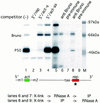
p50 binds both to the 5′ end of osk mRNA and to the repressor element in the 3′ UTR. An RNA competition assay was performed to determine the binding specificity of p50. A radioactive RNA probe consisting of the essential subfragment of the proximal repressor element (region AB; Kim-Ha et al. 1995) was incubated and UV cross-linked to proteins in the oocyte extract in the absence of competitor RNA (lane 1). Competitions were carried out with a 100-fold excess of either repressor fragment (rep, lane 2), EcoRI–BglII fragment containing the 5′ activator in sense orientation (5′/act-s, lane 3), or the same EcoRI–BglII fragment in antisense orientation (5′/act-as, lane 4). Immunoprecipitation of proteins UV cross-linked to radiolabeled repressor fragment (lane 5) by anti-Bruno antiserum (lane 6) or preimmune serum (lane 7). Simultaneous binding of p50 and Bruno was tested by treating with RNase A only after the immunoprecipitation. (Lane 8) Anti-Bruno antiserum; (lane 9) preimmune serum. We noted that in some cases (as shown here) the intensity of the upper band of the p50 doublet was reduced after coprecipitation. The locations of the 5′ and 3′ competitors in the osk transcript are indicated below. The radiolabeled probe is indicated by an asterisk. The reverse experiment, with activator element RNA as a radioactive probe, and cold activator and repressor RNA elements as unlabeled competitors, yielded the same result with respect to p50 (data not shown).
Figure 6
Figure 6
Reduction of p50 binding correlates with premature translation. (A) A truncated EcoRI–DraI repressor RNA fragment shows a >10-fold decrease in p50 binding but Bruno binding is unaffected. A radioactive probe consisting of either the full-length repressor region (lanes 1–3) or a truncated form with 24 nucleotides deleted from the 5′ and 25 nucleotides from the 3′ (lanes 4–6) were incubated with oocyte extract in the presence of 100-fold molar excess of specific competitor (EcoRI–DraI fragment, lanes 2,5) or nonspecific competitor (polylinker of pSP72, lanes 3,6) and subsequently UV cross-linked. The same deletion mutation in the context of the first 380 nucleotides of the osk 3′ UTR had essentially the same effect on p50 and Bruno binding. The locations of the Bruno-binding consensus sequences with respect to the 5′ and 3′ truncations (light gray) are indicated below. (B) In vivo analysis of the effect of the 5′Δ24/3′Δ25 truncation of the proximal repressor element in an otherwise wild-type lacZ reporter gene (m1414lac5′Δ24/3′Δ25). The mutation does not affect RNA localization or levels of reporter transcripts (cf. with Fig. 2A, and see Fig. 7, lane 4,9); however, translation is detectable already from stage 6/7 onward.
Figure 7
Figure 7
Comparative quantification of reporter transcripts by RNase protection. The amounts of input mRNA, isolated from ovaries, were first adjusted to equalize endogenous osk mRNA and then quantified with respect to reporter transcripts. (Lane 1) Antisense Ribo probe alone; (lane 2) probe digested; (lane 3) probe digested in the presence of 10 μg total ovarian RNA (Oregon-R); (lane 4) m1414lacwt; (lane 5) m1117lacwt; (lane 6) m1117lacLS5; (lane 7) m1414lacLS5; (lane 8) m1m2lacwt; (lane 9) m1INVm2lacwt; (lane 10) m1414lacBREbcd; (lane 11) m1414lac5′Δ24/3′Δ25.
Figure 8
Figure 8
The derepressor element functions at the posterior but not at the anterior pole of the oocyte. Flies were generated that bear two distinct reporter transgenes: m1414lacBREbcd and m1414lacwt. m1414lacBREbcd has the osk mRNA 5′ end (m1414), including the derepressor element, and the first 370 nucleotides of the osk 3′ UTR, including the proximal repressor element (BRE). The bcd 3′ UTR, which directs the chimeric transcript to the anterior pole of the oocyte was fused downstream of the BRE. m1414lacwt was described in Fig. 2A. It is localized to the posterior pole of the oocyte and repressed translationally until stage 9, because of the presence of a wild-type osk 3′ UTR (wild type). The amount of transcript produced by each transgene was assessed separately (see Fig. 7, lanes 4,10).

Similar articles

Cited by

References

    1. Ainger K, Avossa D, Morgan F, Hill SJ, Barry C, Barbarese E, Carson JH. Transport and localization of exogenous myelin basic protein mRNA microinjected into oligodendrocytes. J Cell Biol. 1993;123:431–441. - PMC - PubMed
    1. Ausubel, F., R. Brent, R.E. Kingston, D.D. Moore, J.G. Seidman, J.A. Smith, and K. Struhl, eds. 1987–1998. Current protocols in molecular biology. Wiley & Sons, New York, NY.
    1. Berleth T, Burri M, Thoma G, Bopp D, Richstein S, Frigerio G, Noll M, Nüsslein-Volhard C. The role of localization of bicoid RNA in organizing the anterior pattern of the Drosophila embryo. EMBO J. 1988;7:1749–1756. - PMC - PubMed
    1. Castrillon D, Gonczy P, Alexander S, Rawson R, Eberhart CG, Viswanathan S, DiNardo S, Wasserman SA. Toward a molecular genetic analysis of spermatogenesis in Drosophila melanogaster: Characterization of male-sterile mutants generated by single P element mutagenesis. Genetics. 1993;135:489–505. - PMC - PubMed
    1. Clark I, Giniger E, Ruohola-Baker H, Jan LY, Jan YN. Transient posterior localization of a kinesin fusion protein reflects anteroposterior polarity of the Drosophila oocyte. Curr Biol. 1994;4:289–300. - PubMed

Publication types

LinkOut - more resources