. 1998 Jun 1;12(11):1638-51.
doi: 10.1101/gad.12.11.1638.

The histone acetylase PCAF is a nuclear receptor coactivator

Affiliations

The histone acetylase PCAF is a nuclear receptor coactivator

J C Blanco et al. Genes Dev. .

Abstract

Whereas the histone acetylase PCAF has been suggested to be part of a coactivator complex mediating transcriptional activation by the nuclear hormone receptors, the physical and functional interactions between nuclear receptors and PCAF have remained unclear. Our efforts to clarify these relationships have revealed two novel properties of nuclear receptors. First, we demonstrate that the RXR/RAR heterodimer directly recruits PCAF from mammalian cell extracts in a ligand-dependent manner and that increased expression of PCAF leads to enhanced retinoid-responsive transcription. Second, we demonstrate that, in vitro, PCAF directly associates with the DNA-binding domain of nuclear receptors, independently of p300/CBP binding, therefore defining a novel cofactor interaction surface. Furthermore, our results show that dissociation of corepressors enables ligand-dependent PCAF binding to the receptors. This observation illuminates how a ligand-dependent receptor function can be propagated to regions outside the ligand-binding domain itself. On the basis of these observations, we suggest that PCAF may play a more central role in nuclear receptor function than previously anticipated.


Figures
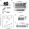
Figure 1
Figure 1
Ligand-dependent recruitment of PCAF by the RXR–RAR heterodimer–DNA complex. (A) Binding assay. Recombinant RXRβ and RARβ were bound to a DNA fragment containing the RARE (DR-5) fixed to agarose beads, and incubated with nuclear extracts. Bound materials were tested in B–E. (B) Immunoblot analysis. Nuclear extracts (3 mg) were incubated with the indicated amounts (μg of protein) of RXR–RAR heterodimer bound to the RARE-conjugated beads in the presence (+) or absence (−) of 1 μm 9-cis RA, and bound materials were detected by immunoblot assay with the appropriate antibodies. (Lane 1) Extracts added to RARE-conjugated beads without receptors. (Input) 40 μg of nuclear extracts. Numbers at the right indicate molecular masses of the immunoreactive proteins. (Bottom) Binding of rRXRβ and rRARβ to the RARE-conjugated beads as detected by immunoblot assay. (C) Histone acetylase activity recovered from the heterodimer–RARE complex. Extract proteins bound to the indicated amounts of heterodimer–RARE complexes in the presence of 1 μm 9-cis RA (⋄) or absence (□) were tested for histone acetylase activity. Values represent the average of four independent assays ±s.d. (D) Histone specificity of acetylase activity tested with free core histones. Approximately 3 μg of bound protein obtained from heterodimer–RARE complexes (3 μg of heterodimer) was tested for acetylation activity with free histones. (Lane 1) Acetylation pattern by rPCAF (50 ng). (Lane 2) Acetylation pattern by eluates from control beads. (E) Histone specificity of acetylase activity tested with nucleosomal histones. Bound materials obtained from 1 μg (+, lanes 4,5) or 2 μg (++, lanes 6,7) of heterodimers were tested for acetylase activity with nucleosomal histones. (Lane 2) Background control without rPCAF or bound materials.
Figure 2
Figure 2
The role of ligand in PCAF recruitment: Dose response and receptor selectivity. (A,B) 9-cis RA concentration dependence. One microgram of heterodimer bound to RARE-conjugated beads was incubated with indicated concentrations of 9-cis RA and tested for recruitment of PCAF and p300 (A) from nuclear extracts as in Fig. 1B or for accumulation of histone acetylase activity (B). (Bottom) RXR–RAR bound to RARE-conjugated beads. (C,D) Receptor selectivity. One microgram of heterodimers bound to RARE-conjugated beads was incubated with 9-cis RA (10−7 and 10−6 m), TTNPB (10−9 and 10−8 m), or sr11237 (10−6 and 10−55 m) and tested for binding of PCAF and p300 (C) or for accumulation of histone acetylase activity (D).
Figure 2
Figure 2
The role of ligand in PCAF recruitment: Dose response and receptor selectivity. (A,B) 9-cis RA concentration dependence. One microgram of heterodimer bound to RARE-conjugated beads was incubated with indicated concentrations of 9-cis RA and tested for recruitment of PCAF and p300 (A) from nuclear extracts as in Fig. 1B or for accumulation of histone acetylase activity (B). (Bottom) RXR–RAR bound to RARE-conjugated beads. (C,D) Receptor selectivity. One microgram of heterodimers bound to RARE-conjugated beads was incubated with 9-cis RA (10−7 and 10−6 m), TTNPB (10−9 and 10−8 m), or sr11237 (10−6 and 10−55 m) and tested for binding of PCAF and p300 (C) or for accumulation of histone acetylase activity (D).
Figure 3
Figure 3
Binding of PCAF to various nuclear hormone receptors in vitro. (A) Direct binding of rPCAF to RXR–RAR heterodimers. rPCAF (2 pmoles, 200 ng) was incubated with increasing amounts of heterodimers (0.5 μg, lanes 3,5 or 1 μg, lanes 4,6) bound to the RARE-conjugated beads in the presence (+) or absence (−) of 1 μm 9-cis RA. Binding of rPCAF was detected by immunoblot analysis with anti-M2-Flag antibody. (Lane 1) 20 ng of rPCAF as input; (lane 2) eluates from RARE-conjugated beads without receptor. (B) Binding of RARβ to rPCAF. Binding of [35S]methionine-labeled RARβ was tested with rPCAF immobilized to the M2 antibody matrix or matrix alone (M2), GST beads, GST–ACTR in the presence (+) or absence (−) of 1 μm 9-cis RA. (Right) Radiolabeled receptor input (30%) tested in B and C. (C) Binding of 35S-labeled ERα or GRα to rPCAF immobilized to the M2 antibody matrix was tested in the presence (+) or absence (−) of 1 μm β-estradiol (for ERα) or 1 μm dexamethasone (for GR). (D) PCAF–receptor interactions detected by yeast two–hybrid assays. Yeast strain Y190 was transformed with indicated GAL4-fusion plasmids. The liquid β-gal assays were performed for the transformants, which were grown in the presence or absence of corresponding hormones (10 nm deoxycorticosterone for GR, 100 nm β-estradiol for ER, and 100 nm dihydrotestosterone for AR). Results represent the average of three independent yeast transformants ± s.d.
Figure 3
Figure 3
Binding of PCAF to various nuclear hormone receptors in vitro. (A) Direct binding of rPCAF to RXR–RAR heterodimers. rPCAF (2 pmoles, 200 ng) was incubated with increasing amounts of heterodimers (0.5 μg, lanes 3,5 or 1 μg, lanes 4,6) bound to the RARE-conjugated beads in the presence (+) or absence (−) of 1 μm 9-cis RA. Binding of rPCAF was detected by immunoblot analysis with anti-M2-Flag antibody. (Lane 1) 20 ng of rPCAF as input; (lane 2) eluates from RARE-conjugated beads without receptor. (B) Binding of RARβ to rPCAF. Binding of [35S]methionine-labeled RARβ was tested with rPCAF immobilized to the M2 antibody matrix or matrix alone (M2), GST beads, GST–ACTR in the presence (+) or absence (−) of 1 μm 9-cis RA. (Right) Radiolabeled receptor input (30%) tested in B and C. (C) Binding of 35S-labeled ERα or GRα to rPCAF immobilized to the M2 antibody matrix was tested in the presence (+) or absence (−) of 1 μm β-estradiol (for ERα) or 1 μm dexamethasone (for GR). (D) PCAF–receptor interactions detected by yeast two–hybrid assays. Yeast strain Y190 was transformed with indicated GAL4-fusion plasmids. The liquid β-gal assays were performed for the transformants, which were grown in the presence or absence of corresponding hormones (10 nm deoxycorticosterone for GR, 100 nm β-estradiol for ER, and 100 nm dihydrotestosterone for AR). Results represent the average of three independent yeast transformants ± s.d.
Figure 4
Figure 4
Corepressors enable ligand-dependent binding of PCAF to the RXR–RAR heterodimer in vitro. (A) The effect of SMRT. One microgram of DNA-bound RXR–RAR heterodimers was incubated with 200 ng of rPCAF and 500 ng of control GST (lanes 1,2) or GST–SMRT (lanes 3,4) in the presence or absence of 1 μm 9-cis RA. Binding of PCAF and GST–SMRT to the heterodimer was detected by anti-M2 antibody and anti-SMRT antibody, respectively. (B) The effect of N-CoR. Binding assays were performed as in Fig. 5A, but with 500 ng of control GST (lane 2–4), GST–N-CoRΔN2 (lane 5–7), or GST–N-CoRΔN6 (lane 8–10) with (+) or without (−) 1 μm 9-cis RA. (Bottom) Binding of N-CoR was detected by anti-N-CoR antibody. (C) Ligand-independent PCAF binding to the hinge mutants of RARα. 35S-Labeled, in vitro-translated hRARα wild-type or hinge region mutants (L187P and 187-188GG) (2 × 105 cpm) were incubated with 2 μg of immobilized rPCAF in the presence of GST or GST–N-CoRΔN2 and with (+) or without (−) 1 μm 9-cis RA. (M2) Binding of radiolabeled RARs to the M2 beads without rPCAF; (INPUT) amounts of RARs (2 × 104 cpm) tested in each reaction. (D) Scheme for corepressor dissociation and PCAF binding.
Figure 4
Figure 4
Corepressors enable ligand-dependent binding of PCAF to the RXR–RAR heterodimer in vitro. (A) The effect of SMRT. One microgram of DNA-bound RXR–RAR heterodimers was incubated with 200 ng of rPCAF and 500 ng of control GST (lanes 1,2) or GST–SMRT (lanes 3,4) in the presence or absence of 1 μm 9-cis RA. Binding of PCAF and GST–SMRT to the heterodimer was detected by anti-M2 antibody and anti-SMRT antibody, respectively. (B) The effect of N-CoR. Binding assays were performed as in Fig. 5A, but with 500 ng of control GST (lane 2–4), GST–N-CoRΔN2 (lane 5–7), or GST–N-CoRΔN6 (lane 8–10) with (+) or without (−) 1 μm 9-cis RA. (Bottom) Binding of N-CoR was detected by anti-N-CoR antibody. (C) Ligand-independent PCAF binding to the hinge mutants of RARα. 35S-Labeled, in vitro-translated hRARα wild-type or hinge region mutants (L187P and 187-188GG) (2 × 105 cpm) were incubated with 2 μg of immobilized rPCAF in the presence of GST or GST–N-CoRΔN2 and with (+) or without (−) 1 μm 9-cis RA. (M2) Binding of radiolabeled RARs to the M2 beads without rPCAF; (INPUT) amounts of RARs (2 × 104 cpm) tested in each reaction. (D) Scheme for corepressor dissociation and PCAF binding.
Figure 5
Figure 5
PCAF domain analysis and the relationship with p300. (A) Heterodimer–RARE complexes (∼2 pmoles) were incubated with 2 pmoles of recombinant N-p300 (amino acids 1–670) alone (lanes 1,2) or along with 1 (+) and 2 (++) pmoles of PCAF (lane 3–6) in the presence (+) or absence (−) of 1 μm 9-cis RA. (B) PCAF domain analysis: 2 pmoles of rPCAF lacking the carboxy-terminal domain (ΔC) or amino-terminal domain (ΔN1 and ΔN2, top) were incubated with heterodimer–RARE complexes (1 μg of heterodimer) in the presence (+) or absence (−) of 1 μm 9-cis RA. Binding was detected with anti-PCAF antibody.
Figure 5
Figure 5
PCAF domain analysis and the relationship with p300. (A) Heterodimer–RARE complexes (∼2 pmoles) were incubated with 2 pmoles of recombinant N-p300 (amino acids 1–670) alone (lanes 1,2) or along with 1 (+) and 2 (++) pmoles of PCAF (lane 3–6) in the presence (+) or absence (−) of 1 μm 9-cis RA. (B) PCAF domain analysis: 2 pmoles of rPCAF lacking the carboxy-terminal domain (ΔC) or amino-terminal domain (ΔN1 and ΔN2, top) were incubated with heterodimer–RARE complexes (1 μg of heterodimer) in the presence (+) or absence (−) of 1 μm 9-cis RA. Binding was detected with anti-PCAF antibody.
Figure 6
Figure 6
Receptor domain analysis. (A) Diagram of full-length and truncated RXRβ and RARα and summary of PCAF binding (+, significant binding; −, no detectable binding). (B) 35S-Labeled receptors were incubated with rPCAF bound to anti-M2-Flag antibody conjugated to beads as in Fig. 4C. (Lane 1) Input (2 × 104 cpm); (lane 2) control M2 beads without PCAF; (lanes 3,4) M2-rPCAF without (−) or with (+) 1 μm 9-cis RA, respectively. 35S-Labeled luciferase was tested as a negative control. (C) Control GST, GST–DBD from RARβ, or GST–DBD from RXRα (500 ng each) was incubated with rPCAF (+, 100 ng; ++, 300 ng) and PCAF binding was detected with anti-PCAF antibody. (Bottom) Loading of the control GST peptide, GST–DBD of RARα, and GST–DBD of RXRβ.
Figure 6
Figure 6
Receptor domain analysis. (A) Diagram of full-length and truncated RXRβ and RARα and summary of PCAF binding (+, significant binding; −, no detectable binding). (B) 35S-Labeled receptors were incubated with rPCAF bound to anti-M2-Flag antibody conjugated to beads as in Fig. 4C. (Lane 1) Input (2 × 104 cpm); (lane 2) control M2 beads without PCAF; (lanes 3,4) M2-rPCAF without (−) or with (+) 1 μm 9-cis RA, respectively. 35S-Labeled luciferase was tested as a negative control. (C) Control GST, GST–DBD from RARβ, or GST–DBD from RXRα (500 ng each) was incubated with rPCAF (+, 100 ng; ++, 300 ng) and PCAF binding was detected with anti-PCAF antibody. (Bottom) Loading of the control GST peptide, GST–DBD of RARα, and GST–DBD of RXRβ.
Figure 7
Figure 7
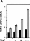
Enhancement of retinoid-dependent promoter activity by transfected PCAF. (A) NIH-3T3 (1.5 × 105) cells were cotransfected with the RARE–Tk–luciferase reporter and pcx–PCAF (+, 50 ng; ++, 200 ng; or +++, 400 ng), and luciferase activity was measured following 24 hr of 1 μm all trans-RA treatment. Shaded and solid bars represent luciferase activity with and without RA, respectively. Values represent the average of triplicate cultures ± s.d. (B) Transfection was performed as in A but with additional expression vectors for RXRβ and RARα (40 ng of each), and with pcx–PCAF (+, 50 ng; ++, 200 ng; and +++, 400 ng) or pcx–PCAF mutants (ΔN2 and ΔC, 400 ng). Values represent the average of four independent assays ±s.d. (C) Transfection was performed as in B, but with 400 ng each of control (pcx), PCAF, or HAT deletions (ΔHAT1 and ΔHAT2). Values represent the average of triplicates ±s.d. (D) Detection of transfected PCAF and endogenous p300. Nuclear extracts from 6 × 105 NIH-3T3 cells transfected with indicated amounts (ng) of pcx–PCAF or deletion PCAFs tested in B and C were immunoblotted with anti-M2-Flag antibody to detect PCAF or with anti-p300 antibody. Asterisks indicate the position of the wild-type or mutant PCAFs. (E) Pool of NIH-3T3 cells stably transfected with the RARE–Tk–luciferase reporter was transiently transfected with pcx–PCAF or pcx–ΔC plus an IL-2R vector, and treated with 1 μm all trans-RA. Cells were sorted by anti-IL-2R antibody panning and luciferase activity was measured as in A. Values represent the average of triplicates ± s.d.
Figure 7
Figure 7
Enhancement of retinoid-dependent promoter activity by transfected PCAF. (A) NIH-3T3 (1.5 × 105) cells were cotransfected with the RARE–Tk–luciferase reporter and pcx–PCAF (+, 50 ng; ++, 200 ng; or +++, 400 ng), and luciferase activity was measured following 24 hr of 1 μm all trans-RA treatment. Shaded and solid bars represent luciferase activity with and without RA, respectively. Values represent the average of triplicate cultures ± s.d. (B) Transfection was performed as in A but with additional expression vectors for RXRβ and RARα (40 ng of each), and with pcx–PCAF (+, 50 ng; ++, 200 ng; and +++, 400 ng) or pcx–PCAF mutants (ΔN2 and ΔC, 400 ng). Values represent the average of four independent assays ±s.d. (C) Transfection was performed as in B, but with 400 ng each of control (pcx), PCAF, or HAT deletions (ΔHAT1 and ΔHAT2). Values represent the average of triplicates ±s.d. (D) Detection of transfected PCAF and endogenous p300. Nuclear extracts from 6 × 105 NIH-3T3 cells transfected with indicated amounts (ng) of pcx–PCAF or deletion PCAFs tested in B and C were immunoblotted with anti-M2-Flag antibody to detect PCAF or with anti-p300 antibody. Asterisks indicate the position of the wild-type or mutant PCAFs. (E) Pool of NIH-3T3 cells stably transfected with the RARE–Tk–luciferase reporter was transiently transfected with pcx–PCAF or pcx–ΔC plus an IL-2R vector, and treated with 1 μm all trans-RA. Cells were sorted by anti-IL-2R antibody panning and luciferase activity was measured as in A. Values represent the average of triplicates ± s.d.
Figure 8
Figure 8
A model for PCAF recruitment: Coupling with the ligand-induced disengagement of the histone deacetylase–corepressor complex. Prior to ligand binding, the heterodimer is associated with histone deacetylases complexed with corepressor–Sin3, providing a repressive chromatin environment. Ligand binding may release the histone deacetylase–Sin3–corepressor complex and allow recruitment of PCAF and p300/CBP, facilitating the generation of transcriptionally active chromatin.

Similar articles

Cited by

References

    1. Alland L, Muhle R, Hou Jr H, Potes J, Chin L, Schreiber-Agus N, DePinho RA. Role for N-CoR and histone deacetylase in Sin3-mediated transcriptional repression. Nature. 1997;387:49–55. - PubMed
    1. Bannister AJ, Kouzarides T. The CBP coactivator is a histone acetyltransferase. Nature. 1996;384:641–643. - PubMed
    1. Beato M. Gene regulation by steroid hormones. Cell. 1989;56:335–344. - PubMed
    1. Berger SL, Pina B, Silverman N, Marcus GA, Agapite J, Regier JL, Triezenberg SJ, Guarente L. Genetic isolation of ADA2: A potential transcriptional adaptor required for function of certain acidic activation domains. Cell. 1992;70:251–265. - PubMed
    1. Blanco JCG, Wang I-M, Tsai S-Y, Tsai M-J, O’Malley BW, Jurutka PW, Haussler MR, Ozato K. Transcription factor TFIIB and the vitamin D receptor cooperatively activate ligand-dependent transcription. Proc Natl Acad Sci. 1995;92:1535–1539. - PMC - PubMed

MeSH terms

LinkOut - more resources