

Incidence and functional consequences of hMLH1 promoter hypermethylation in colorectal carcinoma
- PMID: 9618505
- PMCID: PMC22665
- DOI: 10.1073/pnas.95.12.6870
Incidence and functional consequences of hMLH1 promoter hypermethylation in colorectal carcinoma
Abstract
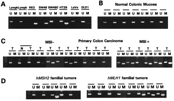
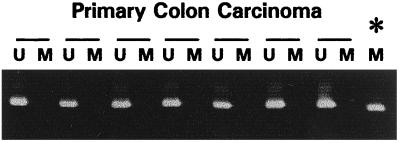
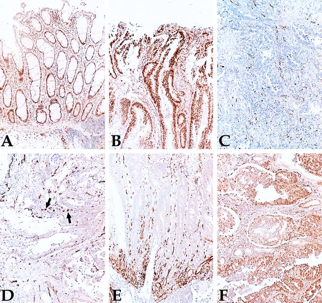
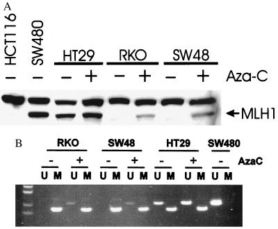
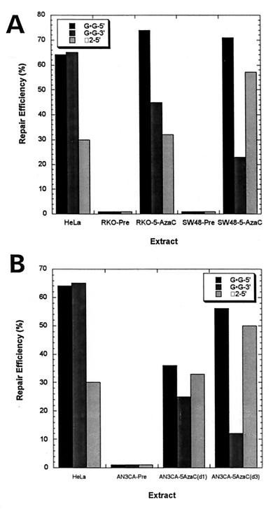
Inactivation of the genes involved in DNA mismatch repair is associated with microsatellite instability (MSI) in colorectal cancer. We report that hypermethylation of the 5' CpG island of hMLH1 is found in the majority of sporadic primary colorectal cancers with MSI, and that this methylation was often, but not invariably, associated with loss of hMLH1 protein expression. Such methylation also occurred, but was less common, in MSI- tumors, as well as in MSI+ tumors with known mutations of a mismatch repair gene (MMR). No hypermethylation of hMSH2 was found. Hypermethylation of colorectal cancer cell lines with MSI also was frequently observed, and in such cases, reversal of the methylation with 5-aza-2'-deoxycytidine not only resulted in reexpression of hMLH1 protein, but also in restoration of the MMR capacity in MMR-deficient cell lines. Our results suggest that microsatellite instability in sporadic colorectal cancer often results from epigenetic inactivation of hMLH1 in association with DNA methylation.
Figures
Similar articles
-
Microsatellite instability in inflammatory bowel disease-associated neoplastic lesions is associated with hypermethylation and diminished expression of the DNA mismatch repair gene, hMLH1.Cancer Res. 2000 Sep 1;60(17):4864-8. Cancer Res. 2000. PMID: 10987299
-
hMLH1 promoter methylation and lack of hMLH1 expression in sporadic gastric carcinomas with high-frequency microsatellite instability.Cancer Res. 1999 Jan 1;59(1):159-64. Cancer Res. 1999. PMID: 9892201
-
Mismatch repair gene expression defects contribute to microsatellite instability in ovarian carcinoma.Cancer. 2003 Nov 15;98(10):2199-206. doi: 10.1002/cncr.11770. Cancer. 2003. PMID: 14601090
-
Diagnostic application of hMLH1 methylation in hereditary non-polyposis colorectal cancer.Dis Markers. 2004;20(4-5):277-82. doi: 10.1155/2004/371941. Dis Markers. 2004. PMID: 15528793 Free PMC article. Review.
-
Emerging pathways in colorectal-cancer development.Lancet Oncol. 2002 Feb;3(2):83-8. doi: 10.1016/s1470-2045(02)00649-6. Lancet Oncol. 2002. PMID: 11905459 Review.
Cited by
-
Prdx1 deficiency in mice promotes tissue specific loss of heterozygosity mediated by deficiency in DNA repair and increased oxidative stress.Mutat Res. 2012 Jul 1;735(1-2):39-45. doi: 10.1016/j.mrfmmm.2012.04.004. Epub 2012 May 11. Mutat Res. 2012. PMID: 22583657 Free PMC article.
-
Kras gene mutation and RASSF1A, FHIT and MGMT gene promoter hypermethylation: indicators of tumor staging and metastasis in adenocarcinomatous sporadic colorectal cancer in Indian population.PLoS One. 2013;8(4):e60142. doi: 10.1371/journal.pone.0060142. Epub 2013 Apr 3. PLoS One. 2013. PMID: 23573237 Free PMC article.
-
Mechanism of mismatch recognition revealed by human MutSβ bound to unpaired DNA loops.Nat Struct Mol Biol. 2011 Dec 18;19(1):72-8. doi: 10.1038/nsmb.2175. Nat Struct Mol Biol. 2011. PMID: 22179786 Free PMC article.
-
The role of aberrant DNA methylation in cancer initiation and clinical impacts.Ther Adv Med Oncol. 2024 Jan 28;16:17588359231220511. doi: 10.1177/17588359231220511. eCollection 2024. Ther Adv Med Oncol. 2024. PMID: 38293277 Free PMC article.
-
Wedding of Molecular Alterations and Immune Checkpoint Blockade: Genomics as a Matchmaker.J Natl Cancer Inst. 2021 Nov 29;113(12):1634-1647. doi: 10.1093/jnci/djab067. J Natl Cancer Inst. 2021. PMID: 33823006 Free PMC article. Review.
References
-
- Thomas D C, Umar A, Kunkel T A. Mutat Res. 1996;350:201–205. - PubMed
-
- Modrich P, Lahue R. Annu Rev Biochem. 1996;65:101–33. , 101–133. - PubMed
-
- Aaltonen L A, Peltomaki P, Leach F S, Sistonen P, Pylkkanen L, Mecklin J P, Jarvinen H, Powell S M, Jen J, Hamilton S R, et al. Science. 1993;260:812–816. - PubMed
-
- Thibodeau S N, Bren G, Schaid D. Science. 1993;260:816–819. - PubMed
-
- Peltomaki P, de la Chapelle A. Adv Cancer Res. 1997;71:93–119. - PubMed
Publication types
MeSH terms
Substances
Grants and funding
LinkOut - more resources
Full Text Sources
Other Literature Sources
Medical
