Assembly, subunit composition, and footprint of human DNA repair excision nuclease
- PMID: 9618470
- PMCID: PMC22593
- DOI: 10.1073/pnas.95.12.6669
Assembly, subunit composition, and footprint of human DNA repair excision nuclease
Abstract
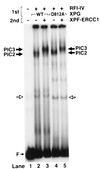
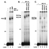
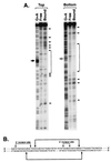
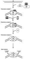
The assembly and composition of human excision nuclease were investigated by electrophoretic mobility shift assay and DNase I footprinting. Individual repair factors or any combination of up to four repair factors failed to form DNA-protein complexes of high specificity and stability. A stable complex of high specificity can be detected only when XPA/RPA, transcription factor IIH, XPC.HHR23B, and XPG and ATP are present in the reaction mixture. The XPF.ERCC1 heterodimer changes the electrophoretic mobility of the DNA-protein complex formed with the other five repair factors, but it does not confer additional specificity. By using proteins with peptide tags or antibodies to the repair factors in electrophoretic mobility shift assays, it was found that XPA, replication protein A, transcription factor IIH, XPG, and XPF.excision repair cross-complementing 1 but not XPC.HHR23B were present in the penultimate and ultimate dual incision complexes. Thus, it appears that XPC.HHR23B is a molecular matchmaker that participates in the assembly of the excision nuclease but is not present in the ultimate dual incision complex. The excision nuclease makes an assymmetric DNase I footprint of approximately 30 bp around the damage and increases the DNase I sensitivity of the DNA on both sides of the footprint.
Figures


Similar articles
-
Mechanism of open complex and dual incision formation by human nucleotide excision repair factors.EMBO J. 1997 Nov 3;16(21):6559-73. doi: 10.1093/emboj/16.21.6559. EMBO J. 1997. PMID: 9351836 Free PMC article.
-
Order of assembly of human DNA repair excision nuclease.J Biol Chem. 1999 Jun 25;274(26):18759-68. doi: 10.1074/jbc.274.26.18759. J Biol Chem. 1999. PMID: 10373492
-
Strong functional interactions of TFIIH with XPC and XPG in human DNA nucleotide excision repair, without a preassembled repairosome.Mol Cell Biol. 2001 Apr;21(7):2281-91. doi: 10.1128/MCB.21.7.2281-2291.2001. Mol Cell Biol. 2001. PMID: 11259578 Free PMC article.
-
DNA damage recognition during nucleotide excision repair in mammalian cells.Biochimie. 1999 Jan-Feb;81(1-2):39-44. doi: 10.1016/s0300-9084(99)80036-4. Biochimie. 1999. PMID: 10214908 Review.
-
Xeroderma pigmentosum and molecular cloning of DNA repair genes.Anticancer Res. 1996 Mar-Apr;16(2):693-708. Anticancer Res. 1996. PMID: 8687116 Review.
Cited by
-
Mechanism of release and fate of excised oligonucleotides during nucleotide excision repair.J Biol Chem. 2012 Jun 29;287(27):22889-99. doi: 10.1074/jbc.M112.374447. Epub 2012 May 9. J Biol Chem. 2012. PMID: 22573372 Free PMC article.
-
DNA damage in the nucleosome core is refractory to repair by human excision nuclease.Mol Cell Biol. 2000 Dec;20(24):9173-81. doi: 10.1128/MCB.20.24.9173-9181.2000. Mol Cell Biol. 2000. PMID: 11094069 Free PMC article.
-
The SWI/SNF chromatin-remodeling factor stimulates repair by human excision nuclease in the mononucleosome core particle.Mol Cell Biol. 2002 Oct;22(19):6779-87. doi: 10.1128/MCB.22.19.6779-6787.2002. Mol Cell Biol. 2002. PMID: 12215535 Free PMC article.
-
Replication protein A safeguards genome integrity by controlling NER incision events.J Cell Biol. 2011 Feb 7;192(3):401-15. doi: 10.1083/jcb.201006011. Epub 2011 Jan 31. J Cell Biol. 2011. PMID: 21282463 Free PMC article.
-
The sequence dependence of human nucleotide excision repair efficiencies of benzo[a]pyrene-derived DNA lesions: insights into the structural factors that favor dual incisions.J Mol Biol. 2009 Mar 13;386(5):1193-203. doi: 10.1016/j.jmb.2008.12.082. Epub 2009 Jan 8. J Mol Biol. 2009. PMID: 19162041 Free PMC article.
References
-
- Sancar A. Annu Rev Biochem. 1996;65:43–81. - PubMed
-
- Mu D, Park C H, Matsunaga T, Hsu D S, Reardon J T, Sancar A. J Biol Chem. 1995;270:2415–2418. - PubMed
-
- Mu D, Hsu D S, Sancar A. J Biol Chem. 1996;271:8285–8294. - PubMed
-
- Moggs J G, Yarema K J, Essigmann J M, Wood R D. J Biol Chem. 1996;271:7177–7186. - PubMed
Publication types
MeSH terms
Substances
Grants and funding
LinkOut - more resources
Full Text Sources

