

CYR61, a product of a growth factor-inducible immediate early gene, promotes angiogenesis and tumor growth
- PMID: 9600969
- PMCID: PMC27701
- DOI: 10.1073/pnas.95.11.6355
CYR61, a product of a growth factor-inducible immediate early gene, promotes angiogenesis and tumor growth
Abstract
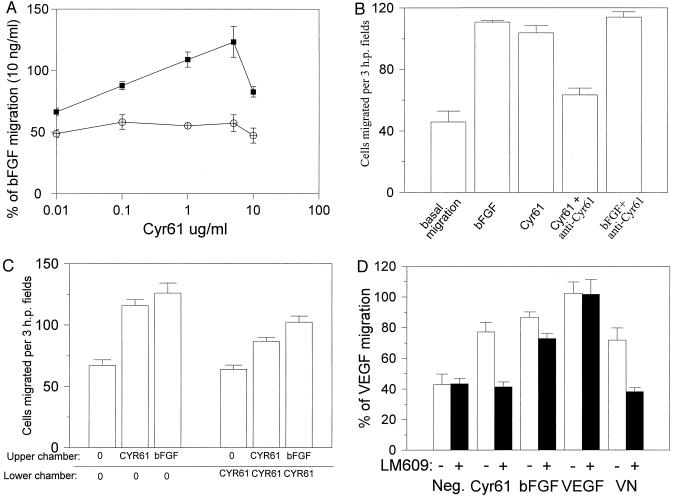
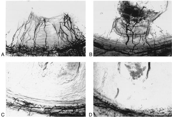
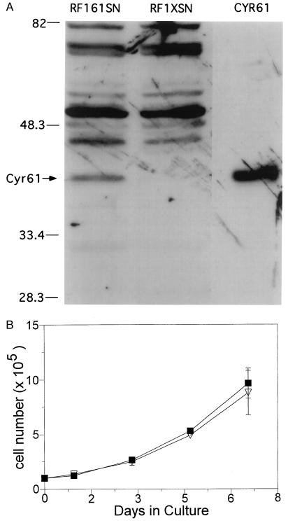
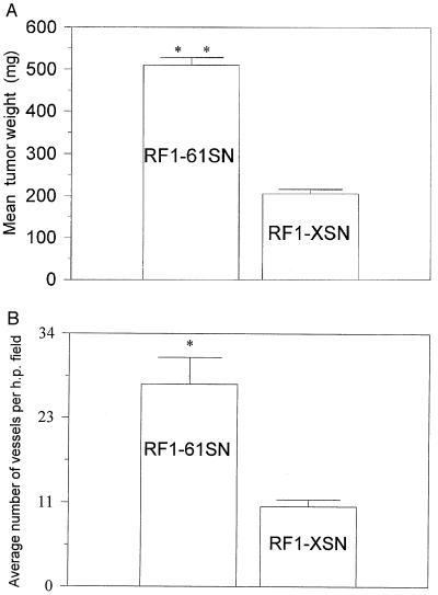
CYR61 is a secreted, cysteine-rich, heparin-binding protein encoded by a growth factor-inducible immediate-early gene. Acting as an extracellular, matrix-associated signaling molecule, CYR61 promotes the adhesion of endothelial cells through interaction with the integrin alphaVbeta3 and augments growth factor-induced DNA synthesis in the same cell type. In this study, we show that purified CYR61 stimulates directed migration of human microvascular endothelial cells in culture through an alphaV beta3-dependent pathway and induces neovascularization in rat corneas. Both the chemotactic and angiogenic activities of CYR61 can be blocked by specific anti-CYR61 antibodies. Whereas most human tumor-derived cell lines tested express CYR61, the gastric adenocarcinoma cell line RF-1 does not. Expression of the CYR61 cDNA under the regulation of a constitutive promoter in RF-1 cells significantly enhances the tumorigenicity of these cells as measured by growth in immunodeficient mice, resulting in tumors that are larger and more vascularized than those produced by control RF-1 cells. Taken together, these results identify CYR61 as an angiogenic inducer that can promote tumor growth and vascularization; the results also suggest potential roles for CYR61 in physiologic and pathologic neovascularization.
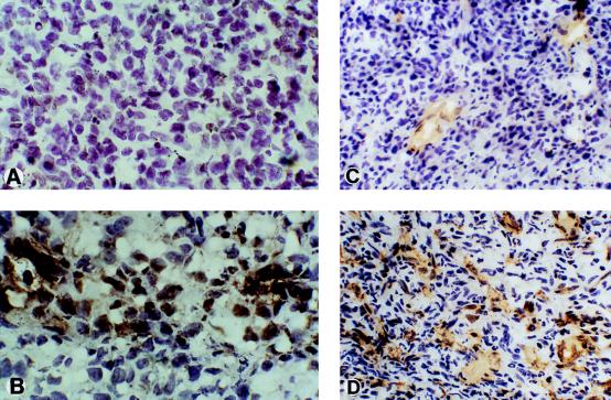
Figures
Similar articles
-
The angiogenic factor cysteine-rich 61 (CYR61, CCN1) supports vascular smooth muscle cell adhesion and stimulates chemotaxis through integrin alpha(6)beta(1) and cell surface heparan sulfate proteoglycans.Endocrinology. 2002 Apr;143(4):1441-50. doi: 10.1210/endo.143.4.8731. Endocrinology. 2002. PMID: 11897702
-
Cyr61, a product of a growth factor-inducible immediate-early gene, promotes cell proliferation, migration, and adhesion.Mol Cell Biol. 1996 Apr;16(4):1326-34. doi: 10.1128/MCB.16.4.1326. Mol Cell Biol. 1996. PMID: 8657105 Free PMC article.
-
The angiogenic factor Cyr61 activates a genetic program for wound healing in human skin fibroblasts.J Biol Chem. 2001 Dec 14;276(50):47329-37. doi: 10.1074/jbc.M107666200. Epub 2001 Oct 2. J Biol Chem. 2001. PMID: 11584015
-
CYR61 stimulates human skin fibroblast migration through Integrin alpha vbeta 5 and enhances mitogenesis through integrin alpha vbeta 3, independent of its carboxyl-terminal domain.J Biol Chem. 2001 Jun 15;276(24):21943-50. doi: 10.1074/jbc.M100978200. Epub 2001 Apr 3. J Biol Chem. 2001. PMID: 11287419
-
The angiogenic factor CYR61 in breast cancer: molecular pathology and therapeutic perspectives.Endocr Relat Cancer. 2003 Jun;10(2):141-52. doi: 10.1677/erc.0.0100141. Endocr Relat Cancer. 2003. PMID: 12790776 Review.
Cited by
-
Cancer-associated Fibroblast-specific Expression of the Matricellular Protein CCN1 Coordinates Neovascularization and Stroma Deposition in Melanoma Metastasis.Cancer Res Commun. 2024 Feb 27;4(2):556-570. doi: 10.1158/2767-9764.CRC-23-0571. Cancer Res Commun. 2024. PMID: 38363129 Free PMC article.
-
Differential gene expression in chemically induced mouse lung adenomas.Neoplasia. 2003 Jan-Feb;5(1):41-52. doi: 10.1016/s1476-5586(03)80016-7. Neoplasia. 2003. PMID: 12659669 Free PMC article.
-
Inhibition of glioma cell growth and tumorigenic potential by CCN3 (NOV).Mol Pathol. 2001 Oct;54(5):293-9. doi: 10.1136/mp.54.5.293. Mol Pathol. 2001. PMID: 11577170 Free PMC article.
-
Molecular responses to hypoxia in tumor cells.Mol Cancer. 2003 Apr 17;2:23. doi: 10.1186/1476-4598-2-23. Mol Cancer. 2003. PMID: 12740039 Free PMC article. Review.
-
The angiogenic factor Cyr61 is induced by the progestin R5020 and is necessary for mammary adenocarcinoma cell growth.Endocrine. 2002 Jul;18(2):147-59. doi: 10.1385/ENDO:18:2:147. Endocrine. 2002. PMID: 12374462
References
Publication types
MeSH terms
Substances
Grants and funding
LinkOut - more resources
Full Text Sources
Other Literature Sources
