Expression of integrin-associated protein gene associated with memory formation in rats
- PMID: 9592107
- PMCID: PMC6792811
- DOI: 10.1523/JNEUROSCI.18-11-04305.1998
Expression of integrin-associated protein gene associated with memory formation in rats
Abstract
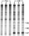
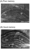
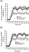
The present study has adopted the PCR differential display method to identify cDNA clones associated with memory formation in rats. The one-way inhibitory avoidance learning task was used as the behavioral paradigm. Total RNA isolated from the hippocampus of poor-memory (<80 sec) and good-memory (600 sec) rats 3 hr after training was used for comparison. Three cDNA fragments corresponding to different spliced forms of integrin-associated protein (IAP) mRNA were found to be differentially expressed in the hippocampus of good-memory rats. Quantitative reverse transcription-PCR revealed approximately four fold higher of IAP mRNA level in good-memory rats. This result was confirmed further by in situ hybridization analysis, and the major difference was in the dentate gyrus. It has been demonstrated that this difference in IAP mRNA expression is not attributable to different sensitivities of individual rats to electric shock. Rapid amplification of cDNA ends obtained the full-length IAP cDNA, which is 1192 bp in length excluding the poly(A+) tail. The IAP mRNA expression was significantly upregulated by NMDA and amphetamine injections to the dentate gyrus of the hippocampus. On the other hand, injection of antisense oligonucleotide complementary to the IAP transcript markedly impaired memory retention in rats and decreased the amplitude and slope of EPSP in the in vivo long-term potentiation paradigm. These results together suggest that IAP gene expression plays an important role in memory formation and synaptic plasticity in rat hippocampus.
Figures


Similar articles
-
Induction of integrin-associated protein (IAP) mRNA expression during memory consolidation in rat hippocampus.Eur J Neurosci. 2000 Mar;12(3):1105-12. doi: 10.1046/j.1460-9568.2000.00985.x. Eur J Neurosci. 2000. PMID: 10762341
-
Brain-derived neurotrophic factor antisense oligonucleotide impairs memory retention and inhibits long-term potentiation in rats.Neuroscience. 1998 Feb;82(4):957-67. doi: 10.1016/s0306-4522(97)00325-4. Neuroscience. 1998. PMID: 9466420
-
Impaired memory retention and decreased long-term potentiation in integrin-associated protein-deficient mice.Learn Mem. 1999 Sep-Oct;6(5):448-57. doi: 10.1101/lm.6.5.448. Learn Mem. 1999. PMID: 10541465 Free PMC article.
-
Memory formation: the sequence of biochemical events in the hippocampus and its connection to activity in other brain structures.Neurobiol Learn Mem. 1997 Nov;68(3):285-316. doi: 10.1006/nlme.1997.3799. Neurobiol Learn Mem. 1997. PMID: 9398590 Review.
-
[Long-term potentiation of the NMDA-dependent component of the EPSP in the hippocampus].Usp Fiziol Nauk. 1998 Oct-Dec;29(4):6-23. Usp Fiziol Nauk. 1998. PMID: 9883495 Review. Russian.
Cited by
-
Novel role and mechanism of protein inhibitor of activated STAT1 in spatial learning.EMBO J. 2011 Jan 5;30(1):205-20. doi: 10.1038/emboj.2010.290. Epub 2010 Nov 19. EMBO J. 2011. PMID: 21102409 Free PMC article.
-
IAP-Based Cell Sorting Results in Homogeneous Transplantable Dopaminergic Precursor Cells Derived from Human Pluripotent Stem Cells.Stem Cell Reports. 2017 Oct 10;9(4):1207-1220. doi: 10.1016/j.stemcr.2017.08.016. Epub 2017 Sep 21. Stem Cell Reports. 2017. PMID: 28943253 Free PMC article.
-
The absence of CD47 promotes nerve fiber growth from cultured ventral mesencephalic dopamine neurons.PLoS One. 2012;7(9):e45218. doi: 10.1371/journal.pone.0045218. Epub 2012 Sep 26. PLoS One. 2012. PMID: 23049778 Free PMC article.
-
sgk, a primary glucocorticoid-induced gene, facilitates memory consolidation of spatial learning in rats.Proc Natl Acad Sci U S A. 2002 Mar 19;99(6):3990-5. doi: 10.1073/pnas.062405399. Epub 2002 Mar 12. Proc Natl Acad Sci U S A. 2002. PMID: 11891330 Free PMC article.
-
Gene expression changes in the medial prefrontal cortex and nucleus accumbens following abstinence from cocaine self-administration.BMC Neurosci. 2010 Feb 26;11:29. doi: 10.1186/1471-2202-11-29. BMC Neurosci. 2010. PMID: 20187946 Free PMC article.
References
-
- Bading H, Greenberg ME. Stimulation of protein tyrosine phosphorylation by NMDA receptor activation. Science. 1991;253:912–914. - PubMed
-
- Bahr BA, Sheppard A, Lynch G. Fibronectin binding by brain synaptosomal membranes may not involve conventional integrins. NeuroReport. 1991a;2:13–16. - PubMed
-
- Bahr BA, Sheppard A, Vanderklish PW, Bakus BL, Capaldi D, Lynch G. Antibodies to the alpha v beta 3 integrin label a protein concentrated in brain synaptosomal membranes. NeuroReport. 1991b;2:321–324. - PubMed
Publication types
MeSH terms
Substances
LinkOut - more resources
Full Text Sources
Other Literature Sources
Medical
Molecular Biology Databases
Research Materials
