. 1998 Jun 1;18(11):4029-41.
doi: 10.1523/JNEUROSCI.18-11-04029.1998.

Endocytotic formation of vesicles and other membranous structures induced by Ca2+ and axolemmal injury

Affiliations

Endocytotic formation of vesicles and other membranous structures induced by Ca2+ and axolemmal injury

C S Eddleman et al. J Neurosci. .

Abstract

Vesicles and/or other membranous structures that form after axolemmal damage have recently been shown to repair (seal) the axolemma of various nerve axons. To determine the origin of such membranous structures, (1) we internally dialyzed isolated intact squid giant axons (GAs) and showed that elevation of intracellular Ca2+ >100 microM produced membranous structures similar to those in axons transected in Ca2+-containing physiological saline; (2) we exposed GA axoplasm to Ca2+-containing salines and observed that membranous structures did not form after removing the axolemma and glial sheath but did form in severed GAs after >99% of their axoplasm was removed by internal perfusion; (3) we examined transected GAs and crayfish medial giant axons (MGAs) with time-lapse confocal fluorescence microscopy and showed that many injury-induced vesicles formed by endocytosis of the axolemma; (4) we examined the cut ends of GAs and MGAs with electron microscopy and showed that most membranous structures were single-walled at short (5-15 min) post-transection times, whereas more were double- and multi-walled and of probable glial origin after longer (30-150 min) post-transection times; and (5) we examined differential interference contrast and confocal images and showed that large and small lesions evoked similar injury responses in which barriers to dye diffusion formed amid an accumulation of vesicles and other membranous structures. These and other data suggest that Ca2+ inflow at large or small axolemmal lesions induces various membranous structures (including endocytotic vesicles) of glial or axonal origin to form, accumulate, and interact with each other, preformed vesicles, and/or the axolemma to repair the axolemmal damage.


Figures
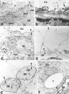
Fig. 1.
Fig. 1.
Electron micrographs of GAs (A,C, E, F) and MGAs (B, D). A, Normal squid (Sepioteuthis) axonal–glial interface.Ax, Axoplasm; bl, basal lamina;gp, gliaplasm; gcn, glial cell nucleus;gol, Golgi body; m, mitochondrion;rer, rough endoplasmic reticulum; s, smooth endoplasmic reticulum; tl, transverse tubular lattice. Apposing arrowheads indicate double-layered membranous structures identified as the axolemma and glialemma, except for E. ∗1–∗5, Double- or multi-walled membranous structures. B, Normal crayfish MGA glial interface. C, Discontinuous axolemma and intermingling of axonal and glial elements ∼20 μm from the cut end of a transected GA fixed at 5 min after transection. D, Invaginating axolemma (Figure legend continued) (downward arrowhead not facing an upward arrowhead) within 50 μm of the cut end of a crayfish MGA fixed at 20 min after transection. Glialemma (apposing arrowheads) does not evaginate at this site of axolemmal invagination. E, Double-walled vesicles containing gliaplasm ∼20 μm from the cut end of a transected GA fixed at 5 min after transection. F, Large (10 μm) single-walled vesicle (V) in axoplasm and highly vesiculated gliaplasm ∼150 μm from the cut end of a GA fixed at 30 min after transection. Scale bars: A–C, E, 0.45 μm;D, 1.2 μm; F, 2.0 μm.
Fig. 2.
Fig. 2.
DIC images showing extent of invagination and vesiculation in the dialyzed region of a squid GA bathed in control internal saline after changes in the ionic composition of the solution used to internally microdialyze the axoplasm. A, No invagination or vesiculation after dialysis with control internal saline (buffered K glutamate). B, Small amount of invagination or vesiculation (arrowheads) after dialysis with the internal saline in A modified by Cl replacement of glutamate. C, Moderate amount of invagination or vesiculation after dialysis with the internal saline in B modified by Na+replacement of K+. D, Extensive invagination or vesiculation (V) after dialysis with the internal saline used in C modified to ensure that the free concentration of Ca2+ equaled 1 mm. All images were acquired 30 min after introducing a new microdialysate solution to ensure equilibration of the axoplasmic ionic concentrations with those of the dialysate. Scale bar (inA), 25 μm.
Fig. 3.
Fig. 3.
DIC images of squid GAs showing Ca2+- or injury-induced invagination and vesiculation. A, Extensive invagination and vesiculation (arrows) ∼400 μm from the cut end in GA axoplasm 30 min after transecting the GA in control external saline containing 10 mm Ca2+. Inset, No invagination or vesiculation of the intact GA in control external saline before its transection. B, No invagination or vesiculation 30 min after adding 1 mmCa2+ to the control internal saline that bathed the isolated desheathed axoplasm (see text). C, No invagination or vesiculation in a GA after microdialysis for 1 hr with buffered KI that removed >99% of the axoplasm. D, Invagination and vesiculation (arrows) ∼350 μm from the cut end induced 15 min after severance in control external saline (containing 10 mm Ca2+) of the same axon shown in C. This result suggests that vesiculation or invagination requires a plasmalemma but does not require the presence of axoplasm. Scale bar (in D): A–D, 25 μm; A, inset, 100 μm.
Fig. 4.
Fig. 4.
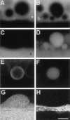
Confocal images taken 50 μm from the cut ends of lesioned MGAs exposed to fluorescent dyes.A, B, Membranous structures identified in DIC (data not shown) in the axoplasm of an MGA injected with 0.01% FITC-dextran before transection and viewed 10 min after transection. Absence of fluorescence (black holes) indicates membranous structures, the contents of which do not include FITC-dextran. Fluorescent area (white region) at the top ofA and B delineates the axoplasm (a) just interior to the axolemma; theblack region at the bottom is the extracellular space. Black holes were not observed in the axoplasm before transecting the MGA. C, Same axoplasmic region shown in A but imaged for fluorescence of Texas Red-dextran placed in the bath after black holes had formed. Many of the black holes in A remained unlabeled, indicating that they are axoplasmic vesicles. Fluorescent area delineates the extracellular bath (b). D, Same axoplasmic region shown in B but imaged for fluorescence of Texas Red-dextran placed in the bath after black holes had formed. The black holes in B are dye-filled in D, indicating that they are invaginations that are connected to the extracellular space. E, Ring-shaped fluorescence of injury-induced membranous structure in axoplasm 10 min after transection of an MGA that was pulse-labeled with FM 4-64 (25 μm for 10 min) before transection. F, Same axoplasmic region shown in E but imaged for fluorescence of FITC-dextran placed in the bath 2 min after transection and washed off at 10 min after transection. After dye washout from the bath, the membranous structure retained the dye, indicating its isolation from the bath saline; i.e., this structure was an axoplasmic vesicle. The images in E and F are consistent with a vesicle that formed from FM-labeled plasmalemmal membrane after an axolemmal invagination which subsequently budded off to become an axoplasmic vesicle. G, Image of a membranous structure invaginating into the axoplasm. The contents of this structure filled with Texas Red-dextran that was added to the bath saline 10 min after transection. H, Same optical section as inG, but imaged for calcein (glial cytosolic marker) (Eddleman et al., 1995) showing no evagination of the glial cell associated with the axoplasmic invagination in G. The images in G and H are consistent with an axolemmal invagination not associated with a glialemmal evagination. Scale bar (in H): A–D, 5 μm;E, F, 1 μm; G, H, 15 μm.
Fig. 5.
Fig. 5.
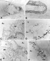
Light (A, B) and electron micrographs (C–F) of a transected squid GA (A, C,E) and a transected crayfish MGA (B, D, F).A–F, The cut axonal end is oriented to theright. Electron micrographs are enlargements of a representative portion of the circles inA and B, which in turn are representative of cortical axoplasmic and adaxonal glial regions within 200 μm of the cut axonal ends. Ax, Axoplasm; bl, basal lamina; c, collagen layer; gp, gliaplasm; gcn, glial cell nucleus. A, Midsagittal section of squid (Sepioteuthis) GA fixed at 150 min after transection. B, Midsagittal section of a crayfish MGA fixed at 60 min after transection. Thearrowhead marks a vesicle in the axoplasmic space near the cut end. C, Magnified view of the large circle in A where the axolemma and glialemma are no longer identifiable, and the gliaplasm mixes with the axoplasm ∼220 μm from the cut end. The axoplasm (Ax) contains no identifiable cytoplasmic structures. D, Magnified view of the large circle in B where the axolemma and glialemma are no longer present, and the adaxonal layer (brackets) has been replaced by electron-dense bodies and vesicles. V, Part of the large vesicle labeled with an arrowhead in B ∼15 μm from the cut end. E, Magnified view of the small circle in A ∼180 μm from the cut end. The axolemma and glialemma are no (Figure legend continued) longer identifiable. The gliaplasm contains many single-layered (∗1) and multi-layered (∗2) membranous structures. F, Magnified view of thesmall circle in B ∼30 μm from the cut end. The axolemma and glialemma are no longer identifiable. The adaxonal layer (brackets) contains single- and multi-layered membranous structures and electron-dense bodies, rather than its normal complement of cytoskeletal structures (e.g., microtubules, ER, and transverse tubular lattice). Scale bar (inF): A, 63 μm; B, 25 μm; C, E, 0.77 μm; D, F, 9.1 μm.
Fig. 6.
Fig. 6.
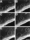
Series of time-lapse confocal fluorescence images in the same optical midsection ∼100 μm from the cut end showing stages of injury-induced endocytosis beginning with an invagination of FM-labeled membrane and ending with a vesicle moving in the axoplasm toward the cut end of a transected MGA. A–F, The axon is at the top, the bath is at the bottom, and the cut end is toward the right. The plasmalemmal membranes of an intact MGA were pulse-labeled with FM 1-43. The MGA was transected and imaged for FM 1-43 fluorescence starting at 11 min after transection without changing the confocal plane or the position of the micrometer stage. A–F, Successive images were acquired at 18 sec intervals and taken from a larger set of images acquired every 6 sec. Asterisks mark the same vesicle in every frame. Note that vesicles are sometimes joined by a fluorescent line, presumably a tether of membranous material. Scale bar (inF), 10 μm.
Fig. 7.
Fig. 7.
Series of time-lapse fluorescence images in the same optical section of a severed MGA (A–C) and at the site of a micropuncture in the axolemma of an MGA (D–F) showing the movement toward, and accumulation at, the cut end and small hole, respectively, of vesicles formed by endocytosis (see Fig. 6) of the axolemma. The MGA was pulse-labeled with FM 1-43 and then transected. The cut end of the MGA is oriented to the left in A–C.A–C, Successive images at 3, 8, and 13 min after transection, respectively, representative of a larger set of images acquired every 6 sec. D–F, Successive images at the site of a micropuncture in an MGA at 5, 10, and 25 min, respectively, after the physiological saline, in which the MGA was punctured, was replaced by a saline containing Texas Red-dextran (1%) at 5 min after the micropuncture. The interior of the axon is at thetop of each panel. The black holes are confocal images of vesicles that contain no dye and are surrounded by Texas Red, which entered the axon through the micropuncture (arrow) after dye was added to the bath. Scale bar (in D):A–C, 40 μm; D–F, 20 μm.
Fig. 8.
Fig. 8.
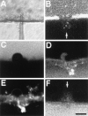
DIC (A) and confocal (B–F) images of MGAs micropunctured in Ca2+-containing salines (A–E) or Ca2+-free salines (F) showing dye exclusion amid an accumulation of Ca2+-induced vesicles and other membranous structures (C–E) or showing dye uptake (B, F). All MGAs were incubated in calcein AM for 5–30 min before being micropunctured. A–F, The axon is at the top, the bath is at thebottom, and the micropuncture is approximately in thecenter. A, Image of a micropipette penetrating an MGA. B, Image of calcein hydrophilic dye leaking out of an MGA at the micropuncture site (arrow) at 3 min after puncture, i.e., before formation of a barrier (seal) to hydrophilic dyes. C, Image of calcein fluorescence showing membranous structures identified with DIC (data not shown) at the micropuncture site in an MGA at 25 min after puncture.D, Same confocal plane as in C but imaged at 25 min after puncture for Texas Red-dextran, which was added to the bath at 20 min after puncture. E, Same confocal plane asC and D but imaged at 50 min after puncture for FM 1-43, which was added to the bath at 40 min after puncture. The styryl dye incorporated into the membranes of the structures at the injury site and did not label membranes in the interior of the MGA; i.e., a barrier to the FM dye formed amid a collection of vesicles at the micropuncture site. F, MGA micropunctured and maintained in Ca2+-free saline. Cy5-dextran hydrophilic dye was added to the bath at 20 min after puncture and then imaged at 25 min after puncture. Note uptake of Cy5-dextran into the axoplasm at the micropuncture site (arrow). Scale bar (in F): 15 μm.

Similar articles

Cited by

References

    1. Ashery U, Penner R, Spira ME. Acceleration of membrane recycling by axotomy of cultured Aplysia neurons. Neuron. 1996;16:641–651. - PubMed
    1. Ballinger ML, Bittner GD. Ultrastructural studies of severed giant and other CNS axons in crayfish. Cell Tissue Res. 1980;208:123–133. - PubMed
    1. Ballinger ML, Blanchette AR, Krause TL, Smyers ME, Fishman HM, Bittner GD. Delaminating myelin membranes help seal the cut ends of severed earthworm giant axons. J Neurobiol. 1997;33:945–960. - PubMed
    1. Baumgold J, Terakawa S, Iwasa K, Gainer H. Membrane-associated cytoskeletal proteins in squid giant axons. J Neurochem. 1981;36:759–764. - PubMed
    1. Betz WJ, Mao F, Bewick GS. Activity-dependent staining and destaining of living vertebrate motor nerve terminals. J Neurosci. 1992;12:363–375. - PMC - PubMed

Publication types

MeSH terms

LinkOut - more resources