

Extrasynaptic vesicular transmitter release from the somata of substantia nigra neurons in rat midbrain slices
- PMID: 9570786
- PMCID: PMC6793140
- DOI: 10.1523/JNEUROSCI.18-10-03548.1998
Extrasynaptic vesicular transmitter release from the somata of substantia nigra neurons in rat midbrain slices
Abstract
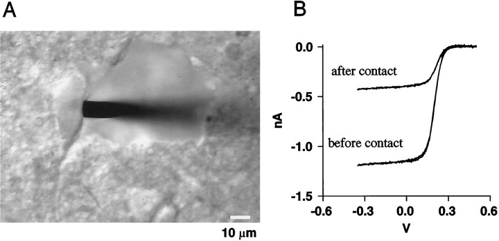
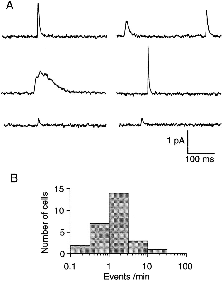
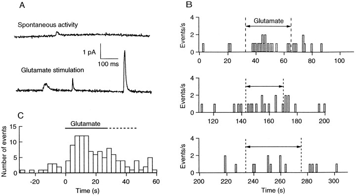
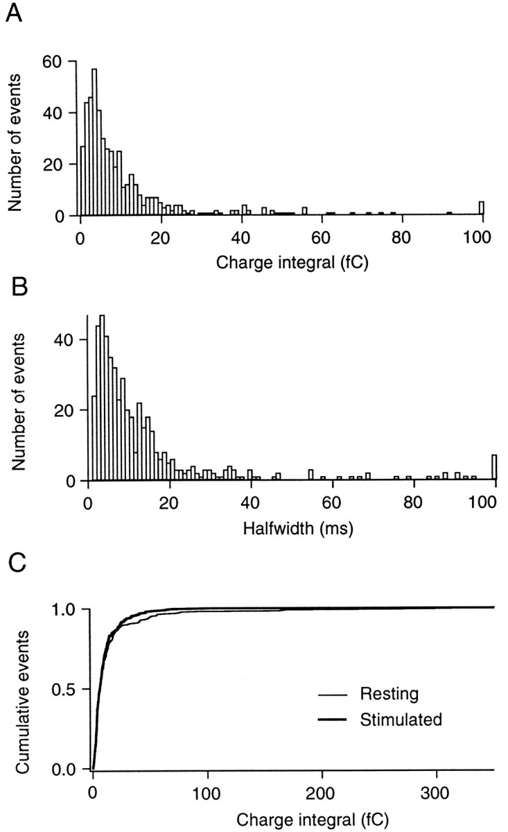
Substantia nigra neurons release dopamine from their somatodendritic regions. A long-unresolved question is whether this release occurs by exocytosis or by a nonvesicular mechanism. We used carbon fiber microelectrodes in a brainstem slice to assay secretion from single cell bodies that had been cleared of connective tissue. Amperometry at the carbon fiber microelectrodes revealed unitary events in approximately 90% of cells in resting conditions. These events had charge integrals ranging from a few femtocoulombs to several hundred femtocoulombs (fC). Local glutamate application enhanced the event frequency by 3.5-fold on average and up to 10-fold in highly responsive cells, although the mean charge integral was not modified. Local application of a high K+-containing saline had effects similar to those of glutamate. The frequency of resting and stimulated amperometric events was much lower at 21-22 degreesC than at 32-35 degreesC. The addition of Cd2+ (50 microM), a blocker of voltage-dependent Ca2+ channels, to the bath solution blocked the stimulatory effects of glutamate. These results suggest that dopamine is released from the somata of substantia nigra neurons by exocytosis and that this mechanism is regulated by neuronal electrical activity. More generally, this study demonstrates the applicability of carbon fiber microelectrodes to the measurement of quantal monoamine secretion in brain slices.
Figures
Similar articles
-
Limited regulation of somatodendritic dopamine release by voltage-sensitive Ca channels contrasted with strong regulation of axonal dopamine release.J Neurochem. 2006 Feb;96(3):645-55. doi: 10.1111/j.1471-4159.2005.03519.x. Epub 2006 Jan 9. J Neurochem. 2006. PMID: 16405515
-
Voltage-operated Ca2+ channels regulate dopamine release from somata of dopamine neurons in the substantia nigra pars compacta.Biochem Biophys Res Commun. 2008 Sep 5;373(4):665-9. doi: 10.1016/j.bbrc.2008.06.099. Epub 2008 Jul 2. Biochem Biophys Res Commun. 2008. PMID: 18601902
-
Glutamate-mediated [Ca2+]c dynamics in spontaneously firing dopamine neurons of the rat substantia nigra pars compacta.J Cell Sci. 2003 Jul 1;116(Pt 13):2665-75. doi: 10.1242/jcs.00481. Epub 2003 May 13. J Cell Sci. 2003. PMID: 12746490
-
Somatodendritic dopamine release: recent mechanistic insights.Philos Trans R Soc Lond B Biol Sci. 2015 Jul 5;370(1672):20140185. doi: 10.1098/rstb.2014.0185. Philos Trans R Soc Lond B Biol Sci. 2015. PMID: 26009764 Free PMC article. Review.
-
Synaptic and extrasynaptic secretion of serotonin.Cell Mol Neurobiol. 2005 Mar;25(2):297-312. doi: 10.1007/s10571-005-3061-z. Cell Mol Neurobiol. 2005. PMID: 16047543 Review.
Cited by
-
Estimating the effect of endogenous dopamine on baseline [(11) C]-(+)-PHNO binding in the human brain.Synapse. 2016 Nov;70(11):453-60. doi: 10.1002/syn.21920. Epub 2016 Jul 11. Synapse. 2016. PMID: 27341789 Free PMC article.
-
Extrasynaptic exocytosis and its mechanisms: a source of molecules mediating volume transmission in the nervous system.Front Physiol. 2012 Sep 4;3:319. doi: 10.3389/fphys.2012.00319. eCollection 2012. Front Physiol. 2012. PMID: 22969726 Free PMC article.
-
Long-term depression of a dopamine IPSC.J Neurosci. 2007 Feb 21;27(8):2074-80. doi: 10.1523/JNEUROSCI.3251-06.2007. J Neurosci. 2007. PMID: 17314302 Free PMC article.
-
Spontaneous inhibitory synaptic currents mediated by a G protein-coupled receptor.Neuron. 2013 Jun 5;78(5):807-12. doi: 10.1016/j.neuron.2013.04.013. Neuron. 2013. PMID: 23764286 Free PMC article.
-
The real catecholamine content of secretory vesicles in the CNS revealed by electrochemical cytometry.Sci Rep. 2013;3:1447. doi: 10.1038/srep01447. Sci Rep. 2013. PMID: 23486177 Free PMC article.
References
-
- Björklund A, Lindvall O. Dopamine in the dendrites of substantia nigra neurons: suggestions for a role in dendritic terminals. Brain Res. 1975;83:531–537. - PubMed
-
- Bruns D, Jahn R. Real-time measurement of transmitter release from single synaptic vesicles. Nature. 1995;377:62–65. - PubMed
-
- Cheramy A, Leviel V, Glowinski J. Dendritic release of dopamine in the substantia nigra. Nature. 1981;289:537–542. - PubMed
-
- Chow RH, Klingauf J, Heinemann C, Zucker RS, Neher E. Mechanisms determining the time course of secretion in neuroendocrine cells. Neuron. 1996;16:369–376. - PubMed
Publication types
MeSH terms
Substances
LinkOut - more resources
Full Text Sources
Miscellaneous