. 1998 May 4;141(3):779-89.
doi: 10.1083/jcb.141.3.779.

The juxtamembrane region of the cadherin cytoplasmic tail supports lateral clustering, adhesive strengthening, and interaction with p120ctn

Affiliations

The juxtamembrane region of the cadherin cytoplasmic tail supports lateral clustering, adhesive strengthening, and interaction with p120ctn

A S Yap et al. J Cell Biol. .

Abstract

Cadherin cell-cell adhesion molecules form membrane-spanning molecular complexes that couple homophilic binding by the cadherin ectodomain to the actin cytoskeleton. A fundamental issue in cadherin biology is how this complex converts the weak intrinsic binding activity of the ectodomain into strong adhesion. Recently we demonstrated that cellular cadherins cluster in a ligand-dependent fashion when cells attached to substrata coated with the adhesive ectodomain of Xenopus C-cadherin (CEC1-5). Moreover, forced clustering of the ectodomain alone significantly strengthened adhesiveness (Yap, A.S., W.M. Brieher, M. Pruschy, and B.M. Gumbiner. Curr. Biol. 7:308-315). In this study we sought to identify the determinants of the cadherin cytoplasmic tail responsible for clustering activity. A deletion mutant of C-cadherin (CT669) that retained the juxtamembrane 94-amino acid region of the cytoplasmic tail, but not the beta-catenin-binding domain, clustered upon attachment to substrata coated with CEC1-5. Like wild-type C-cadherin, this clustering was ligand dependent. In contrast, mutant molecules lacking either the complete cytoplasmic tail or just the juxtamembrane region did not cluster. The juxtamembrane region was itself sufficient to induce clustering when fused to a heterologous membrane-anchored protein, albeit in a ligand-independent fashion. The CT669 cadherin mutant also displayed significant adhesive activity when tested in laminar flow detachment assays and aggregation assays. Purification of proteins binding to the juxtamembrane region revealed that the major associated protein is p120(ctn). These findings identify the juxtamembrane region of the cadherin cytoplasmic tail as a functionally active region supporting cadherin clustering and adhesive strength and raise the possibility that p120(ctn) is involved in clustering and cell adhesion.


Figures

Figure 1
Figure 1
C-cadherin mutant molecules. A schematic depiction of the full-length C-cadherin molecule consists of the ectodomain (gray), transmembrane region, the 94–amino acid juxtamembrane region of the cytoplasmic tail (black), and catenin-binding site (distal 56 amino acids; open oval). CT consists of the ectodomain and transmembrane regions of C-cadherin alone, the complete cytoplasmic tail being replaced by a myc tag (hatched). The predicted catenin-binding site is deleted in CT669 that retains the 94–amino acid juxtamembrane region of the cytoplasmic tail (filled rectangle) followed by a 6×-myc tag (hatched). In CT-CAT the juxtamembrane region is deleted and replaced by a 5×-myc tag (hatched). IL2R669 consists of the 94–amino acid juxtamembrane region of the C-cadherin cytoplasmic tail fused to the ectodomain and transmembrane regions of the interleukin 2 receptor α-subunit (cross-hatched).
Figure 2
Figure 2
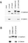
Expression of C-cadherin and mutant cadherin proteins in CHO cells. (A) Comparison between cadherin expression and cellular β-catenin levels. Western blots of lysates from parental CHO cells and CHO cells stably expressing wild-type C-cadherin (C-CHO, clone 12), tailless cadherin mutant (CT-CHO, clone 8), a deletion mutant lacking the catenin-binding region (CT669-CHO, clone A1) and a deletion mutant lacking the juxtamembrane region of the cytoplasmic tail (CT-CAT-CHO, clone 13) were probed for the C-cadherin ectodomain (top), then stripped and reprobed for β-catenin (bottom). Identical amounts of total cellular protein were loaded in each lane. Both the CT669 and CT-CAT mutants display polypeptide bands that run at or slightly higher than wild-type C-cadherin, consistent with the addition of multi-copy myc-epitope tags. Total cellular β-catenin levels were increased in cells expressing wild-type C-cadherin and CT-CAT, but not in cells expressing the CT or CT669 mutants. (B) β-catenin coimmunoprecipitates with wild-type C-cadherin and CT-CAT, but not with CT669. Lysates from C-CHO, CT669-CHO, and CT-CAT-CHO cells were immunoprecipitated with a pAb directed against the C-cadherin ectodomain, transferred to nitrocellulose and probed for C-cadherin (top) or β-catenin (bottom). C-cadherin immunoblots identify mature and precursor forms of the wild-type and mutant cadherins.
Figure 3
Figure 3
Clustering activity of wild-type and mutant C-cadherin molecules. CHO cells stably expressing C-cadherin or mutant molecules were allowed to attach for 1 h to glass substrata coated with CEC1-5 (A–E, G, and H) or poly-l-lysine (F) and then fixed and stained for the cadherin molecule (A, C, D, F, G, and H) or β-catenin (B, C, and E) by simultaneous dual-label immunofluorescence microscopy. Wild-type C-cadherin was detected using a polyclonal antibody raised against the whole cytoplasmic tail of mouse E-cadherin; mutants CT, CT669, and CT-CAT were detected by staining for the myc epitope tag. Specimens A–C were examined by confocal laser scanning microscopy with the plane of focus at the cell– substrate interface; all other samples were examined by epi-illumination microscopy. (A–C) Wild-type C-cadherin localized in clusters (A) that colocalized with β-catenin (B), as seen by the yellow fluorescence in the overlay image (C). Some β-catenin that did not colocalize with C-cadherin was detected at borders where the cells were close but not directly touching; this is likely to be in a cytoplasmic pool. (D and E) CT669 clustered in cells attached to substrata coated with CEC1-5 (D), but not with poly-l-lysine (F). CT669 clusters did not colocalize with β-catenin that remained diffusely distributed within cells (E). The image in E was overexposed for clarity in reproduction and therefore may give a misleading impression of β-catenin expression in CT669-CHO cells, which was similar to untransfected CHO cells (Fig. 2). Neither CT (G) nor CT-CAT (H) clustered upon attachment to CEC1-5–coated substrata. Note that CT-CHO and CT-CAT-CHO cells spread less on CEC1-5 than either C-CHO or CT669-CHO cells. Bars: (A–C) 25 μm; (D–H) 10 μm.
Figure 4
Figure 4
Surface localization of a chimeric molecule bearing the juxtamembrane region of the cadherin cytoplasmic tail alone. IL2R-669 and the parental IL2R molecule were transiently expressed in CHO cells and cells attached to poly-l-lysine–coated coverslips. Surface staining of the IL2R ectodomain in fixed, unpermeabilized cells was studied by immunofluorescence microscopy. IL2R-669, bearing the juxtamembrane region of the cadherin cytoplasmic tail stained in prominent clusters (A), whereas staining of the parent IL2R molecule was diffuse (B). Bar, 10 μm.
Figure 5
Figure 5
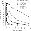
Tail-less C-cadherin mutant displays adhesive binding activity but not temporal strengthening of adhesion. CHO cells stably expressing similar levels of C-cadherin (C-CHO clone 21) and tail-less C-cadherin, CT (CT-CHO clone 8) were allowed to attach to glass capillaries coated with CEC1-5 for 10 or 40 min, then adhesive strength measured by resistance to detachment by progressively increasing rates of buffer. Data are means ± SE (n = 3).
Figure 6
Figure 6
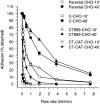
Adhesive activity of C-cadherin and mutants assessed by laminar flow assay. Parental CHO cells, C-CHO cells (clone 12), CT669-CHO cells (clone A1), and CT-CAT-CHO cells (clone 13), which were all matched for similar expression levels, were allowed to attach to glass capillaries for 10 or 40 min before adhesive strength was assessed by resistance to detachment by progressively increasing buffer flow rates. Data are means ± SE (n = 3).
Figure 7
Figure 7
Adhesive activity of C-cadherin and mutant cadherin molecules assessed by aggregation in suspension. Parental CHO cells, C-CHO cells (clone 12), CT-CHO cells (clone 11), CT669-CHO cells (clone A1) and CT-CAT-CHO cells (clone 13), which were all matched for similar expression levels, were isolated by trypsinization in the presence of Ca2+, then allowed to aggregate in suspension. After 45 min the number of individual cells remaining in suspension (Nt) was counted and expressed as a percentage of the number of cells counted in the freshly isolated suspension (N0). Increased aggregation is reflected in a fall in the Nt/ N0 ratio. Data are means ± SE (n = 3).
Figure 8
Figure 8
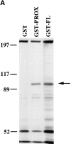
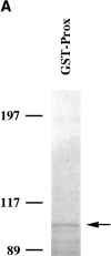
Identification of polypeptides that bind to GST-fusion proteins containing the cadherin cytoplasmic tail. (A) Binding of metabolically labeled proteins to GST fusion proteins. Lysates of 35S-labeled HCT116 cells were incubated with either GST, GST-Prox, or GST-FL. Bound proteins were separated by SDS-PAGE. A 92-kD protein binds to both GST-Prox and GST-FL but not to GST alone. (B) β-catenin bind to GST-FL but not to GST or GST-Prox. A Western blot of proteins bound to the GST fusion proteins was probed with a mAb to β-catenin.
Figure 8
Figure 8
Identification of polypeptides that bind to GST-fusion proteins containing the cadherin cytoplasmic tail. (A) Binding of metabolically labeled proteins to GST fusion proteins. Lysates of 35S-labeled HCT116 cells were incubated with either GST, GST-Prox, or GST-FL. Bound proteins were separated by SDS-PAGE. A 92-kD protein binds to both GST-Prox and GST-FL but not to GST alone. (B) β-catenin bind to GST-FL but not to GST or GST-Prox. A Western blot of proteins bound to the GST fusion proteins was probed with a mAb to β-catenin.
Figure 9
Figure 9
The 92-kD protein bound by GST-Prox is identical to p120ctn. (A) Detection of the 92-kD protein isolated from HCT116 in a large scale experiment. Coomassie brilliant blue staining of a large scale experiment isolating the 92-kD protein from HCT116 lysates using GST-Prox. Arrow marks the 92-kD protein. (B) Peptides identified in the protein sequence of p120ctn. Mass spectrometry analysis identified 27 major fragments that are underlined in the sequence. In bold are the sequences that were found in more than one peptide fragment. The two fragments that were subjected to NH2-terminal sequence analysis are shown in italics.
Figure 9
Figure 9
The 92-kD protein bound by GST-Prox is identical to p120ctn. (A) Detection of the 92-kD protein isolated from HCT116 in a large scale experiment. Coomassie brilliant blue staining of a large scale experiment isolating the 92-kD protein from HCT116 lysates using GST-Prox. Arrow marks the 92-kD protein. (B) Peptides identified in the protein sequence of p120ctn. Mass spectrometry analysis identified 27 major fragments that are underlined in the sequence. In bold are the sequences that were found in more than one peptide fragment. The two fragments that were subjected to NH2-terminal sequence analysis are shown in italics.
Figure 10
Figure 10
p120ctn associates with the proximal domain of C-cadherin in CHO cells. (A) p120ctn isoforms bind to both GST-Prox and GST-FL. Lysates of CHO cells were incubated with GST, GST-Prox, or GST-FL, transferred to nitrocellulose and probed with a mAb recognizing the major isoforms of p120ctn. (B) Wild-type Cadherin and CT669 but not CT-CAT or CT coimmunoprecipate with p120ctn. Lysates from C-CHO, CT669-CHO, C-CAT-CHO, and CT-CHO cells were immunoprecipitated with a mAb to p120ctn, transferred to nitrocellulose and probed for C-cadherin. In the bottom panel total lysates of these cells were also transferred to nitrocellulose and probed for C-cadherin to compare relative expression levels. C-cadherin immunoblots identify mature and precursor forms of the wild-type and mutant cadherins. Both the CT669 and CT-CAT mutants display polypeptide bands that run at or slightly higher than wild-type C-cadherin, consistent with the addition of muti-copy myc-epitope tags.
Figure 10
Figure 10
p120ctn associates with the proximal domain of C-cadherin in CHO cells. (A) p120ctn isoforms bind to both GST-Prox and GST-FL. Lysates of CHO cells were incubated with GST, GST-Prox, or GST-FL, transferred to nitrocellulose and probed with a mAb recognizing the major isoforms of p120ctn. (B) Wild-type Cadherin and CT669 but not CT-CAT or CT coimmunoprecipate with p120ctn. Lysates from C-CHO, CT669-CHO, C-CAT-CHO, and CT-CHO cells were immunoprecipitated with a mAb to p120ctn, transferred to nitrocellulose and probed for C-cadherin. In the bottom panel total lysates of these cells were also transferred to nitrocellulose and probed for C-cadherin to compare relative expression levels. C-cadherin immunoblots identify mature and precursor forms of the wild-type and mutant cadherins. Both the CT669 and CT-CAT mutants display polypeptide bands that run at or slightly higher than wild-type C-cadherin, consistent with the addition of muti-copy myc-epitope tags.

Similar articles

Cited by

References

    1. Aberle H, Butz S, Stappert J, Weissig H, Kemler R, Hoschuetzky H. Assembly of the cadherin-catenin complex in vitro with recombinant proteins. J Cell Sci. 1994;107:3655–3663. - PubMed
    1. Angres B, Barth A, Nelson WJ. Mechanism for transition from initial to stable cell-cell adhesion: kinetic analysis of E-cadherin–mediated adhesion using a quantitative adhesion assay. J Cell Biol. 1996;134:549–557. - PMC - PubMed
    1. Brieher WM, Gumbiner BM. Regulation of cadherin function during activin induced morphogenesis of Xenopusanimal caps. J Cell Biol. 1994;126:519–527. - PMC - PubMed
    1. Brieher WM, Yap AS, Gumbiner BM. Lateral dimerization is required for the homophilic binding activity of C-cadherin. J Cell Biol. 1996;135:487–489. - PMC - PubMed
    1. Chen H, Paradies NE, Fedor-Chaiken M, Brackenbury R. E-cadherin mediates adhesion and suppresses cell motility via distinct mechanisms. J Cell Sci. 1997;110:345–356. - PubMed

Publication types