. 1998 May 4;141(3):689-701.
doi: 10.1083/jcb.141.3.689.

Characterization of the human homologue of the yeast spc98p and its association with gamma-tubulin

Affiliations

Characterization of the human homologue of the yeast spc98p and its association with gamma-tubulin

A M Tassin et al. J Cell Biol. .

Abstract

A trimeric complex formed by Tub4p, the budding yeast gamma-tubulin, and the two spindle pole body components, Spc98p and Spc97p, has recently been characterized in Saccharomyces cerevisiae. We reasoned that crucial functions, such as the control of microtubule nucleation, could be maintained among divergent species. SPC98-related sequences were searched in dbEST using the BLASTN program. Primers derived from the human expressed sequence tag matching SPC98 were used to clone the 5' and 3' cDNA ends by rapid amplification of cDNA ends (RACE)-PCR. The human Spc98 cDNA presents an alternative splicing at the 3' end. The deduced protein possesses 22% identity and 45% similarity with the yeast homologue. We further report that the human Spc98p, like gamma-tubulin, is concentrated at the centrosome, although a large fraction is found in cytosolic complexes. Sucrose gradient sedimentation of the cytosolic fraction and immunoprecipitation experiments demonstrate that both gamma-tubulin and HsSpc98p are in the same complex. Interestingly, Xenopus sperm centrosomes, which are incompetent for microtubule nucleation before their activation in the egg cytoplasm, were found to contain similar amounts of both Spc98p and gamma-tubulin to human somatic centrosomes, which are competent for microtubule nucleation. Finally, affinity-purified antibodies against Spc98p inhibit microtubule nucleation on isolated centrosomes, as well as in microinjected cells, suggesting that this novel protein is indeed required for the nucleation reaction.


Figures
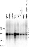
Figure 3
Figure 3
Northern blot analysis on different human tissues (indicated on the top of the figure) using a probe located in the common region (1084–1921). A major messenger at 4.4 kb is observed (band 3). Three minor bands (bands 1, 2, and 4) are also observed. The membrane was further stripped and reprobed with two other probes in the divergent part of the sequence. Probe 2, located in bp 80–645 of clone 02, stained bands 1–3. Probe 3, located in bp 2450–2577 of the initial clone, stained exclusively band 4.
Figure 1
Figure 1
(A) Cloning strategy for identifying the HsSPC98 full-length sequence. The location of the primers used, as well as the length of the fragments obtained by RACE-PCR, are reported. Some ESTs homologous to the human sequence have been also indicated. (B) Predicted protein sequence for the two major forms of HsSpc98p. The arrowheads in the sequence indicate where the sequences differ. The minor form of the protein is written below the major one. These sequence data for HsSpc98p major form and minor form have been submitted to the EMBL Nucleotide Sequence Database under accession numbers AJ003061 and AJ003062.
Figure 2
Figure 2
(A) Alignment sequence of HsSpc98p with the yeast Spc98p and Spc97p (indicated as Sc). The underlined sequence corresponds to the NLS in yeast ScSpc98p. (B) Alignment of the conserved sequence of HsSpc98p with ScSpc98p and with mouse (Mm), zebrafish (zf), and rice ESTs. These comparisons suggest that the protein is highly conserved in this small region. Alignments were performed using the Pileup program (Infobiogen, Villejuif, France). Comparisons to ScSpc98p, ScSpc97p and the ESTs were performed using the Boxshade program (Institut Suisse de Recherche Experimentales sur le Cancer, Lausanne, France). Identical amino acids are boxed in black. Conservative changes are boxed in gray.
Figure 4
Figure 4
Western blot analysis of low-speed, Triton X-100–soluble (S) and -insoluble (I) protein fractions from unsynchronized KE37 cells and of a highly enriched centrosome preparation (CTR). Proteins were probed with affinity-purified HsSpc98p IgG (left). A band at 103 kD is observed in all fractions, while highly enriched in the centrosome fraction. The same blot was subsequently probed with anti–γ-tubulin (right). Note the similar partition of both proteins in all fractions. 10 μg of Triton X-100–soluble and -insoluble proteins representing 2 × 10⁵ and 6 × 10⁵ cells, respectively, and ∼3 × 10⁷ centrosomes were loaded.
Figure 5
Figure 5
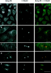
Double immunostaining of HeLa cells with anti-HsSpc98p affinity-purified antibody and a monoclonal antibody CTR453, which recognizes the PCM observed by confocal microscopy. All documents presented are two-dimensional projections of images collected at all relevant z-axes. (A and B) Anti-HsSpc98p antibody recognizes the centrosome in all cells as demonstrated by the colocalization of both staining. The inset in A shows the accumulation of HsSpc98-positive material at the midbody during anaphase. (C–E) Representative blow ups of cells at different stages during the cell cycle. In G2 cells (C), HsSpc98 staining is restricted to the centrioles, while CTR453 presented in addition of the centrioles an accumulation of PCM. During prophase (D), HsSpc98p accumulates to the centrosome concomitant with the increase of microtubule nucleation activity. During metaphase (E), HsSpc98p antibodies recognize the centrosome as well as the polar microtubules, as described elsewhere for γ-tubulin. Bars, 10 μm.
Figure 6
Figure 6
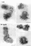
Preembedding immunostaining with affinity-purified anti-Spc98p or anti–γ-tubulin antibodies on isolated centrosomes after a methanol fixation. Note that gold particles are associated with the PCM close to the centrioles regardless of the antibody. Note also the particles at the tip of the subdistal appendages (arrowheads). Bars, 200 μm.
Figure 7
Figure 7
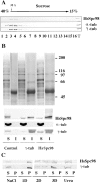
The cytosolic as well as the centrosomal form of HsSpc98p are associated with γ-tubulin. (A) G1/S cytosol from HeLa cells are loaded on a 15–40% sucrose gradient (10 ml in SW41 tubes) and centrifuged at 100,000 g for 16 h. The γ-tubulin– rich fractions were reloaded on the top of another small 15–40% sucrose gradient (4 ml in SW55 tubes). HsSpc98p cosediments with γ-tubulin as well as with α-tubulin. (B) Immunoprecipitation experiments of the γ-tubulin–rich fractions obtained from sucrose gradient as presented in A with preimmune (Control), anti– γ-tubulin (γ-tub), and anti-HsSpc98p (HsSpc98) immunoglobulins coupled to protein G beads. The proteins precipitated with the beads are indicated as “I,” while the proteins not associated with the beads are referred as “S.” The top figure shows the silver staining of the immunoprecipitate. Note that no proteins are detected with the control beads and that anti–γ-tubulin or anti-HsSPc98p antibodies precipitate the same proteins. The lower part shows the immunostaining of the different fractions with γ-tubulin, α-tubulin, and HsSpc98 antibodies. Anti–γ-tubulin as well as anti-HsSpc98 immunoglobulins precipitate both γ-tubulin and HsSpc98p. However, no α-tubulin is detected in the immunoprecipitates, suggesting that α-tubulin is not part of the complex. (C) Biochemical extraction of the centrosomal HsSpc98p. Soluble (S) and insoluble (P) centrosomal protein fractions obtained in different extraction conditions (NaCl, 1 M; 1D, 0.5% NP-40; 2D, 0.5% NP-40 and 0.5% deoxycholate; 3D, 0.5% NP-40, 0.5% deoxycholate, and 0.1% SDS; Urea, 8 M) were immunodetected with anti–γ-tubulin or anti-HsSpc98p antibodies. Note that the same extraction conditions are necessary to solubilize both γ-tubulin and HsSpc98p. Observe in 2D buffer that 50% of the protein is soluble while 50% is insoluble.
Figure 8
Figure 8
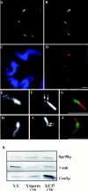
(A–J) Decoration of Xenopus sperm centrosome before activation in Xenopus egg extract. Double labeling with either anti–γ-tubulin antibodies (A and E) or with anti-HsSpc98p (H) and with GT335, a monoclonal antibody recognizing polyglutamylated tubulin (B, F, and I; arrows point to the centrioles). D, G, and J are the superposition of the respective labelings. (C) DAPI staining of the sperm nuclei. (A–D) A field of Xenopus sperm heads. (E–J) High magnification of a single sperm head. Note that both γ-tubulin and Spc98p accumulate around the two centrioles and along the striated rootlets. (K) Western blot analysis of Xenopus egg extract (X E), Xenopus sperm centrosomes (X Sperm CTR), and human somatic centrosomes (KE37 CTR). Proteins were probed with affinity-purified HsSpc98p IgG, anti–γ-tubulin antibody, or anti-HsCen3p antibody. Note the presence of similar amounts of both Spc98p and γ-tubulin in Xenopus sperm centrosomes and human somatic centrosomes. Bars, 5 μm.
Figure 9
Figure 9
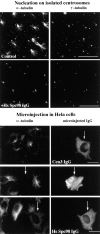
Affinity-purified anti-HsSpc98p antibodies inhibit microtubule nucleation in vitro (top) and in vivo (bottom). (Top) Microtubule nucleating activity of isolated centrosomes was monitored by double immunofluorescence with anti–α-tubulin and with anti–γ-tubulin. Centrosomes were preincubated with preimmune immunoglobulins (Control) or with anti-HsSpc98p immunoglobulins (+HsSpc98 IgG) on ice for 30 min and subsequently incubated in PC-tubulin. Microtubules were allowed to grow for 4 min. Note the specific inhibition of aster formation after 4 min of growth with HsSpc98 immunoglobulins, while the control centrosomes were growing typical microtubule asters. (Bottom) Anti-HsSpc98 immunoglobulins were microinjected into HeLa cells (HsSpc98 IgG). 2 h after microinjection, microtubules were depolymerized for 2 h with 5 × 10⁻⁶ M nocodazole. After washing out the nocodazole, microtubules were allowed to regrow for 10 min. Cells were fixed with methanol and processed for immunofluorescence with monoclonal anti–α-tubulin antibodies followed by the mouse and rabbit secondary antibodies. In this way, only the microinjected cells are detected with the fluorescein anti–rabbit secondary antibody. Note that the nonmicroinjected cells presented the usual centrosome-growing microtubule aster, while heavily microinjected cells did not show any microtubule (arrows). Note that in cells micronjected with an unrelated antibody (anti-Cen3p IgG), microtubule regrowth is not affected. Bars, 10 μm.

Similar articles

Cited by

References

    1. Archer J, Vega LR, Solomon F. Rbl2p, a yeast protein that binds to β-tubulin and participates in microtubule function in vivo. Cell. 1995;82:425–434. - PubMed
    1. Bailly E, Doree M, Nurse P, Bornens M. p34cdc2 is located in both nucleus and cytoplasm; part is centrosomally associated at G2/M and enters vesicles at anaphase. EMBO (Eur Mol Biol Organ) J. 1989;8:3985–3995. - PMC - PubMed
    1. Chrétien D, Buendia B, Fuller DF, Karsenti E. Reconstruction of the centrosome cycle from cryoelectron micrographs. J Struct Biol. 1997;120:117–133. - PubMed
    1. Détraves C, Mazarguil H, Lajoie-Mazenc I, Julian M, Raynaud-Messina B, Wright M. Protein complexes containing γ-tubulin are present in mammalian brain microtubule protein preparations. Cell Motil Cytoskel. 1997;36:179–189. - PubMed
    1. Dingwall, C., and R.A. Laskey. 1991. Nuclear targeting sequences—a consensus? Trends Biochem. Sci. 478–481. - PubMed

Publication types

MeSH terms

Associated data