. 1998 Apr 28;95(9):5287-92.
doi: 10.1073/pnas.95.9.5287.

The BRC repeats in BRCA2 are critical for RAD51 binding and resistance to methyl methanesulfonate treatment

Affiliations

The BRC repeats in BRCA2 are critical for RAD51 binding and resistance to methyl methanesulfonate treatment

P L Chen et al. Proc Natl Acad Sci U S A. .

Abstract

The BRCA2 gene was identified based on its involvement in familial breast cancer. The analysis of its sequence predicts that the gene encodes a protein with 3,418 amino acids but provides very few clues pointing to its biological function. In an attempt to address this question, specific antibodies were prepared that identified the gene product of BRCA2 as a 390-kDa nuclear protein. Furthermore, direct binding of human RAD51 to each of the four single 30-amino acid BRC repeats located at the 5' portion of exon 11 of BRCA2 was demonstrated. Such an interaction is significant, as BRCA2 and RAD51 can be reciprocally coimmunoprecipitated by each of the individual, specific antibodies and form complexes in vivo. Inferring from the function of RAD51 in DNA repair, human pancreatic cancer cells, Capan-1, expressing truncated BRCA2 were shown to be hypersensitive to methyl methanesulfonate (MMS) treatment. Exogenous expression of wild-type BRCA2, but not BRC-deleted mutants, in Capan-1 cells confers resistance to MMS treatment. These results suggest that the interaction between the BRC repeats of BRCA2 and RAD51 is critical for cellular response to DNA damage caused by MMS.


Figures

Figure 1
Figure 1
Identification of BRCA2 protein. (A) cDNAs and GST fusion proteins for antibody preparation. Four overlapping cDNAs were used to assemble the full-length cDNA for the BRCA2 coding sequence. Also shown are the two regions of the BRCA2 cDNA used to construct the GST fusion proteins (boxes). These clones were used to express antigens to raise antibodies to BRCA2. The names for each of the antisera are shown in the left margin. (B) Identification of the BRCA2 protein. The indicated GST fusion proteins were used to immunize mice, and sera were used to precipitate BRCA2 protein from 35S-labeled T-24 cells. The position of the size markers and the BRCA2 protein are indicated in the left margin. The antibodies and competitive antigens used in each experiment are indicated above the lanes. Asterisks indicate double immunoprecipitations with the indicated antibodies. (C) Nuclear localization of BRCA2. The letters above each lane denote total extract (T), cytoplasmic fraction (C), membrane fraction (M), and nuclear fraction (N). The BRCA2 protein is indicated. Also shown are the controls, p84 (a known nuclear matrix protein) (36) and GST (a known cytoplasmic protein. Note, in lane 4, the nuclear partitioning of BRCA2 in this assay. (D) Localization of GFP-BRCA2. MCF10A was transiently transfected with expression plasmids, CHPL, containing either GFP or GFP-BRCA2 fusions. Thirty hours after transfection, cells were fixed and stained with DAPI (a and b). GFP fluorescence is shown (b and d). Note the nuclear localization of GFP-BRCA2 (d).
Figure 2
Figure 2
BRC repeats in BRCA2 mediate binding to RAD51. (A) The indicated regions of BRCA2 (solid rectangles) were fused to the DNA binding domain of GAL4 in pAS1. Rad51 was fused to the activation domain of GAL4 in pGAD10. These plasmids, used to cotransform yeast strain Y153, were scored for colony color and assayed for β-galactosidase activity as indicated. (B) GST pull-down assay for the in vitro binding of BRCA2 to RAD51. (Left) A schematic drawing of full-length BRCA2 and the regions fused to GST for bacterial expression. The stippled area denotes exon 11. (Right) SDS/polyacrylamide gel stained with Coomassie blue for the purification of the GST-BRCA2 proteins after binding to glutathione-Sepharose beads. Asterisks indicate the full-length GST fusion proteins. The GST-BRCA2 beads were then used to bind in vitro transcribed and translated 35S-labeled RAD51. The bound proteins were analyzed by SDS/PAGE and autoradiography. Lane 5 demonstrates that the NCB fragment of BRCA2 binds to RAD51. Lane 1 shows the input RAD51. (C) A specific BRC repeat binds to RAD51. (Left) A schematic drawing of the relative positions of the BRC repeats (solid rectangles) in the NCB fragment of BRCA2 and the various truncated and internally deleted constructs fused to GST. (Right) The purification of the GST-NCB proteins after binding to glutathione beads. Asterisks indicate the full-length fusion proteins. The GST beads were used to bind in vitro translated 35S-labeled RAD51 and analyzed as above. Lanes 3–5 and 7–9 demonstrate that the fragments of BRCA2 shown above each lane bind to Rad51. Lane 1 shows the input RAD51 protein. (D) Bacterially expressed and purified human RAD51 was used as the input protein (lane 1) for binding the GST-NCB fusion proteins described in C and indicated above each lane. Note that the same fragments that bind purified RAD51 also bind in vitro translated RAD51.
Figure 3
Figure 3
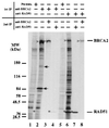
Reciprocal coimmunoprecipitation of BRCA2 and RAD51. [35S]Methionine-labeled T-24 cellular lysates were first precipitated with the indicated antibodies or preimmune serum. After dissolution of the immunoprecipitates, the proteins were precipitated a second time with the indicated antibodies (lanes 3, 4, 7, and 8). The precipitated proteins were then analyzed by SDS/PAGE and autoradiographed. Carets (for BRCA2) and asterisks (for RAD51) mark the bands of interest. Note in lanes 2 and 4 that RAD51 (indicated in the right margin) is precipitated by BRCA2, and conversely, in lanes 6 and 8, that BRCA2 (indicated in the right margin) is coprecipitated by RAD51 antibodies. Arrows indicate unknown proteins specifically coimmunoprecipitated with BRCA2.
Figure 4
Figure 4
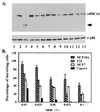
BRCA2-deficient Capan-1 cells are hypersensitive to MMS treatment. (A) Identification of Capan-1 with a truncated BRCA2 protein. Cell lines surveyed for altered BRCA2 protein expression were as follows: lanes 1 and 2, HBL100; lane 3, Capan-1; lane 4, MB361; lane 5, MB175–7; lane 6, MB435; lane 7, MB468; lane 8, MB231; lane 9, BT483; lane 10, SKBR3; lane 11, MCF7; lane 12, ZR75; lane 13, T47D. Full-length BRCA2 is indicated in the right margin. The truncated BRCA2 protein expressed in Capan-1 cells (lane 3) is indicated by the arrow. p84 is a nuclear matrix protein (36) that served as internal control for immunoprecipitations. Cellular lysates were immunoprecipitated by using an equal mixture of anti-p400 and anti-BBA. After separation by SDS/PAGE and blotting, BRCA2 was detected by using rabbit polyclonal anti-BBA antibodies. (B) Hypersensitivity of Capan-1 to MMS treatment. Four different cell lines were treated with various concentrations of MMS (indicated under the x axis), and total surviving cells were counted by trypan blue exclusion assay using hematocytometry. These experiments were repeated three times. Note that, relative to others tested, Capan-1 cells seem to be very sensitive to MMS treatment.
Figure 5
Figure 5
Ectopic expression of BRCA2 in Capan-1 rescues MMS resistance. (A) Schematic diagrams of the BRCA2 cDNAs used to rescue MMS hypersensitivity of Capan-1 cells in B. The black region denotes exon 11, and white bars are BRC repeats. Constructs named with a “CNF” prefix indicate an N-terminal fusion with the flag epitope (open box). Two of the constructs containing missense mutations, V1283G and ΔT1302, in the vicinity of the BRC repeats are indicated. (B) Graphic summary of cell survival in response to 0.075% MMS treatment. Parallel cultures of transfected or untransfected cells (lane 1) were treated with 0.075% of MMS. Each construct was transfected into 2 × 10⁶ cells. The number of the surviving cells was counted by trypan blue exclusion assay. The experiment was repeated three times, and the transfection efficiency was normalized with β-galactosidase activity from the cotransfected pSV2-Gal. The average number and SD were plotted on the y axis. Note transfection of the cells with wild-type BRCA2 (CNF-2hFL) (lane 3), but none of the mutants (V1283G, ΔT1302, P1, P13, and X13, lanes 4–8, respectively) or the vector CNF alone (lane 2) restored resistance to treatment with MMS. (C) Detection of exogenous BRCA2 mRNA in transfected cells. Total cellular RNA from parallel cell cultures transfected with BRCA2 cDNAs after 48 h was extracted and served as templates for RT-PCR as described in Materials and Methods. The expected PCR product, 536 bp, was analyzed in a 1.5% agarose gel. Lane 1, reaction with all PCR reagents except RNA or cDNA template. Lane 2, reaction with 1 ng of CNF-2hFL DNA as template. RNA was used as templates in the PCR reaction of the following odd lanes and cDNA as templates in even lanes. The templates were prepared from either untransfected cells (lanes 3 and 4) or cells transfected with CNF (lanes 5 and 6), CNF-2hFL (lanes 7 and 8), CNF-V1283G (lanes 9 and 10), CNF-ΔT1302 (lanes 11 and 12), CNF-P1 (lanes 13 and 14), CNF-P13 (lanes 15 and 16), and CNF-X13 (lanes 17 and 18).

Similar articles

Cited by

References

    1. Wooster R, Bignell G, Lancaster J, Swift S, Seal S, Mangion J, Collins N, Gregory S, Gumbs C, Micklem G, et al. Nature (London) 1995;378:789–792. - PubMed
    1. Tavtigian S V, Simard J, Rommens J, Couch F, Shattuckeidens D, Neuhausen S, Merajver S, Thorlacius S, Offit K, Stoppalyonnet D, et al. Nat Genet. 1996;12:333–337. - PubMed
    1. Wooster R, Neuhausen S L, Mangion J, Quirk Y, Ford D, Collins N, Nguyen K, Seal S, Tran T, Averill D, et al. Science. 1994;265:2088–2090. - PubMed
    1. Gudmundsson J, Johannesdottir G, Arason A, Bergthorsson J T, Ingvarsson S, Egilsson V, Barkardottir R B. Am J Hum Genet. 1996;58:749–756. - PMC - PubMed
    1. Thorlacius S, Olafsdottir G, Tryggvadottir L, Neuhausen S, Jonasson J G, Tavtigian S V, Tulinius H, Ogmundsdottir H M, Eyfjörd J E. Nat Genet. 1996;13:117–119. - PubMed

Publication types

MeSH terms

LinkOut - more resources