Chemokine receptor utilization by human immunodeficiency virus type 1 isolates that replicate in microglia
- PMID: 9557714
- PMCID: PMC109654
- DOI: 10.1128/JVI.72.5.4243-4249.1998
Chemokine receptor utilization by human immunodeficiency virus type 1 isolates that replicate in microglia
Abstract
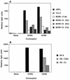
The role of human immunodeficiency virus (HIV) strain variability remains a key unanswered question in HIV dementia, a condition affecting around 20% of infected individuals. Several groups have shown that viruses within the central nervous system (CNS) of infected patients constitute an independently evolving subset of HIV strains. A potential explanation for the replication and sequestration of viruses within the CNS is the preferential use of certain chemokine receptors present in microglia. To determine the role of specific chemokine coreceptors in infection of adult microglial cells, we obtained a small panel of HIV type 1 brain isolates, as well as other HIV strains that replicate well in cultured microglial cells. These viruses and molecular clones of their envelopes were used in infections, in cell-to-cell fusion assays, and in the construction of pseudotypes. The results demonstrate the predominant use of CCR5, at least among the major coreceptors, with minor use of CCR3 and CXCR4 by some of the isolates or their envelope clones.
Figures


Similar articles
-
Apoptosis induced by infection of primary brain cultures with diverse human immunodeficiency virus type 1 isolates: evidence for a role of the envelope.J Virol. 1999 Feb;73(2):897-906. doi: 10.1128/JVI.73.2.897-906.1999. J Virol. 1999. PMID: 9882290 Free PMC article.
-
Isolated human astrocytes are not susceptible to infection by M- and T-tropic HIV-1 strains despite functional expression of the chemokine receptors CCR5 and CXCR4.Glia. 2001 May;34(3):165-77. Glia. 2001. PMID: 11329179
-
Differential CD4/CCR5 utilization, gp120 conformation, and neutralization sensitivity between envelopes from a microglia-adapted human immunodeficiency virus type 1 and its parental isolate.J Virol. 2001 Apr;75(8):3568-80. doi: 10.1128/JVI.75.8.3568-3580.2001. J Virol. 2001. PMID: 11264346 Free PMC article.
-
Chemokine receptors and mechanisms of cell death in HIV neuropathogenesis.J Neurovirol. 2000 May;6 Suppl 1:S24-32. J Neurovirol. 2000. PMID: 10871762 Review.
-
Chemokine receptors and virus entry in the central nervous system.J Neurovirol. 1999 Dec;5(6):643-58. doi: 10.3109/13550289909021293. J Neurovirol. 1999. PMID: 10602405 Review.
Cited by
-
HIV-infected microglia mediate cathepsin B-induced neurotoxicity.J Neurovirol. 2015 Oct;21(5):544-58. doi: 10.1007/s13365-015-0358-7. Epub 2015 Jun 20. J Neurovirol. 2015. PMID: 26092112 Free PMC article.
-
TRAIL-mediated apoptosis in HIV-1-infected macrophages is dependent on the inhibition of Akt-1 phosphorylation.J Immunol. 2006 Aug 15;177(4):2304-13. doi: 10.4049/jimmunol.177.4.2304. J Immunol. 2006. PMID: 16887991 Free PMC article.
-
Genetic and functional analysis of full-length human immunodeficiency virus type 1 env genes derived from brain and blood of patients with AIDS.J Virol. 2003 Nov;77(22):12336-45. doi: 10.1128/jvi.77.22.12336-12345.2003. J Virol. 2003. PMID: 14581570 Free PMC article.
-
Apoptosis induced by infection of primary brain cultures with diverse human immunodeficiency virus type 1 isolates: evidence for a role of the envelope.J Virol. 1999 Feb;73(2):897-906. doi: 10.1128/JVI.73.2.897-906.1999. J Virol. 1999. PMID: 9882290 Free PMC article.
-
Patterns of chemokine receptor fusion cofactor utilization by human immunodeficiency virus type 1 variants from the lungs and blood.J Virol. 1999 Aug;73(8):6680-90. doi: 10.1128/JVI.73.8.6680-6690.1999. J Virol. 1999. PMID: 10400765 Free PMC article.
References
-
- Albright A V, Strizki J, Harouse J M, Lavi E, O’Connor M, González-Scarano F. HIV-1 infection of cultured human adult oligodendrocytes. Virology. 1996;217:211–219. - PubMed
-
- Alkahatib G, Liao F, Berger E A, Farber J M, Peden K W C. A new SIV co-receptor STRL33. Nature. 1997;388:238. - PubMed
-
- Bagasara O, Lavi E, Bobroski L, Tawadros R, Pestaner J P, Pomerantz R J. Cellular reservoirs of HIV-1 in the central nervous system of infected-individuals: identification by the combination of in situ PCR and immunohistochemistry. AIDS. 1996;10:573–585. - PubMed
-
- Berger, E. A. 1997. HIV entry and tropism: the chemokine receptor connection. AIDS 11(Suppl. A):S3–S16. - PubMed
-
- Carne C A, Teddr R S, Smith A. Acute encephalopathy coincident with seroconversion for anti-HTLV-III. Lancet. 1985;ii:1206–1208. - PubMed
Publication types
MeSH terms
Substances
Grants and funding
LinkOut - more resources
Full Text Sources
Molecular Biology Databases

