. 1998 May 1;18(9):3147-57.
doi: 10.1523/JNEUROSCI.18-09-03147.1998.

Calcium-dependent regulation of rab3 in short-term plasticity

Affiliations

Calcium-dependent regulation of rab3 in short-term plasticity

F Doussau et al. J Neurosci. .

Abstract

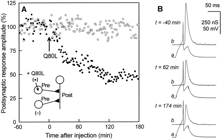
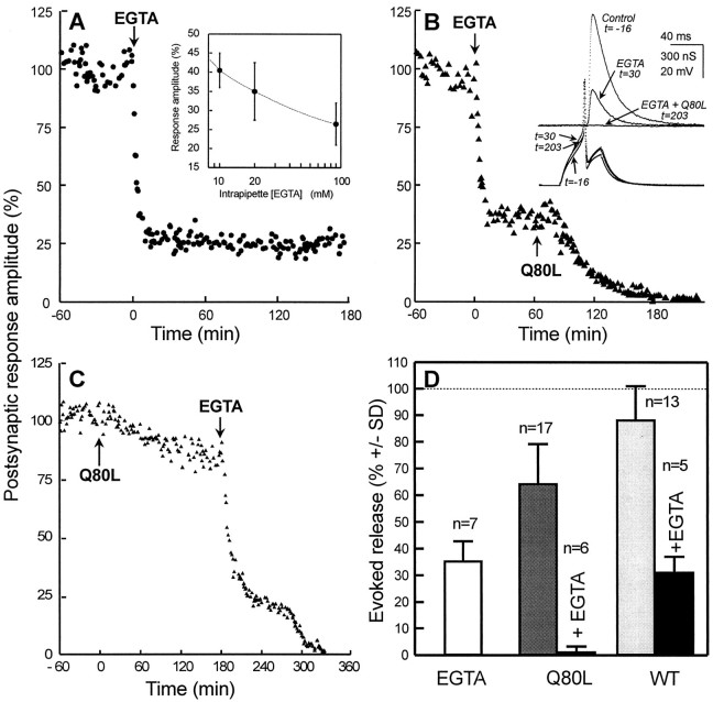
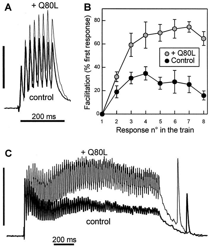
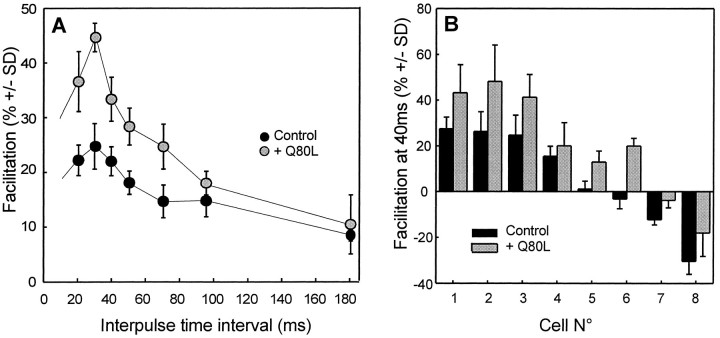
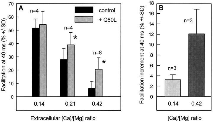
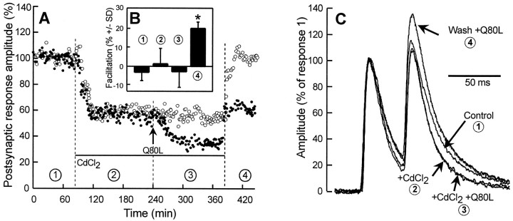
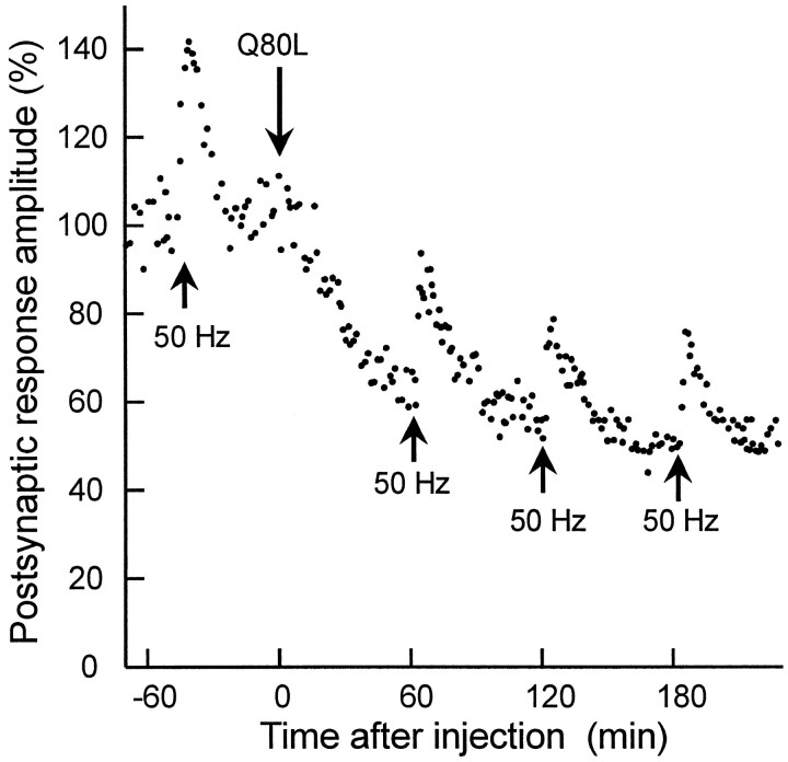
The Rab3 proteins are monomeric GTP-binding proteins associated with secretory vesicles. In their active GTP-bound state, Rab3 proteins are involved in the regulation of hormone secretion and neurotransmitter release. This action is thought to involve specific effectors, including two Ca2+-binding proteins, Rabphilin and Rim. Rab3 acts late in the exocytotic process, in a cell domain in which the intracellular Ca2+ concentration is susceptible to rapid changes. Therefore, we examined the possible Ca2+-dependency of the regulatory action of GTP-bound Rab3 and wild-type Rab3 on neuroexocytosis at identified cholinergic synapses in Aplysia californica. The effects of recombinant GTPase-deficient Aplysia-Rab3 (apRab3-Q80L) or wild-type apRab3 were studied on evoked acetylcholine release. Intraneuronal application of apRab3-Q80L in identified neurons of the buccal ganglion of Aplysia led to inhibition of neurotransmission; wild-type apRab3 was less effective. Intracellular chelation of Ca2+ ions by EGTA greatly potentiated the inhibitory action of apRab3-Q80L. Train and paired-pulse facilitation, two Ca2+-dependent forms of short-term plasticity induced by a rise in intraterminal Ca2+ concentration, were increased after injection of apRab3-Q80L. This result suggests that the inhibition exerted by GTP-bound Rab3 on neuroexocytosis is reduced during transient augmentations of intracellular Ca2+ concentration. Therefore, a Ca2+-dependent modulation of GTP-bound Rab3 function may contribute to short-term plasticity.


Figures

Fig. 1.
Fig. 1.
Inhibition of evoked acetylcholine release by recombinant GTPase-deficient Aplysia-Rab3.A, ACh release was evoked at identified synapses in the buccal ganglion of Aplysia californica. Postsynaptic response amplitude (%) is plotted against time (after injection). After the control recording was made, recombinantAplysia-Rab3 deficient in its GTPase activity (apRab3-Q80L, abbreviated as Q80L in the figure) was pressure-injected into one of the two presynaptic cholinergic neurons (see inset for a schematic drawing of the neuronal connections). The final concentration of apRab3-Q80L was ∼0.3–0.5 μm in the cell body; the other presynaptic neuron (○) was kept for internal control of release stability. B, Recordings of presynaptic action potentials (a) and postsynaptic responses (b), before and after injection of apRab3-Q80L. Note that amplitude of postsynaptic response is expressed as a change in postsynaptic conductance (nS). For further details see experimental procedures.
Fig. 2.
Fig. 2.
Intracellular removal of Ca2+increases apRab3-Q80L efficacy. All experiments were similar to that described in Figure 1A. A, EGTA mixed with Tris buffer, pH 7.4 (20/40 mm respective concentrations in the injection pipette), was injected intracellularly (arrow; ∼1% of the cell body volume) giving rise to a final intrasomatic [EGTA] of 200 μm.Inset, Mean amplitude of postsynaptic responses as a function of intraneuronal [EGTA] (±SD; n = 3–5). B, A typical experiment of three is shown. EGTA was first injected (arrow) to give an intracellular concentration of ∼200 μm. After removal of the injection micropipette and stabilization of the inhibition of evoked release, a second micropipette containing apRab3-Q80L (Q80L) was impaled. Then, apRab3-Q80L was pressure-injected (arrow; ∼0.3–0.5 μmfinal in the soma). Inset, Typical recordings during the experiment. C, Similar experiment as in Bbut in the reversed order. After EGTA injection, note the typical plateau (at ∼20%) at which postsynaptic responses transiently stabilized (it was seen in the 3 experiments performed).D, Comparison of the mean release (±SD) observed 3 hr after injection of EGTA–Tris alone (∼200/400 μm final intrasomatic concentration), apRab3-Q80L alone (∼0.3–0.5 μm), or after sequential injection of both EGTA and apRab3-Q80L, wild-type apRab3 (WT), alone (∼0.3–0.5 μm) or combined with EGTA. Significance: for all comparisons, p < 0.001, except WT alone (light gray bar) versus control, p< 0.01, and EGTA alone (white bar) versus WT + EGTA (black bar), p > 0.2.
Fig. 3.
Fig. 3.
Evoked ACh release under repetitive stimulation is potentiated after injection of apRab3-Q80L. A,B, Trains of eight action potentials at 50 Hz were elicited both in control conditions (control) and after stabilization of the inhibition induced by apRab3-Q80L (+ Q80L). In this experiment, the inhibition induced by apRab3-Q80L was 51%, 3 hr after injection. A shows the superposition of two postsynaptic recordings made in these two conditions; they were normalized against the amplitude of the first response in the train. Recording in control condition is denoted by thethick line. Vertical calibration (100% of first response in the train): 485 nS (control, before injection of apRab3-Q80L) and 250 nS (after injection of apRab3-Q80L and stabilization of the inhibition). B, Facilitation was determined for each of the eight stimulations in the train. In this typical experiment from a series of five, the plot shows the averaged facilitation values (mean ± SD; p < 0.001) determined for six trains in the absence or the presence of apRab3-Q80L. C, Effect of apRab3-Q80L on the facilitation observed during a 1.5 sec train of action potentials at 50 Hz. Same presentation as in A. In this experiment, the mean inhibition produced by apRab3-Q80L was 42%. Vertical calibration (100% of first response in the train): 1020 nS (before injection) and 620 nS (after injection).
Fig. 4.
Fig. 4.
Intracellular injection of apRab3-Q80L potentiates paired-pulse facilitation. Paired stimuli were given at various interpulse time intervals in the control medium, before and after intraneuronal injection of recombinant apRab3-Q80L. A, In this representative experiment, the extent of paired-pulse facilitation was calculated in control conditions and after stabilization of apRab3-Q80L-induced inhibition of evoked ACh release (here of 46%). The mean facilitation (± SD, from 25–30 recordings at each time interval) is significantly potentiated by apRab3-Q80L (p < 0.001) except at an interval of 90 msec (p ∼ 0.01) and 180 msec (p > 0.05). B, Summary of eight distinct experiments. For comparison, the mean facilitation observed at a 40 msec interpulse interval is reported for eight neurons (±SD, calculated from at least 25 recordings). Note that whatever the initial facilitation, positive or negative, apRab3-Q80L application was followed by an increased facilitation. For all cells,p < 0.001.
Fig. 5.
Fig. 5.
Ca2+ dependency of the effect of apRab3-Q80L on paired-pulse facilitation. A, In separate experiments, neurotransmitter release was evoked in the presence of a physiological medium with [Ca2+]/[Mg2+] ratios of 0.14, 0.21, or 0.42 to change extracellular [Ca2+]. When referred to the release measured at a 0.42 [Ca2+]/[Mg2+] ratio, ACh release was depressed to 72% (±7; n = 4) at 0.21 [Ca2+]/[Mg2+] ratio and to 35% (±8; n = 4) at 0.14 [Ca2+]/[Mg2+] ratio. Paired-pulse facilitation was determined (as described in Fig. 4) at these different ionic conditions (black bars). Then apRab3-Q80L was injected into the neurons, and after stabilization of the inhibition induced by apRab3-Q80L, paired-pulse facilitation was determined (gray bars). Note that the paired-pulse facilitation values are reported for an interpulse interval of 40 msec. Asterisk denotes thatp < 0.001. At [Ca2+]/[Mg2+] ratio = 0.14, p > 0.05. B, Similar experiment as in A, except paired-pulse facilitation was determined for each neuron at the unique 40 msec interval in control medium ([Ca2+]/[Mg2+] ratio of 0.42) and then in low Ca2+ medium ([Ca2+]/[Mg2+] ratio of 0.14). Similar measurements were made after stabilization of the inhibition induced by apRab3-Q80L. At each [Ca2+]/[Mg2+] ratio, results are expressed as the mean difference between the facilitation observed before and after injection of the mutated Rab3 protein;p < 0.001.
Fig. 6.
Fig. 6.
Effect of CdCl2 on paired-pulse facilitation before and after injection of apRab3-Q80L.A, In this typical experiment, neurotransmitter release from two neurons was followed in the presence of control medium (condition 1), in presence of 200 μmCdCl2 (denoted by the horizontal bar in conditions 2 and 3), and after washing out CdCl2 (condition 4). In addition, one neuron (•) was injected with apRab3-Q80L (arrow,Q80L; conditions 3 and4). B, For the neuron identified by • (i.e., the one injected by apRab3-Q80L), paired-pulse facilitation (mean ± SD) was determined at a 40 msec interpulse interval in the four experimental conditions described inA. Asterisk denotes p< 0.001 when the amplitude of facilitation is compared with any other condition. C, Typical recordings of paired postsynaptic responses recorded in the four experimental conditions described inA. For comparison, postsynaptic responses were normalized against the amplitude of the first response in the pair. Note that decay times of postsynaptic responses were shorter in CdCl2; this was taken into account for determining paired-pulse facilitation (see Materials and Methods).
Fig. 7.
Fig. 7.
Post-tetanic potentiation is not affected by apRab3-Q80L. This experiment was similar to that described in Figure 1, and in addition, during control period or after injection of apRab3-Q80L, post-tetanic potentiation (PTP) was elicited (arrows) (see Materials and Methods). For simplification, only one PTP is reported during the control period. The magnitude of PTP was not significantly different before and after injection of apRab3-Q80L.

Similar articles

Cited by

References

    1. Adler EM, Augustine GJ, Duffy SN, Charlton MP. Alien intracellular calcium chelators attenuate neurotransmitter release at the squid giant synapse. J Neurosci. 1991;11:1496–1507. - PMC - PubMed
    1. Bao J-X, Kandel ER, Hawkins RD. Involvement of pre- and postsynaptic mechanisms in posttetanic potentiation at Aplysia synapses. Science. 1997;275:969–973. - PubMed
    1. Bean AJ, Scheller RH. Better late than never: a role for Rabs late in exocytosis. Neuron. 1997;19:751–754. - PubMed
    1. Bittner MA, Holz RW. Kinetic analysis of secretion from permeabilized adrenal chromaffin cells reveals distinct components. J Biol Chem. 1992;267:16219–16225. - PubMed
    1. Borst JGG, Sakmann B. Calcium influx and transmitter release at fast CNS synapse. Nature. 1996;383:431–434. - PubMed

Publication types

LinkOut - more resources