. 1998 Apr 6;141(1):101-14.
doi: 10.1083/jcb.141.1.101.

Dynamin at the neck of caveolae mediates their budding to form transport vesicles by GTP-driven fission from the plasma membrane of endothelium

Affiliations

Dynamin at the neck of caveolae mediates their budding to form transport vesicles by GTP-driven fission from the plasma membrane of endothelium

P Oh et al. J Cell Biol. .

Abstract

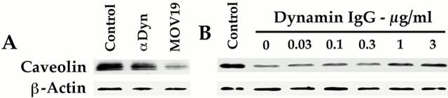
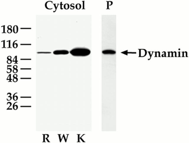
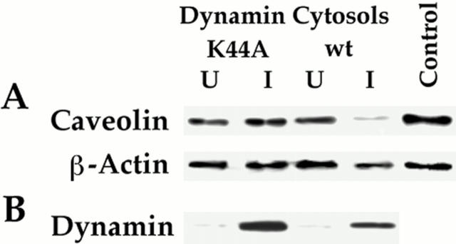
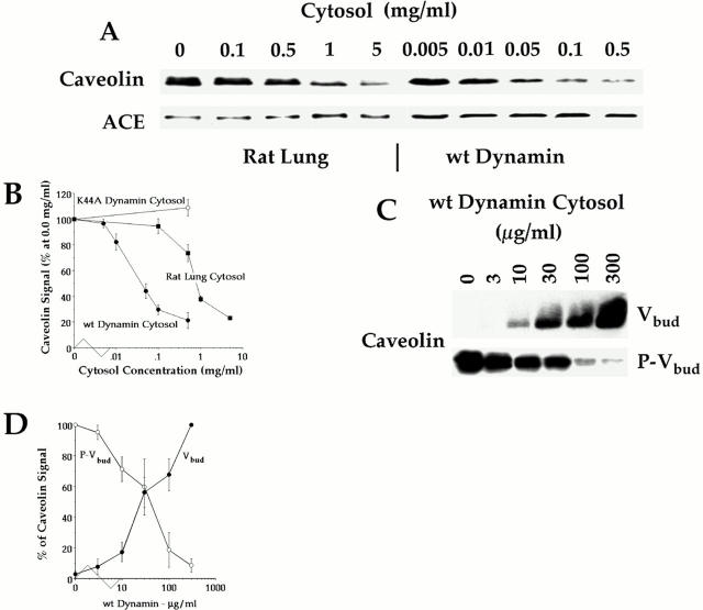
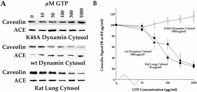
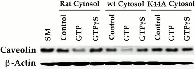
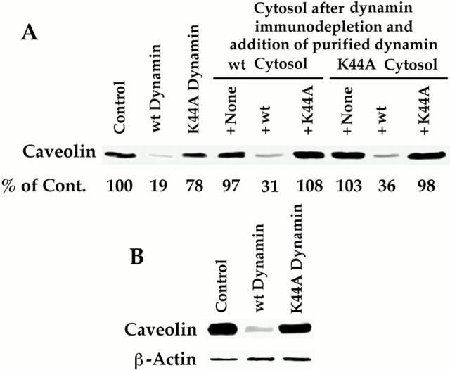
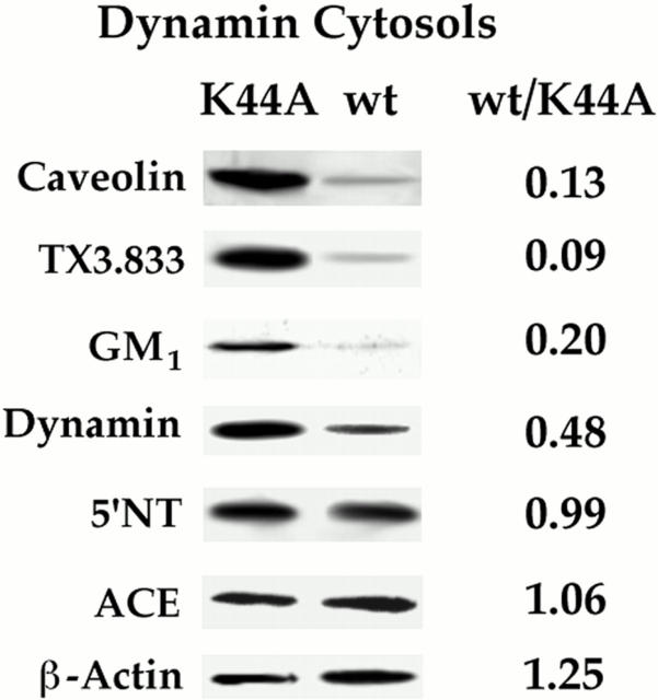
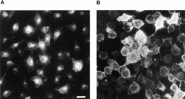
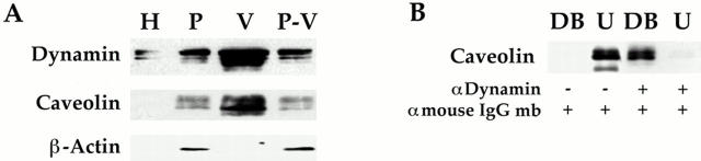
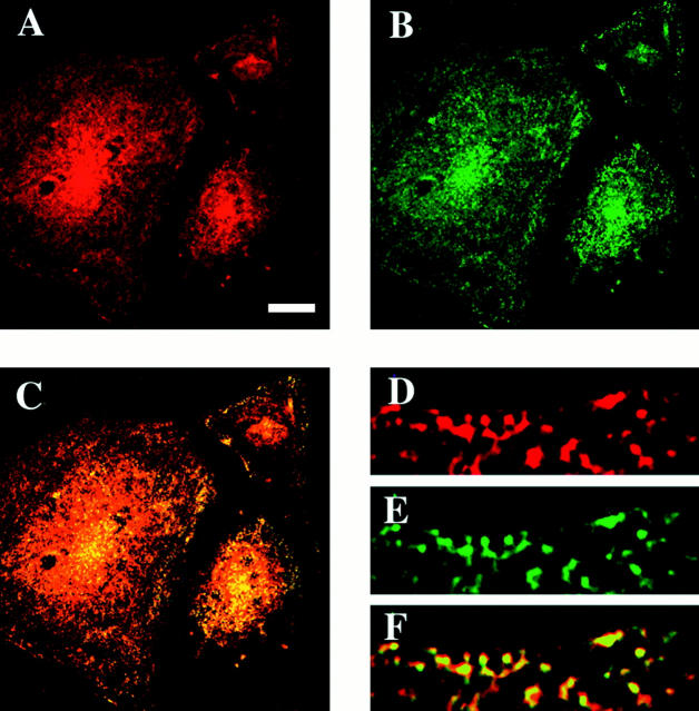
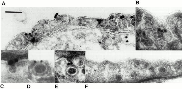
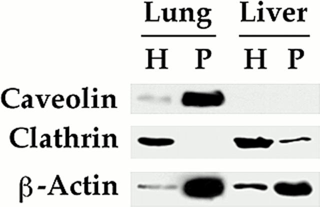
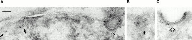
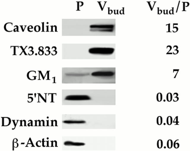
The molecular mechanisms mediating cell surface trafficking of caveolae are unknown. Caveolae bud from plasma membranes to form free carrier vesicles through a "pinching off" or fission process requiring cytosol and driven by GTP hydrolysis (Schnitzer, J.E., P. Oh, and D.P. McIntosh. 1996. Science. 274:239-242). Here, we use several independent techniques and functional assays ranging from cell-free to intact cell systems to establish a function for dynamin in the formation of transport vesicles from the endothelial cell plasma membrane by mediating fission at the neck of caveolae. This caveolar fission requires interaction with cytosolic dynamin as well as its hydrolysis of GTP. Expression of dynamin in cytosol as well as purified recombinant dynamin alone supports GTP-induced caveolar fission in a cell-free assay whereas its removal from cytosol or the addition to the cytosol of specific antibodies for dynamin inhibits this fission. Overexpression of mutant dynamin lacking normal GTPase activity not only inhibits GTP-induced fission and budding of caveolae but also prevents caveolae-mediated internalization of cholera toxin B chain in intact and permeabilized endothelial cells. Analysis of endothelium in vivo by subcellular fractionation and immunomicroscopy shows that dynamin is concentrated on caveolae, primarily at the expected site of action, their necks. Thus, through its ability to oligomerize, dynamin appears to form a structural collar around the neck of caveolae that hydrolyzes GTP to mediate internalization via the fission of caveolae from the plasma membrane to form free transport vesicles.


Figures

Figure 1
Figure 1
Anti-dynamin IgG inhibits GTP-induced fission of caveolae from endothelial cell plasma membranes. The cell-free assay reconstituting GTP-dependent caveolar fission was performed on purified, silica-coated endothelial cell plasma membranes under standard conditions with GTP (100 μM) and rat lung cytosol (1 mg/ml) supplemented with anti-dynamin IgG (0–3 μg/ml). Neither GTP nor IgG was added to the samples labeled control. After 1 h, the silica-coated plasma membranes were sedimented by centrifugation and the loss of caveolae was assessed by subjecting all of the membrane pellet to SDS-PAGE and Western analysis using antibodies for caveolin and β-actin. The similar β-actin signal detected in each lane is consistent with the equal aliquots used in the assay and loaded onto the gel lanes. (A) The effect of anti-dynamin IgG was compared with a control IgG (MOV19) (both IgG1 and at 3 μg/ ml). (B) The effect of the indicated concentrations of anti-dynamin IgG was assessed. Results shown in A and B are representative of ⩾2 experiments.
Figure 2
Figure 2
Monospecific immunodetection of dynamin in endothelial cell plasma membranes and various cytosols used in the cell-free assays. Western blot analysis with the monoclonal antibody for dynamin was performed on proteins (10 μg) of the silica-coated endothelial cell plasma membranes purified from rat lungs (P) as well as cytosols derived from either rat lungs (R) or HeLa cells induced to express wild-type dynamin (W) or K44A dynamin (K). Note that by densitometry, the K44A cytosol appeared to have ∼3- and 10-fold more dynamin than the wild-type and rat lung cytosols, respectively (of course, assuming equivalent recognition by the antibody). It also should be noted that the other dynamin antibody (HUDY-1) also was quite specific with a single reactive band detected strongly in P (data not shown).
Figure 3
Figure 3
Cytosol from cells induced to express wild-type (but not K44A) dynamin supports GTP-dependent caveolar fission. The standard cell-free caveolar fission assay was performed on P with Western analysis for caveolin and β-actin as shown. (A) This assay was performed in the presence of buffer without cytosol (Control) or with the indicated cytosols (30 μg/ml) isolated from HeLa cells either uninduced (U) or induced (I) to express either wild-type dynamin or the K44A mutant dynamin. (B) Western analysis of dynamin levels in the induced and uninduced cytosols (10 μg of protein per lane). Results shown in A and B are representative of at least two experiments.
Figure 4
Figure 4
Effect of cytosol concentration and dynamin overexpression on GTP-induced caveolar fission. The standard cell-free caveolar fission assay was performed on P in the presence of indicated cytosols at the indicated concentrations. (A) Western analysis for caveolin and ACE using the repelleted membranes after treatment. (B) Caveolin signal, which is plotted as a function of cytosol concentration was quantified densitometrically and expressed as the percentage of the signal detected in the absence of cytosol. Each point gives the average value with SD from three experiments, one of which is shown in A. (C) Western analysis for the indicated proteins on both the isolated budded caveolae (Vbud) and the repelleted plasma membranes (P–Vbud) after budding in the presence of the indicated concentration of wild-type dynamin cytosol. (D) Caveolin signal quantified in P–Vbud (•) and Vbud (○) as a function of wild-type dynamin cytosol concentration. The densitometric signal is expressed as a percentage of the maximum detected signal: for P–Vbud that is in the absence of cytosol and for Vbud that is at the highest concentration of cytosol used (300 μg/ml). Each point with SD represents the average of three experiments of which one is shown in C.
Figure 5
Figure 5
GTP dependence of caveolar fission in various cytosols. The standard cell-free caveolar fission assay was performed on P with Western analysis for caveolin, ACE, and β-actin as shown in A. P was incubated with GTP at the indicated concentrations plus the indicated cytosols. Unlike caveolin, the signal for both ACE and β-actin remained unchanged, consistent with the equivalent loading of the treated resedimented P onto each gel lane. (B) The caveolin signal in this assay was quantified as in Fig. 4 and then plotted as function of GTP concentration. Each point gives the average value with SD from three or more experiments.
Figure 6
Figure 6
Budding of caveolae from the surface of BLMVEC requires GTP hydrolysis and GTPase-competent dynamin. Caveolar budding was assessed in cultured cells as previously described (Schnitzer et al., 1996). Briefly, BLMVEC were permeabilized with streptolysin O, incubated for 10 min at 37°C with the indicated cytosol (rat lung cytosol [1 mg/ml], wild-type or K44A cytosol [0.5 mg/ml]) in cytosolic buffer alone (Control) or supplemented with either GTP or GTPγS to a final concentration of 100 μM. The BLMVEC plasma membranes were purified for Western analysis using caveolin antibodies to quantify cell surface caveolae and β-actin antibodies as a control. As an additional reference, the plasma membrane was isolated from cells left untreated and not permeabilized (SM). Two experiments showed nearly identical results.
Figure 7
Figure 7
GTPase-active dynamin is necessary and sufficient for GTP-induced fission of caveolae. The standard cell-free caveolar fission assay was performed on P with Western analysis for caveolin. (A) Requirement for dynamin in cytosol. P was incubated with the indicated cytosols: wild-type or K44A dynamin cytosol, either cytosol immunodepleted of dynamin or either immunodepleted cytosol replenished with purified recombinant wild-type or K44A dynamin. The percentage of caveolin quantified by densitometry and calculated relative to the control is indicated below each lane. (B) Dynamin alone can replace cytosol in GTP- induced fission assay. P was incubated with purified recombinant wild-type or K44A dynamin (1 μg/ml). Neither dynamin was added to the sample labeled control. The caveolin signal decreased by 73% in the presence of wild-type but not K44A dynamin. The β-actin signal remained constant. Both experiments were performed twice with nearly identical results.
Figure 8
Figure 8
K44A mutant dynamin inhibits GTP-induced, dynamin-dependent caveolar fission. The standard cell-free caveolar fission assay was performed on P in the presence of no cytosol, wild-type dynamin cytosol, K44A cytosol, or wild-type and K44A dynamin cytosol combined, each at 500 μg/ml. Budding of caveolae was calculated from the loss of caveolin signal, which was quantified densitometrically. Each value represents the mean of three experiments.
Figure 9
Figure 9
GTP in the presence of wild-type but not K44A dynamin depletes the plasma membrane of various caveolar markers but not general plasmalemmal proteins. The standard cell-free caveolar fission assay was performed on P in the presence of wild-type dynamin cytosol or K44A dynamin cytosol (each at 500 μg/ml) followed by Western analysis with antibodies to the indicated proteins.
Figure 10
Figure 10
Mutant dynamin prevents CT-B internalization in intact cells. BLMVEC were transfected with either wild-type (A) or K44E (B) dynamin cDNA before examining by fluorescence microscopy the internalization of cell surface-bound CT-B–FITC upon warming to 37°C for 30 min. Bar, 5 μm.
Figure 11
Figure 11
Dynamin associated with the endothelial cell plasma membrane in vivo concentrates in caveolae. (A) Western analysis with antibodies to dynamin and caveolin was performed on the proteins (5 μg) from the following membrane fractions: rat lung homogenate (H), purified silica-coated plasma membranes (P), purified caveolae (V), and plasma membrane stripped of caveolae (P–V). (B) Immunoaffinity isolation of caveolae with dynamin antibody. The purified caveolae (V) were incubated with anti–mouse IgG Dynabeads either alone or prebound with anti-dynamin IgG. The Dynabeads plus any bound material (DB) were separated magnetically from the unbound material (U) before Western analysis of both fractions. Results shown in A and B are representative of at least two experiments.
Figure 12
Figure 12
Colocalization of caveolin and dynamin at the endothelial cell surface. Immunofluorescence confocal microscopy was performed on BLMVEC using antibodies against dynamin and caveolin. The signals for dynamin (red) (A and D) and caveolin (green) (B and E) overlap significantly on the cell surface as shown by the orange-yellow signal denoting colocalization in the composite, superimposed image (C and F). Little, if any, staining was detected in the absence of primary antibodies or with other antibodies as negative controls (data not shown). Bar, (A–C) 20 μm; (D–F) 2 μm.
Figure 13
Figure 13
Dynamin localization to the neck of caveolae in endothelium in vivo. Immunogold electron microscopy (15-nm gold) was performed on ultrathin cryosections of rat lung tissue using antibody to dynamin (A–E) and control monoclonal antibody (F). Bar: (A, B, and D–F) 150 nm; (C) 72 nm.
Figure 14
Figure 14
Expression of clathrin and caveolin on endothelial cell plasma membranes isolated from rat lung and liver. Western analysis for caveolin, β-actin, and the heavy chain of clathrin was performed on tissue homogenates (H) and silica-coated endothelial cell plasma membranes (P) isolated from lung and liver.
Figure 15
Figure 15
Dynamin labeling of caveolae and clathrin-coated pits in cultured endothelial cells. Immunogold labeling for dynamin (5-nm gold) was performed on ultrathin cryosections of cultured BAEC. (A) Labeled caveolae (solid arrows) (partial or forming caveola on left of fully formed caveolae) and clathrin-coated invagination (open arrow). (B) Single caveolae with gold label concentrated in the neck region. (C) Single clathrin-coated pit showing labeling over all areas of clathrin coat. Bar: (A and B) 76 nm; (C) 91 nm.
Figure 16
Figure 16
GTP-budded free caveolar vesicles contain specific caveolar markers but lack dynamin. Western analysis with antibodies to the indicated proteins was performed on P (before the budding reaction) and Vbud the caveolar vesicles induced to bud by GTP in wild-type dynamin cytosol and isolated by flotation in a continuous sucrose gradient. The signal was quantified densitometrically and the ratio of Vbud/P is provided as an indication of enrichment under equal protein loads (2 μg).

Similar articles

Cited by

References

    1. Anderson RG. Caveolae: where incoming and outgoing messengers meet. Proc Natl Acad Sci USA. 1993;90:10909–10913. - PMC - PubMed
    1. Artalejo CR, Henley JR, McNiven MA, Palfrey HC. Rapid endocytosis coupled to exocytosis in adrenal chromaffin cells involves Ca2+, GTP and dynamin but not clathrin. Proc Natl Acad Sci USA. 1995;92:8328–8332. - PMC - PubMed
    1. Chen MS, Obar RA, Schroeder CC, Austin TW, Poodry CA, Wadsworth SC, Vallee RB. Multiple forms of dynamin are encoded by shibire, a Drosophilia gene involved in endocytosis. Nature. 1991;351:583–586. - PubMed
    1. Damke H, Baba T, Warnock DE, Schmid SL. Induction of mutant dynamin specifically blocks endocytic coated vesicle formation. J Cell Biol. 1994;127:915–934. - PMC - PubMed
    1. Damke H, Baba T, van der Bliek AM, Schmid SL. Clathrin-independent pinocytosis is induced in cells overexpressing a temperature-sensitive mutant of dynamin. J Cell Biol. 1995a;131:69–80. - PMC - PubMed

Publication types

MeSH terms