. 1998 Apr;18(4):1967-77.
doi: 10.1128/MCB.18.4.1967.

Protein serine/threonine phosphatase Ptc2p negatively regulates the unfolded-protein response by dephosphorylating Ire1p kinase

Affiliations

Protein serine/threonine phosphatase Ptc2p negatively regulates the unfolded-protein response by dephosphorylating Ire1p kinase

A A Welihinda et al. Mol Cell Biol. 1998 Apr.

Abstract

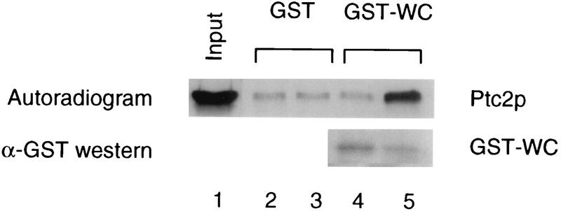
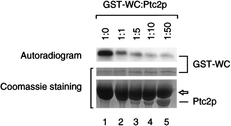
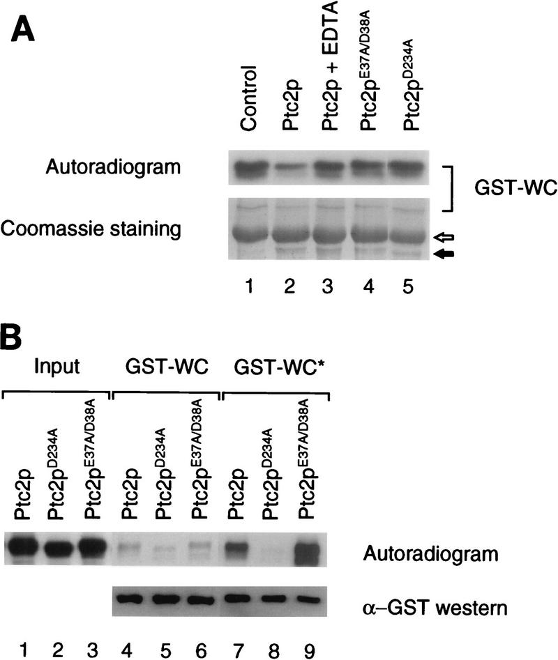
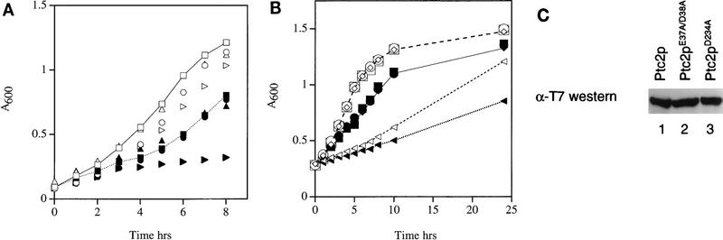
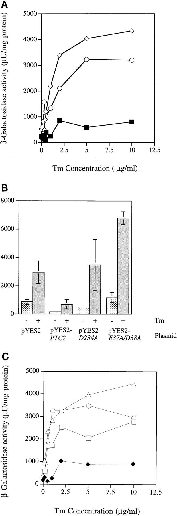
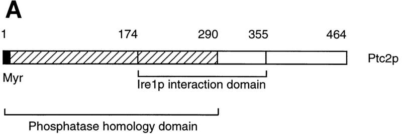
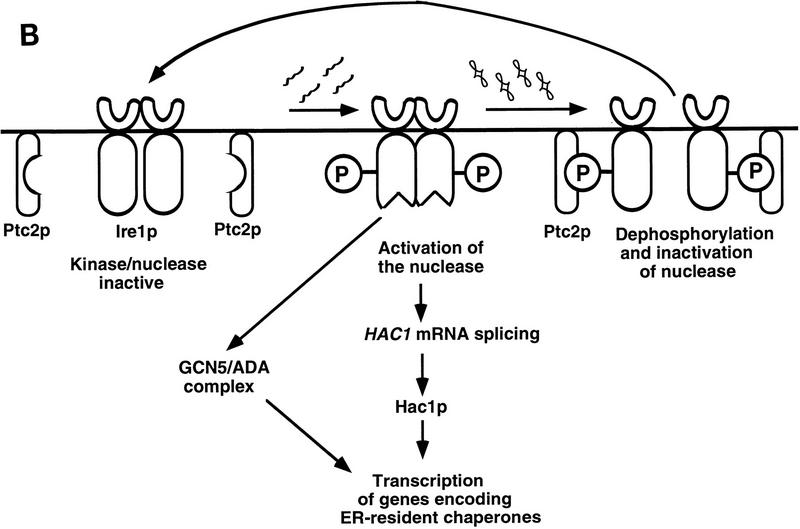
Cells respond to the accumulation of unfolded proteins in the endoplasmic reticulum (ER) by increasing the transcription of the genes encoding ER-resident chaperone proteins. Ire1p is a transmembrane protein kinase that transmits the signal from unfolded proteins in the lumen of the ER by a mechanism that requires oligomerization and trans-autophosphorylation of its cytoplasmic-nucleoplasmic kinase domain. Activation of Ire1p induces a novel spliced form of HAC1 mRNA that produces Hac1p, a transcription factor that is required for activation of the transcription of genes under the control of the unfolded-protein response (UPR) element. Searching for proteins that interact with Ire1p in Saccharomyces cerevisiae, we isolated PTC2, which encodes a serine/threonine phosphatase of type 2C. The Ptc2p interaction with Ire1p is specific, direct, dependent on Ire1p phosphorylation, and mediated through a kinase interaction domain within Ptc2p. Ptc2p dephosphorylates Ire1p efficiently in an Mg2+-dependent manner in vitro. PTC2 is nonessential for growth and negatively regulates the UPR pathway. Strains carrying null alleles of PTC2 have a three- to fourfold-increased UPR and increased levels of spliced HAC1 mRNA. Overexpression of wild-type Ptc2p but not catalytically inactive Ptc2p reduces levels of spliced HAC1 mRNA and attenuates the UPR, demonstrating that the phosphatase activity of Ptc2p is required for regulation of the UPR. These results demonstrate that Ptc2p downregulates the UPR by dephosphorylating Ire1p and reveal a novel mechanism of regulation in the UPR pathway upstream of the HAC1 mRNA splicing event.


Figures

FIG. 1
FIG. 1
Analysis of Ire1p interaction with Ptc2p and mapping of interaction domains. (A) Two-hybrid analysis. LexA fusion proteins with intact and truncated forms of the cytoplasmic domain of Ire1p (bait) were tested for interaction with the original clone B42-HA–Ptc2p (aa 174 to 464), B42-HA–Ptc2p containing aa 1 to 312, or B42-HA–Ptc2p containing aa 174 to 355. Transformants harboring IRE1 and PTC2 fusions were patched onto His Trp plates and replica plated onto His Trp Leu plates containing either glucose or galactose. The growth of strain EGY48 harboring different regions of the Ire1p cytoplasmic domain as LexA fusions (in pEG202) and as B42-HA fusions (in pJG4-5) on His Trp medium (control) and His Trp Leu medium containing either galactose plus raffinose (Gal) or glucose (Glu) was monitored. (B) Schematic representation of LexA-Ire1p fusion proteins (bait) and their interactions with B42-HA–Ptc2p (prey) as detected by two-hybrid analysis. WC, wild-type cytoplasmic-nucleoplasmic domain; NK, N linker plus kinase domain; WK, wild-type kinase domain; KC, kinase domain plus C-terminal tail; CT, C-terminal tail.
FIG. 2
FIG. 2
Direct physical association of Ire1p and Ptc2p in vitro. Aliquots of an in vitro translation reaction mixture containing 35S-labeled Ptc2p were incubated with glutathione-Sepharose beads impregnated with either phosphorylated (lane 5) or nonphosphorylated (lane 4) GST-WC. As a control, beads coated with GST alone and either with (lane 3) or without (lane 2) prior incubation in in vitro kinase buffer (41) were used to adsorb Ptc2p. Beads were collected, washed, and boiled to release the bound proteins. Equal proportions of the samples were analyzed by SDS-PAGE, Western blotting, and fluorography. A portion (8.3%) of the Ptc2p input is shown in lane 1.
FIG. 3
FIG. 3
Dephosphorylation of Ire1p by Ptc2p. A fixed amount of 32P-labeled GST-WC (1 μg) was incubated with increasing amounts of Ptc2p in dephosphorylation buffer (21). Samples were boiled, and equal portions were analyzed by SDS-PAGE and autoradiography. The GST-WC/Ptc2p ratios used were determined by a protein assay and were confirmed by scanning of the Coomassie blue-stained gel with NIH Image. The migration of bovine serum albumin is indicated by an arrow.
FIG. 4
FIG. 4
Sequence alignment of the catalytic domains of human PP2Cα and Ptc2p. The GenBank accession numbers for the PP2Cα gene and PTC2 nucleotide sequences are S87759 and U18839, respectively. Invariant residues are shaded in gray. Conserved residues are boxed. Residues that coordinate metal ions are indicated by circles. Residues that were mutated in this study are denoted by diamonds. The alignment was made with the Wisconsin sequence analysis package (Genetics Computer Group Inc., Madison, Wis.).
FIG. 5
FIG. 5
Effect of mutations at the predicted Mg2+ binding sites on the Ire1p interaction and phosphatase activity. (A) In vitro dephosphorylation assay. Equal amounts (1 μg) of 32P-labeled GST-WC were incubated with dephosphorylation buffer (control, lane 1), Ptc2p (1 μg, lane 2), Ptc2p (1 μg) with EDTA (lane 3), Ptc2pE37A/D38A (1 μg, lane 4), or Ptc2pD234A (1 μg, lane 5). Samples were boiled, and equal portions were analyzed by SDS-PAGE Coomassie staining, and autoradiography. The migration of the wild-type and mutant phosphatases is indicated by a solid arrow. The open arrow indicates the migration of bovine serum albumin. (B) In vitro binding assay. Beads impregnated with equal amounts of either nonphosphorylated (lanes 4 to 6) or phosphorylated (lanes 7 to 9) GST-WC were tested for interaction with equal amounts of in vitro-translated Ptc2p (lanes 4 and 7), Ptc2pD234A (lanes 5 and 8), or Ptc2pE37A/D38A (lanes 6 and 9). Bound proteins were analyzed by SDS-PAGE, Western blotting, and fluorography. Ten percent of the Ptc2p input is shown in lanes 1 to 3. The phosphorylated form of GST-WC is indicated by an asterisk.
FIG. 6
FIG. 6
Effect of PTC2 on the growth of S. cerevisiae in the presence (closed symbols) or absence (open symbols) of tunicamycin. (A) PTC2 deletion does not affect the growth rate. AWY446 (ptc2-1::kanr) (triangles), AWY500 (PTC2) (squares), AWY506 (ptc2-Δ1::LEU2) (circles), and AWY 516 (ire1Δ1) (arrowheads) were grown in 1% yeast extract–2% peptone–2% dextrose (YPD) medium to an A600 of 0.1, and the cultures were divided into flasks. Tunicamycin (final concentration, 0.25 μg/ml) was added to one set of flasks, and incubation was continued at 30°C. Aliquots were removed at the indicated times, and the A600 was measured. (B) Ptc2p overproduction inhibits cell growth. AWY500 harboring PTC2 (arrowheads), ptc2D234A (circles), or ptc2E37A/D38A (squares) in a 2μm vector, pYES2 (diamonds), was grown in Ura medium containing 2% galactose and 1% raffinose for 10 h. Cultures were divided and treated as in panel A. (C) Wild-type Ptc2p and single and double Ptc2p mutants are expressed equally in S. cerevisiae. AWY500 harboring PTC2 (lane 1), ptc2E37A/D38A (lane 2), and ptc2D234A (lane 3) in the pYES2 vector was grown in Ura medium containing 2% galactose and 1% raffinose for 10 h. Cells were harvested and lysed, and the extracts were analyzed by Western blotting with anti-T7 antibody.
FIG. 7
FIG. 7
PTC2 downregulates the UPR. (A) Null mutants of PTC2 exhibit an elevated UPR. Cultures of strains AWY446 (ptc2-1::kanr) diamonds, AWY500 (PTC2) (squares), and AWY506 (ptc2-Δ1::LEU2) (circles) were grown in YPD to the early log phase and divided, and tunicamycin (Tm) was added to the final concentrations indicated. After 90 min of incubation at 30°C, cells were harvested and lysed and β-galactosidase activity was measured. (B) Overexpression of wild-type Ptc2p and catalytically inactive Ptc2p deregulates the UPR. Strains expressing either wild-type or mutant Ptc2p were grown in Ura medium containing 2% galactose and 1% raffinose for 10 h to the early log phase. Cultures were divided, Tm was added to a final concentration of 2 μg/ml, and incubated was continued for 90 min. Cells were harvested and lysed, and β-galactosidase activity was measured. Specific β-galactosidase activity represents an average of three independent experiments. Bars indicate standard deviations. (C) Tm dose dependence of the UPR in wild-type cells overexpressing wild-type or mutant Ptc2p. The UPR was monitored as described in panel B, except that different concentrations of Tm were used for induction. Symbols: squares, pYES2 vector; diamonds, pYES2 carrying PTC2; circles, pYES2 carrying the D234A allele; triangles, pYES2 carrying the E37A/D38A allele.
FIG. 8
FIG. 8
PTC2 regulates HAC1 mRNA splicing. Cultures of strains AWY500, AWY506, and AWY500 overexpressing either wild-type Ptc2p or Ptc2p mutants were grown in synthetic complete medium with or without uracil to the early log phase, divided, and induced with tunicamycin (Tm) (2 μg/ml) for 90 min. Cells were harvested, RNA was isolated, and cleavage of HAC1 mRNA was assayed by an RNase protection assay with a probe that is colinear with the S. cerevisiae HAC1 gene. The 147-nucleotide (nt) fragment represents the 3′ portion of (HAC1) mRNA that extends to the 3′ cleavage site. The 74-nucleotide fragment derived from the 5′ side of the spliced HAC1 mRNA is not shown in this analysis. The abundance of spliced HAC1 mRNA HAC1i relative to ACT1 mRNA is indicated. Products of higher molecular weights may represent intermediates in the splicing reaction that are observed only in Δptc2 cells (asterisks). 2μ, 2μm.
FIG. 9
FIG. 9
Domain structure of Ptc2p and model for the role of Ptc2p in the UPR pathway. (A) Linear representation of Ptc2p. The myristoylation (Myr) signal and the phosphatase domain are deduced from the sequence of PTC2. The Ire1p interaction domain, as mapped by the two-hybrid analysis, is shown. (B) Model for the activation and inactivation of the UPR pathway in S. cerevisiae. P, covalently attached phosphate.
FIG. 9
FIG. 9
Domain structure of Ptc2p and model for the role of Ptc2p in the UPR pathway. (A) Linear representation of Ptc2p. The myristoylation (Myr) signal and the phosphatase domain are deduced from the sequence of PTC2. The Ire1p interaction domain, as mapped by the two-hybrid analysis, is shown. (B) Model for the activation and inactivation of the UPR pathway in S. cerevisiae. P, covalently attached phosphate.

Similar articles

Cited by

References

    1. Alani E, Cao L, Kleckner N. A method for gene disruption that allows repeated use of URA3 selection in the construction of multiply disrupted yeast strains. Genetics. 1987;116:541–545. - PMC - PubMed
    1. Barford D. Molecular mechanisms of the protein serine/threonine phosphatases. Trends Biochem Sci. 1996;21:407–412. - PubMed
    1. Berben G, Dumont J, Gilliquet V, Bolle P A, Hilger F. The YDp plasmids: a uniform set of vectors bearing versatile gene disruption cassettes for Saccharomyces cerevisiae. Yeast. 1991;7:475–477. - PubMed
    1. Boeke J D, LaCroute F, Fink G R. A positive selection for mutants lacking orotidine-5′-phosphate decarboxylase activity in yeast: 5-fluoro-orotic acid resistance. Mol Gen Genet. 1984;197:345–346. - PubMed
    1. Chang S C, Erwin A E, Lee A S. Glucose-regulated protein GRP94 and GRP78 genes share common regulatory domains and are coordinately regulated by common trans-acting factors. Mol Cell Biol. 1989;9:2153–2162. - PMC - PubMed

Publication types

MeSH terms

Associated data