Hepatitis C virus core from two different genotypes has an oncogenic potential but is not sufficient for transforming primary rat embryo fibroblasts in cooperation with the H-ras oncogene
- PMID: 9525629
- PMCID: PMC109754
- DOI: 10.1128/JVI.72.4.3060-3065.1998
Hepatitis C virus core from two different genotypes has an oncogenic potential but is not sufficient for transforming primary rat embryo fibroblasts in cooperation with the H-ras oncogene
Abstract
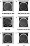
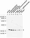
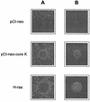
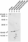
Persistent infection with hepatitis C virus (HCV) is associated with the development of liver cirrhosis and hepatocellular carcinoma. To examine the oncogenic potential of the HCV core gene product, primary rat embryo fibroblasts (REFs) were transfected with the core gene in the presence or absence of the H-ras oncogene. In contrast to a previous report (R. B. Ray, L. M. Lagging, K. Meyer, and R. Ray, J. Virol. 70:4438-4443, 1996), HCV core proteins from two different genotypes (type 1a and type 1b) were not found to transform REFs to tumorigenic phenotype in cooperation with the H-ras oncogene, although the core protein was successfully expressed 20 days after transfection. In addition, REFs transfected with E1A- but not core-expressing plasmid showed the phenotype of immortalized cells when selected with G418. The biological activity was confirmed by observing the transcription activation from two viral promoters, Rous sarcoma virus long terminal repeat and simian virus 40 promoter, which are known to be activated by the core protein from HCV-1 isolate. In contrast to the result with primary cells, the Rat-1 cell line, stably expressing HCV core protein, exhibited focus formation, anchorage-independent growth, and tumor formation in nude mice. HCV core protein was able to induce the transformation of Rat-1 cells with various efficiencies depending on the expression level of the core protein. These results indicate that HCV core protein has an oncogenic potential to transform the Rat-1 cell line but is not sufficient to either immortalize primary REFs by itself or transform primary cells in conjunction with the H-ras oncogene.
Figures


Similar articles
-
Hepatitis C virus core protein cooperates with ras and transforms primary rat embryo fibroblasts to tumorigenic phenotype.J Virol. 1996 Jul;70(7):4438-43. doi: 10.1128/JVI.70.7.4438-4443.1996. J Virol. 1996. PMID: 8676467 Free PMC article.
-
Induction of apoptosis by the transactivating domains of the hepatitis B virus X gene leads to suppression of oncogenic transformation of primary rat embryo fibroblasts.Oncogene. 2000 Feb 24;19(9):1173-80. doi: 10.1038/sj.onc.1203417. Oncogene. 2000. PMID: 10713705
-
Induction and progression of the transformed phenotype in cloned rat embryo fibroblast cells: studies employing type 5 adenovirus and wild-type and mutant Ha-ras oncogenes.Mol Carcinog. 1992;5(2):118-28. doi: 10.1002/mc.2940050207. Mol Carcinog. 1992. PMID: 1554410
-
The p53 tumor suppressor gene and gene product.Princess Takamatsu Symp. 1989;20:221-30. Princess Takamatsu Symp. 1989. PMID: 2488233 Review.
-
Mechanisms of oncogene cooperation: activation and inactivation of a growth antagonist.Environ Health Perspect. 1991 Jun;93:97-103. doi: 10.1289/ehp.919397. Environ Health Perspect. 1991. PMID: 1837777 Free PMC article. Review.
Cited by
-
Hepatitis C virus NS5A physically associates with p53 and regulates p21/waf1 gene expression in a p53-dependent manner.J Virol. 2001 Feb;75(3):1401-7. doi: 10.1128/JVI.75.3.1401-1407.2001. J Virol. 2001. PMID: 11152513 Free PMC article.
-
Access of viral proteins to mitochondria via mitochondria-associated membranes.Rev Med Virol. 2009 May;19(3):147-64. doi: 10.1002/rmv.611. Rev Med Virol. 2009. PMID: 19367604 Free PMC article. Review.
-
The roles of hepatitis C virus proteins in modulation of cellular functions: a novel action mechanism of the HCV core protein on gene regulation by nuclear hormone receptors.Cancer Sci. 2003 Nov;94(11):937-43. doi: 10.1111/j.1349-7006.2003.tb01381.x. Cancer Sci. 2003. PMID: 14611668 Free PMC article. Review.
-
Ectopic expression of hepatitis C virus core protein differentially regulates nuclear transcription factors.J Virol. 1998 Dec;72(12):9722-8. doi: 10.1128/JVI.72.12.9722-9728.1998. J Virol. 1998. PMID: 9811706 Free PMC article.
-
Polymorphisms in the hepatitis C virus core and its association with development of hepatocellular carcinoma.J Biosci. 2017 Sep;42(3):509-521. doi: 10.1007/s12038-017-9695-4. J Biosci. 2017. PMID: 29358564 Review.
References
-
- Aach R D, Stevens C E, Hollinger F B, Mosley J W, Peterson D A, Taylor P E, Johnson R G, Barbosa L H, Nemo G J. Hepatitis C virus infection in post-transfusion hepatitis. An analysis with first- and second-generation assays. N Engl J Med. 1991;325:1325–1329. - PubMed
-
- Alter H J, Purcell R, Shih J, Melpolder J, Choo Q L, Kuo G. Detection of antibody to hepatitis C virus in prospectively followed transfusion recipients with acute and chronic non-A, non-B hepatitis. N Engl J Med. 1989;321:1494–1500. - PubMed
Publication types
MeSH terms
Substances
LinkOut - more resources
Full Text Sources
Other Literature Sources
Research Materials
Miscellaneous

