. 1998 Apr;72(4):2567-76.
doi: 10.1128/JVI.72.4.2567-2576.1998.

Characterization of the DNA-binding domain of the bovine papillomavirus replication initiator E1

Affiliations

Characterization of the DNA-binding domain of the bovine papillomavirus replication initiator E1

G Chen et al. J Virol. 1998 Apr.

Abstract

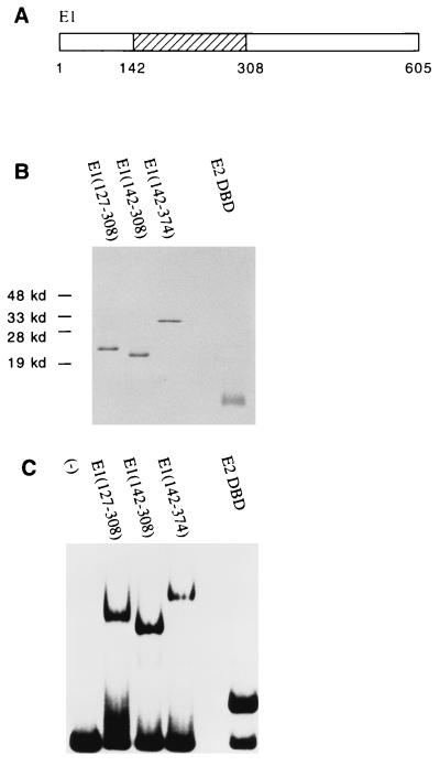
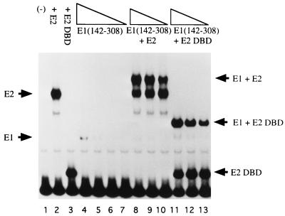
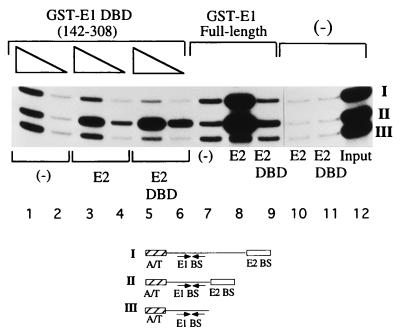
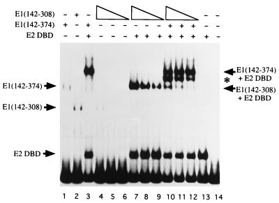
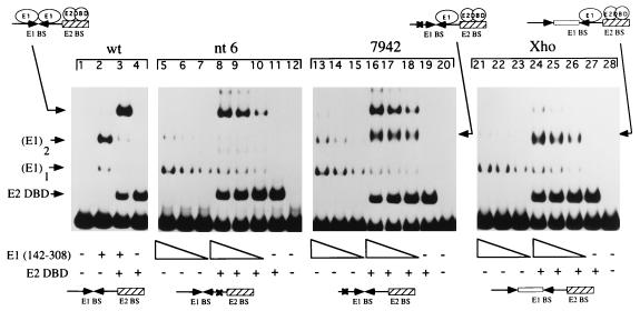
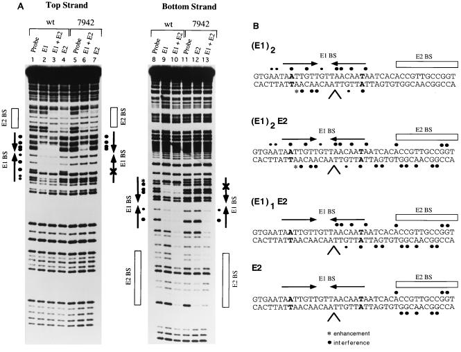
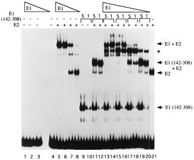
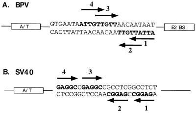
The bovine papillomavirus replication initiator protein E1 is an origin of replication (ori)-binding protein absolutely required for viral DNA replication. In the presence of the viral transcription factor E2, E1 binds to the ori and initiates DNA replication. To understand how the E1 initiator recognizes the ori and how E2 assists in this process, we have expressed and purified a 166-amino-acid fragment which corresponds to the minimal E1 DNA-binding domain (DBD). DNA binding studies using this protein demonstrate that the E1 DBD can bind to the palindromic E1 binding site in several forms but that binding of two monomers, each recognizing one half-site of the E1 palindrome, is the predominant form. This is reminiscent of the binding of the T-antigen DBD to the SV40 ori, and interestingly, the arrangement of E1 binding sites shows striking similarities to the arrangement of T-antigen binding sites in the SV40 ori even though the recognition sequences are unrelated. The E1 DBD is capable of interacting cooperatively with E2; however, the E2 DBD and not the E2 activation domain mediates this interaction. Furthermore, the E2 DBD stimulates binding of two monomers of the E1 DBD to the ori by binding cooperatively with one E1 monomer. Finally, we show that our results concerning the DNA-binding properties of the E1 DBD can be extended to full-length E1.


Figures

FIG. 1
FIG. 1
DNA-binding activities of the E1 and E2 DBDs. (A) The E1 DBD maps to residues 142 to 308. (B) Analysis by SDS-PAGE (15% gel) and Coomassie staining of 1 μg of protein after the final step of purification. E1(127–308), E1(142–308), and E1(142–374) were expressed as N-terminal GST fusions in E. coli. After affinity purification using glutathione-agarose beads, the E1 fragments were treated with thrombin to remove the GST. The E2 DBD(323–410) was also expressed in E. coli but without any GST fusion. Extracts were then loaded directly onto an S-Sepharose column. Peak fractions were pooled and further purified on a Mono-S column. (C) DNA-binding activity of the purified E1 or E2 DBD protein. Gel mobility shift assays were performed with a probe containing the BPV minimal ori with a high-affinity E2 binding site (BS12H). Ten-microliter binding reaction mixtures containing 12 ng of E1(127–308), 2 ng of E1(142–308), 5 ng of E1(142–374), or 36 pg of E2 DBD(325–410) were incubated for 30 min at room temperature and analyzed on a 5% polyacrylamide gel.
FIG. 2
FIG. 2
The E1 DBD binds cooperatively with E2. In 10-μl binding reactions, 0.8, 0.4, 0.2, and 0.1 ng of E1(142–308) were incubated alone (lanes 4 to 7) or in the presence of either 0.1 ng of full-length E2 (lanes 8 to 10) or 18 pg of the E2 DBD (lanes 11 to 13). The probe used in the binding reactions contained the BPV minimal ori with a high-affinity E2 binding site (BS12H). After 30 min of incubation at room temperature, the reaction mixtures were loaded directly onto a 5% polyacrylamide gel.
FIG. 3
FIG. 3
The E1 DBD interacts with the E2 DBD but not with the E2 activation domain. Three different probes containing a high-affinity E2 binding site distal to the E1 binding site (I), a high-affinity E2 binding site proximal to the E1 binding site (II), and only an E1 binding site (III) were mixed with either 0.5 (lane 1) or 0.25 (lane 2) ng of GST-E1(142–308) in 10-μl binding reactions; 2.0 ng of E2 (lanes 3 and 4) or 0.80 ng of the E2 DBD (lanes 5 and 6) was incubated with GST-E1(142–308) in the binding reaction. Probes bound by GST-E1 DBD were recovered by using glutathione-agarose beads. The recovered probes were analyzed on a 6% urea gel. Control reactions containing GST-E1 (full length) (6 ng) alone (lane 7) or together with full-length E2 (lane 8) or the E2 DBD (lane 9) were performed simultaneously.
FIG. 4
FIG. 4
The E1 DBD binds as two monomers to the minimal ori. To determine if the E1 DBD binds as two monomers on the E1 palindrome, three quantities of E1(142–308), 0.4, 0.2, and 0.1 ng, were incubated together with 0.5 ng of E1(142–374), 18 pg of the E2 DBD, and probe containing the BPV minimal ori with a high-affinity E2 binding site in 10-μl binding reactions (lanes 10 to 12). The asterisk marks the mobility of an intermediate complex formed when E1(142–374) and E1(142–308) were mixed in the presence of the E2 DBD (lanes 3 and 7 to 9, respectively). The levels of binding by E1(142–308) in the absence of the E2 DBD and E1(142–374) are shown in lanes 4 to 6. Lanes 1 and 2 are markers for the binding of E1(142–374) and E1(142–308), respectively.
FIG. 5
FIG. 5
Gel shift analysis of complexes formed on mutant ori. Assays to determine the ability to form complexes in the presence of the E1 DBD alone or in the presence of both the E1 and E2 DBDs were performed with a probe containing the wild-type (wt) minimal ori (lanes 1 to 4), a probe containing a single-point mutation in the E1 half-site proximal to the E2 binding site (nt 6; lanes 5 to 12), a probe containing a single-point mutation in the E1 half-site distal to the E2 binding site (7942; lanes 13 to 20), and probe in which the two half-sites are separated by an 8 bp XhoI linker (Xho; lanes 21 to 28). All four probes contained a high-affinity E2 binding site (BS9). Lane 2 contains 1 ng of E1(142–308); lane 3 contains 0.5 ng of E1(142–308) and 18 pg of the E2 DBD. For the mutant probes, three twofold dilutions of E1(142–308), corresponding to 5, 2.5, and 1.3 ng, were used both in the presence and in the absence of the E2 DBD; 18 pg of the E2 DBD was used in all E2-containing reactions. Binding reactions were in a final volume of 10 μl and incubated at room temperature for 30 min. The reactions were subsequently loaded on a 6% native polyacrylamide gel.
FIG. 6
FIG. 6
(A) One monomer of the E1 DBD binds to one half-site of the E1 palindrome. DEPC interference analysis was performed on both strands of the probe containing the wild-type minimal ori (wt) and probe containing the ori with a single-point mutation in nucleotide position 7942, which is distal to the E2 binding site. The wt and 7942 probes were treated with DEPC, which modifies A and G residues, and then used in binding reactions. Complexes corresponding to the binding of two E1 monomers (lanes 2 and 9) and two E1 monomers together with the E2 DBD (lanes 3 and 10) were generated on the wild-type probe, as well as the monomer of E1 complexed with the E2 DBD on the 7942 probe (lanes 6 and 13). Complexes containing only the E2 DBD were also generated for both probes (lanes 4, 7, and 12). Complexes were separated on a 6% polyacrylamide gel, the recovered probes were cleaved with piperidine, and the products were analyzed on an 8% sequencing gel. (B) Summary of the DEPC interference analysis. Interferences are shown for the complex containing two E1 monomers, the E1 monomer and E2 DBD, and the E2 DBD alone. The position of the mutation present in either the 7942 probe or nt 6 probe is shown in bold. The caret indicates the position of the XhoI linker insertion.
FIG. 7
FIG. 7
Full-length E1 also binds as two monomers to the minimal ori. A mixing experiment similar to that shown in Fig. 4 was performed with full-length E1. Four reactions containing 5, 1.3, 0.6, and 0.3 ng of full-length E1 were incubated alone (lanes 1 to 4), together with 0.1 ng of full-length E2 (lanes 5 to 8), or with two different quantities of truncated E1(142–308) (2 and 0.4 ng) in the presence of 0.1 ng of full-length E2 (lanes 13 to 20) in 10-μl binding reactions containing probe with the BS12H high-affinity E2 binding site. The asterisk indicates the position of the intermediate complex which formed when E1 and truncated E1(142–308) were mixed together in the presence of E2. Lanes 9 and 10 contain truncated E1(142–308) alone at the two concentrations used in the mixing experiment; lanes 11 and 12 contain the two concentrations of E1(142–308) in the presence of the E2 DBD. After incubation for 30 min at room temperature, the reactions were analyzed by SDS-PAGE (6% gel).
FIG. 8
FIG. 8
Comparison of the arrangements of T-antigen binding sites in the SV40 ori with that of the proposed E1 binding sites in the BPV minimal ori. (A) The BPV minimal ori may contain four E1 binding sites. Binding sites 2 and 4, arranged in a head-to-head orientation, are used for the binding of two E1 monomers in the E1E2-ori complex. Binding sites 2 and 4, which are also oriented head to head, overlap binding sites 1 and 3 by three nucleotides. (B) The SV40 ori consists of four T-antigen binding sites of which T antigen binds primarily to sites 1 and 3, which are also oriented head to head.

Similar articles

Cited by

References

    1. Aurora R, Herr W. Segments of the POU domain influence one another’s DNA-binding specificity. Mol Cell Biol. 1992;12:455–467. - PMC - PubMed
    1. Berg M, Stenlund A. Functional interactions between papillomavirus E1 and E2 proteins. J Virol. 1997;71:3853–3863. - PMC - PubMed
    1. Blitz I L, Laimins L. The 68-kilodalton E1 protein of bovine papillomavirus is a DNA-binding phosphoprotein which associates with the E2 transcriptional activator in vitro. J Virol. 1991;65:649–656. - PMC - PubMed
    1. Bonne-Andrea C, Santucci S, Clertant P. Bovine papillomavirus E1 protein can, by itself, efficiently drive multiple rounds of DNA synthesis in vitro. J Virol. 1995;69:3201–3205. - PMC - PubMed
    1. Chen, G., and A. Stenlund. Unpublished results.

Publication types

LinkOut - more resources