

Estradiol increases dendritic spine density by reducing GABA neurotransmission in hippocampal neurons
- PMID: 9502814
- PMCID: PMC6793090
- DOI: 10.1523/JNEUROSCI.18-07-02550.1998
Estradiol increases dendritic spine density by reducing GABA neurotransmission in hippocampal neurons
Abstract
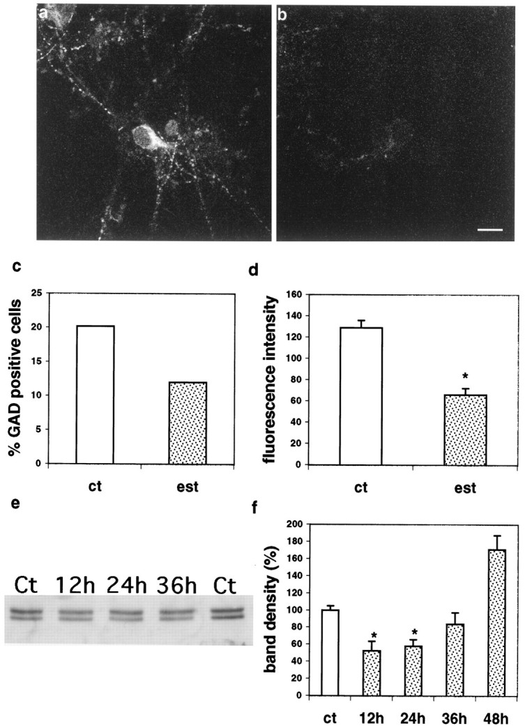
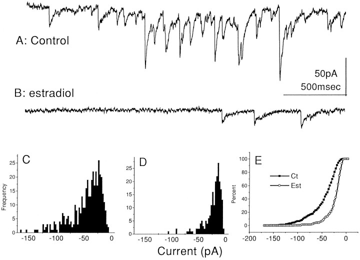
We have previously shown that estradiol causes a twofold rise in dendritic spine density in cultured rat hippocampal neurons, as it does in vivo. More recently, estrogen receptors have been localized to aspiny inhibitory hippocampal interneurons, indicating that their effect on spiny pyramidal neurons may be indirect. We therefore examined the possibility that estradiol affects spine density by regulating inhibition in cultured hippocampal interneurons. Immunocytochemically, estrogen receptors were found to be co-localized with glutamate decarboxylase (GAD)-positive neurons (approximately 21% of total neurons in the culture). Exposure of cultures to estradiol for 1 d caused a marked decrease (up to 80%) in the GAD content of the interneurons, measured both by immunohistochemistry and Western blotting. Also, the number of GAD-positive neurons in the cultures decreased to 12% of the total cell population. Moreover, GABAergic miniature IPSCs were reduced in both size and frequency by estradiol, whereas miniature EPSCs increased in frequency. We then mimicked the proposed effects of estradiol by blocking GABA synthesis with mercaptopropionic acid (MA). Cultures treated with MA expressed a dose-dependent decrease in GABA immunostaining that mimicked that seen with estradiol. MA-treated cultures displayed a significant 50% increase in dendritic spine density over controls, similar to that produced by estradiol. These results indicate that estradiol decreases GABAergic inhibition in the hippocampus, which appears to effectively increase the excitatory drive on pyramidal cells, and thus may provide a mechanism for formation of new dendritic spines.
Figures


Similar articles
-
Quantitative analysis of ER alpha and GAD colocalization in the hippocampus of the adult female rat.J Comp Neurol. 2001 Nov 12;440(2):144-55. doi: 10.1002/cne.1376. J Comp Neurol. 2001. PMID: 11745614
-
Kalirin-7, an important component of excitatory synapses, is regulated by estradiol in hippocampal neurons.Hippocampus. 2011 Jun;21(6):661-77. doi: 10.1002/hipo.20780. Epub 2010 Mar 23. Hippocampus. 2011. PMID: 20333733 Free PMC article.
-
Brain-derived neurotrophic factor mediates estradiol-induced dendritic spine formation in hippocampal neurons.Proc Natl Acad Sci U S A. 1998 Sep 15;95(19):11412-7. doi: 10.1073/pnas.95.19.11412. Proc Natl Acad Sci U S A. 1998. PMID: 9736750 Free PMC article.
-
Estradiol induces formation of dendritic spines in hippocampal neurons: functional correlates.Horm Behav. 2001 Sep;40(2):156-9. doi: 10.1006/hbeh.2001.1688. Horm Behav. 2001. PMID: 11534976 Review.
-
Estradiol and GABAergic transmission in the hippocampus.Vitam Horm. 2010;82:279-300. doi: 10.1016/S0083-6729(10)82015-1. Vitam Horm. 2010. PMID: 20472144 Review.
Cited by
-
Sex hormone levels in females of different ages suffering from depression.BMC Womens Health. 2021 May 22;21(1):215. doi: 10.1186/s12905-021-01350-0. BMC Womens Health. 2021. PMID: 34022874 Free PMC article.
-
Estrogen-induced increase in the magnitude of long-term potentiation occurs only when the ratio of NMDA transmission to AMPA transmission is increased.J Neurosci. 2005 Aug 24;25(34):7780-91. doi: 10.1523/JNEUROSCI.0762-05.2005. J Neurosci. 2005. PMID: 16120779 Free PMC article.
-
Estradiol targets synaptic proteins to induce glutamatergic synapse formation in cultured hippocampal neurons: critical role of estrogen receptor-alpha.J Neurosci. 2007 Jun 27;27(26):6903-13. doi: 10.1523/JNEUROSCI.0909-07.2007. J Neurosci. 2007. PMID: 17596438 Free PMC article.
-
Ovariectomy of adult rats leads to increased expression of astrocytic basic fibroblast growth factor in the ventral tegmental area and in dopaminergic projection regions of the entorhinal and prefrontal cortex.J Neurosci. 1999 Oct 1;19(19):8665-73. doi: 10.1523/JNEUROSCI.19-19-08665.1999. J Neurosci. 1999. PMID: 10493767 Free PMC article.
-
Hippocampal synapses depend on hippocampal estrogen synthesis.J Neurosci. 2004 Jun 30;24(26):5913-21. doi: 10.1523/JNEUROSCI.5186-03.2004. J Neurosci. 2004. PMID: 15229239 Free PMC article.
References
-
- Berninger B, Marty S, Zafra F, Berzaghi M, Thoenen H, Lindholm D. GABAergic stimulation switches from enhancing to repressing BDNF expression in rat hippocampal neurons during maturation in vitro. Development. 1995;121:2327–2335. - PubMed
-
- Brinton RD, Tran J, Proffitt P, Montoya M. 17-Beta-estradiol enhances the outgrowth and survival of neocortical neurons in culture. Neurochem Res. 1997;22:1339–1351. - PubMed
-
- Buterbaugh GG, Hudson GM. Estradiol replacement to female rats facilitates dorsal hippocampal but not ventral hippocampal kindled seizure acquisition. Exp Neurol. 1991;111:55–64. - PubMed
-
- Davis AM, Grattan DR, Selmanoff M, McCarthy MM. Sex differences in glutamic acid decarboxylase mRNA in neonatal rat brain: implications for sexual differentiation. Horm Behav. 1996;30:538–552. - PubMed
Publication types
MeSH terms
Substances
LinkOut - more resources
Full Text Sources
Other Literature Sources