. 1998 Mar 1;12(5):679-91.
doi: 10.1101/gad.12.5.679.

Arginine methylation facilitates the nuclear export of hnRNP proteins

Affiliations

Arginine methylation facilitates the nuclear export of hnRNP proteins

E C Shen et al. Genes Dev. .

Abstract

Eukaryotic mRNA processing and export is mediated by various heterogeneous nuclear ribonucleoproteins (hnRNPs). Many of these hnRNPs are methylated on arginine residues. In the yeast, Saccharomyces cerevisiae, the predominant enzyme responsible for arginine methylation is Hmt1p. Hmt1p methylates both Npl3p and Hrp1p, which are shuttling hnRNPs involved in mRNA processing and export. Here, we employ an in vivo nuclear export assay to show that arginine methylation is important for the nuclear export of these hnRNPs. Both Npl3p and Hrp1p fail to exit the nucleus in cells lacking Hmt1p, and overexpression of Hmt1p enhances Npl3p export. The export of a novel hnRNP-like protein, Hrb1p, which does not bind poly(A)+ RNA, however, is not affected by the lack of methylation. Furthermore, we find a genetic relationship between Hmt1p and cap-binding protein 80 (CBP80). Together, these findings establish that one biological role for arginine methylation is in facilitating the export of certain hnRNPs out of the nucleus.


Figures

Figure 1
Figure 1
Export of Npl3p and Hrp1p is blocked in the absence of Hmt1p. Either nup49-313 cells (A–D and I–L) or the nup49-313 strain in which HMT1 has been deleted (E–H and M–P) were incubated in the export assay. The reporter used was GFP–Npl3p (pPS811; A–H) or GFP–Hrp1p (pPS1358; I–P). Cells were incubated at 25°C (A,B,E,F,I,J,M,N) or at 36.5°C (C,D,G,H,K, L,O,P) for 5 hr. Cells were photographed by use of Nomarski optics (A,C,E,G,I,K,M,O) or for GFP fluorescence (B,D,F, H,J,L,N,P).
Figure 2
Figure 2
Npl3p, Hrp1p and Hrb1p are methylated by Hmt1p. Recombinant substrates Npl3p (lanes 1,5), Hrp1p (lanes 2,6), Hrb1p (lanes 3,7) were purified from E. coli and BSA was used as a negative control (lanes 4,8). Substrates were incubated with [methyl3H]-SAM in the presence (lanes 1–4) or absence (lanes 5–8) of Hmt1p purified from E. coli. Samples were resolved by SDS-PAGE. Proteins were detected by Coomassie staining (B) and labeling was detected by fluorography (A). Protein molecular weights are indicated in kilodaltons.
Figure 3
Figure 3
Hrb1p is a novel nuclear hnRNP. Cells harboring plasmids expressing Npl3p–Myc expressed from its endogenous promoter (A–C), or Hrb1p (D–F) and Hrb1p–Myc (G–I) under control of the inducible GAL1 promoter were subjected to immunofluorescence microscopy with the 9E10 monoclonal antibody. The Myc-tagged proteins were visualized with FITC labeled anti-mouse antibody (C,F,I). DNA was visualized using DAPI (B,E,H). Cells were photographed by use of Nomarski optics (A,D,G).
Figure 4
Figure 4
Hrb1p export is unaffected by the absence of Hmt1p. Wild-type cells (A–D), nup49-313 cells (E–H), and nup49-313 ΔHMT1 double-mutant cells (I–L) containing the reporter plasmid pPS1341 expressing GFP–Hrb1p were subjected to the export assay. Cells were incubated at 25°C (A,B,E,F,I,J) or 36.5°C (C,D,G,H,K,L). Cells were photographed either using Nomarski optics (A,C,E,G,I,K) or for the GFP signal (B,D,F,H,J,L).
Figure 5
Figure 5
Export is blocked in the absence of RNA Pol II transcription. Double-mutant nup49-313 rpb1-1 cells carrying reporter plasmids expressing GFP–Npl3p (A–D), GFP–Hrp1p (E–H), or GFP–Hrb1p (I–L) were subjected to the export assay. Cells were incubated at 25°C (A,B,E,F,I,J) or 36.5°C (C,D,G,H,K,L) for 5 hr. Living cells were photographed by Nomarski optics (A,C,E,G,I,K) or for the GFP signal (B,D,F,H,J,L).
Figure 6
Figure 6
Overproduction of Hmt1p rescues the export defects of npl3-17. (a) A npl3-17 strain was transformed with the following plasmids and streaked on synthetic complete plates lacking leucine and containing glucose as the carbon source: CEN vector alone (pRS315; panels A,C), CEN HMT1 (pPS1305; panels B,D), 2μ vector alone (YEp351; panels E,G), 2μ HMT1 (pPS1308; panels F,H). Plates were incubated at either 25°C (panels A,B,E,F) or 36°C (panels C,D,G,H) for 3 days. (b) npl3-17 cells harboring GAL1 vector pPS311 (panels A–D) or GAL1 HMT1 pPS1141 (panels E–H). Transformants were induced for protein expression for 2 hr then incubated at 36°C for 4 hr, fixed with formaldehyde and processed for in situ hybridization and immunofluorescence microscopy to detect Npl3p (panels C,G) and poly(A)+ RNA (panels D,H). DNA was visualized by DAPI staining (panels B,F), and cells were photographed by use of Nomarski optics (panels A,E). (c) nup49-313 cells containing GFP–Npl3p (pPS811; panels A,B), GFP–Npl3p with HMT1 (pPS1348; panels C,D), GFP–F160L (pPS879; panels E,F), and GFP–F160L with HMT1 (pPS1349; panels G,H) were subjected to the export assay. Cells shifted to 36.5°C for 5 hr are shown. Cells were photographed by use of Nomarski optics (panels A,C,E,G) and for the GFP signal (panels B,D,F,H).
Figure 6
Figure 6
Overproduction of Hmt1p rescues the export defects of npl3-17. (a) A npl3-17 strain was transformed with the following plasmids and streaked on synthetic complete plates lacking leucine and containing glucose as the carbon source: CEN vector alone (pRS315; panels A,C), CEN HMT1 (pPS1305; panels B,D), 2μ vector alone (YEp351; panels E,G), 2μ HMT1 (pPS1308; panels F,H). Plates were incubated at either 25°C (panels A,B,E,F) or 36°C (panels C,D,G,H) for 3 days. (b) npl3-17 cells harboring GAL1 vector pPS311 (panels A–D) or GAL1 HMT1 pPS1141 (panels E–H). Transformants were induced for protein expression for 2 hr then incubated at 36°C for 4 hr, fixed with formaldehyde and processed for in situ hybridization and immunofluorescence microscopy to detect Npl3p (panels C,G) and poly(A)+ RNA (panels D,H). DNA was visualized by DAPI staining (panels B,F), and cells were photographed by use of Nomarski optics (panels A,E). (c) nup49-313 cells containing GFP–Npl3p (pPS811; panels A,B), GFP–Npl3p with HMT1 (pPS1348; panels C,D), GFP–F160L (pPS879; panels E,F), and GFP–F160L with HMT1 (pPS1349; panels G,H) were subjected to the export assay. Cells shifted to 36.5°C for 5 hr are shown. Cells were photographed by use of Nomarski optics (panels A,C,E,G) and for the GFP signal (panels B,D,F,H).
Figure 7
Figure 7
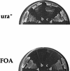
Deletion of both CBP80 and HMT1 is lethal. One tetrad from a cross of ΔHMT1 covered with wild-type HMT1 and ΔCBP80 is shown. Two spores are wildtype for both alleles (A,D), and two contain both deletion mutants (B,C). Spores were streaked on synthetic complete media lacking uracil (top) or containing 5-FOA (bottom). Plates were incubated at 25°C for 2 days.
Figure 8
Figure 8
Models for HMT1 function in pre-mRNA metabolism, packaging, and export. (A) Genetic data suggest that HMT1 affects diverse functions in RNA metabolism including capping, polyadenylation, and export. (B) Hmt1p methylates at least two nuclear RNA binding proteins that play a role in the export of RNA, possibly providing a signal to facilitate association of properly packaged RNAs with the export machinery. Once in the cytoplasm, RNA binding proteins are released and re-enter the nucleus.

Similar articles

Cited by

References

    1. Adachi Y, Yanagida M. Higher order chromosome structure is affected by cold-sensitive mutations in a Schizosaccharomyces pombe gene CRM1+, which encodes a 115-kD protein preferentially localized in the nucleus and its periphery. J Cell Biol. 1989;108:1195–1207. - PMC - PubMed
    1. Baudin A, Ozier-Kalogeropoulou O, Denouel A, Lacroute F, Cullin C. A simple and efficient method of direct gene deletion in Saccharomyces cerevisiae. Nucleic Acids Res. 1993;21:3329–3330. - PMC - PubMed
    1. Beyer AL, Christensen ME, Walker BW, LeStourgeon WM. Identification and characterization of the packaging proteins of core 40S hnRNP particles. Cell. 1977;11:127–138. - PubMed
    1. Birney E, Kumar S, Krainer AR. Analysis of the RNA-recognition motif and RS and RGG domains: Conservation in metazoan pre-mRNA splicing factors. Nucleic Acids Res. 1993;21:5803–5816. - PMC - PubMed
    1. Boffa LC, Karn J, Vidali G, Allfrey VG. Distribution of NG-NG-dimethylarginine in nuclear protein fractions. Biochem Biophys Res Commun. 1977;74:969–976. - PubMed

Publication types

MeSH terms