. 1998 Mar 1;12(5):640-53.
doi: 10.1101/gad.12.5.640.

Critical residues for histone acetylation by Gcn5, functioning in Ada and SAGA complexes, are also required for transcriptional function in vivo

Affiliations

Critical residues for histone acetylation by Gcn5, functioning in Ada and SAGA complexes, are also required for transcriptional function in vivo

L Wang et al. Genes Dev. .

Abstract

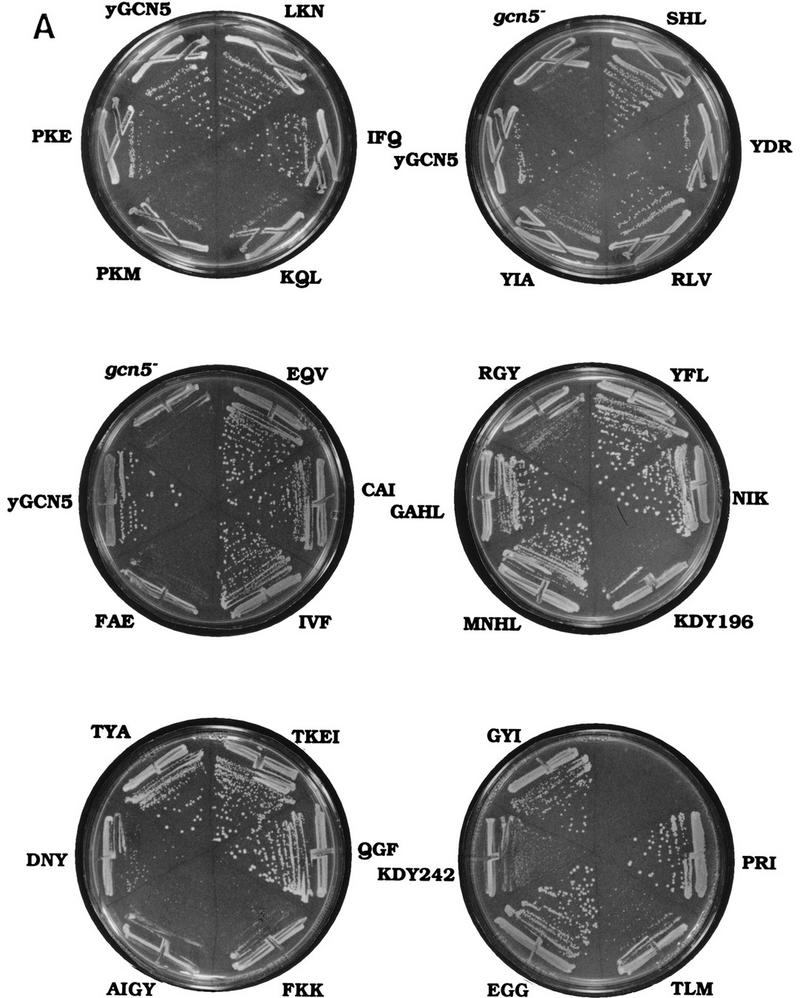
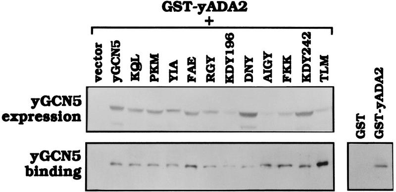
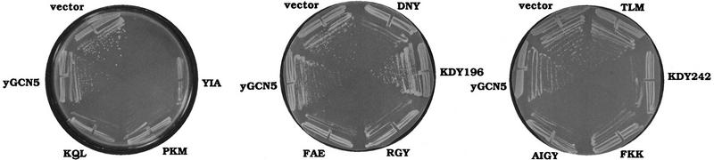
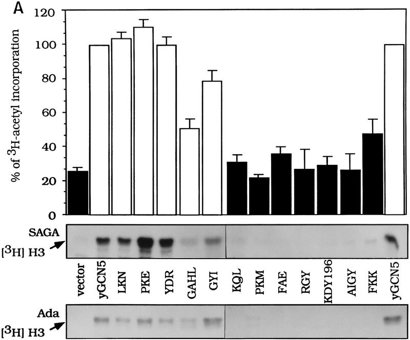
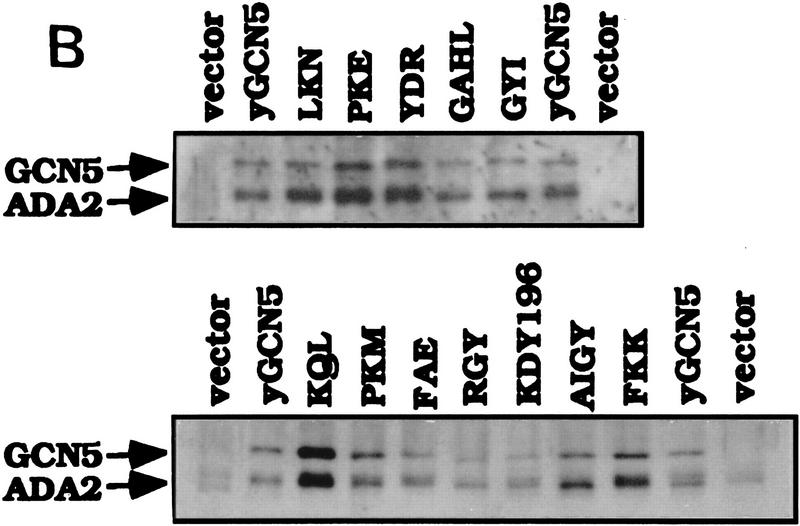
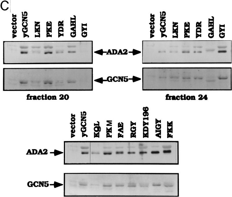
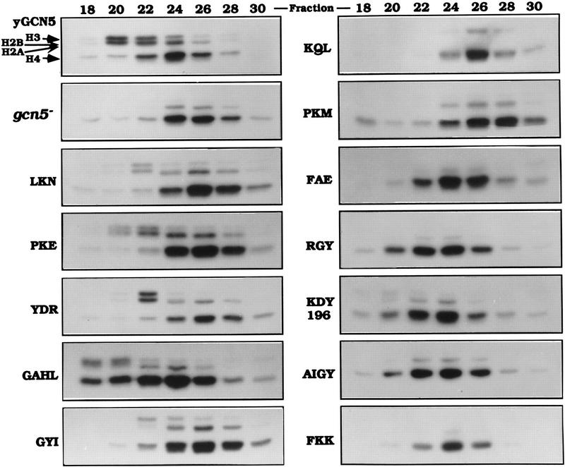
Several previously known transcription cofactors have been demonstrated in vitro recently to be histone acetyltransferases and deacetyltransferases, suggesting that remodeling of chromatin through histone acetylation plays a fundamental role in gene regulation. Clear evidence has not yet been obtained, however, to demonstrate that histone acetylation is required for gene activation in vivo. In this study we performed an alanine-scan mutagenesis through the HAT (histone acetyltransferase) domain identified previously by deletion mapping in recombinant yeast Gcn5. We identified multiple substitution mutations that eliminated completely Gcn5's ability to potentiate transcriptional activation in vivo. Strikingly, each of these mutations was also critical for free and nucleosomal histone acetylation by Gcn5 functioning within the native yeast HAT complexes, Ada, and SAGA. Moreover, the growth phenotypes of these mutations as measured by colony size and liquid growth assay closely tracked transcription and HAT activities. In contrast, mutations that did not affect in vivo function of Gcn5 were able to acetylate histones. These data argue strongly that acetylation is required for gene regulation by Gcn5 in vivo, and support previous arguments that nucleosomal histones are among the physiological substrates of acetylation by Gcn5.


Figures

Figure 1
Figure 1
Amino acid sequence within the putative HAT domains of the Gcn5 family. (A) Sequence comparison between the putative HAT domains of yeast Gcn5, human P/CAF, human Gcn5 and Tetrahymena p55. The protein sequence within the putative HAT domains are shown by single amino acid letter-code. Numbers at left indicate amino acid residues. The highly conserved regions I–IV (Brownell et al. 1996) are indicated at the top. Solid boxes indicate amino acid identity and shaded boxes indicate similarity. (B) Yeast Gcn5 substitution mutants created in the study. The substitution mutants are named according to the group of amino acids mutated to alanine, each of which is indicated by brackets.
Figure 2
Figure 2
Growth phenotypes of GCN5 substitution mutants in the gcn5 strain. Wild-type GCN5, each of the GCN5 mutants, or vector alone were introduced into gcn5 strain. (A) Colony growth assay. Transformants were streaked onto synthetic minimal medium. Colony growth was assessed after 2 days of growth at 30°C. (B) Doubling time of the Gcn5 substitution mutants. Aliquots of cultures in liquid synthetic minimal medium were taken every 2 hr and the A600 of the cultures was measured. The generation time was calculated as the OD doubling time during exponential growth.
Figure 2
Figure 2
Growth phenotypes of GCN5 substitution mutants in the gcn5 strain. Wild-type GCN5, each of the GCN5 mutants, or vector alone were introduced into gcn5 strain. (A) Colony growth assay. Transformants were streaked onto synthetic minimal medium. Colony growth was assessed after 2 days of growth at 30°C. (B) Doubling time of the Gcn5 substitution mutants. Aliquots of cultures in liquid synthetic minimal medium were taken every 2 hr and the A600 of the cultures was measured. The generation time was calculated as the OD doubling time during exponential growth.
Figure 3
Figure 3
Stability and Ada2 interaction of Gcn5 substitution mutants in yeast. Wild-type GCN5, mutant GCN5, or vector alone were cotransformed with GST–ADA2, and proteins were induced using the GAL1-10 promoter. (Top) Gcn5 protein levels in the yeast extracts were determined by Western blot analysis using Gcn5 antisera. (Bottom left) The yeast extracts were incubated with GST beads to allow GST–Ada2 to bind. Wild-type or mutant Gcn5 bound to GST–Ada2 was determined by Western analysis using Gcn5 antiserum. (Bottom right) Co-expression of wild-type Gcn5 with GST alone was used as a negative control.
Figure 4
Figure 4
Transcriptional activation in the presence of Gcn5 substitution mutants. GAL4–VP16 (left panel) and LexA–GCN4 (right panel) were cotransformed with wild-type GCN5, each of the GCN5 mutants or vector alone, along with the appropriate LacZ reporters. β-Gal activities of the mutants are shown as a percentage of wild-type Gcn5, which was set at 100%. (Solid bars) Mutants that were defective in the growth and transcription assays; (dark gray bars) mutants having intermediate phenotypes; (light gray bars) mutants having activity similar to wild-type Gcn5. Error bars represent the standard error about the mean from three independent experiments.
Figure 5
Figure 5
Dominant-negative effects of Gcn5 HAT mutants. Wild-type GCN5, the GCN5 mutants, or vector alone were transformed into yeast strain bearing wild-type GCN5. The transformants were restreaked onto galactose plates to induce expression of transformed Gcn5 proteins and grown at 25°C for 6 days.
Figure 6
Figure 6
Free histone HAT activity of Gcn5 substitution mutants in SAGA and Ada complexes. (A) SAGA and Ada complexes prepared from Gcn5 mutants displaying wild-type activity in functional assays (light gray bars in Fig. 4) and Gcn5-defective mutants (solid bars in Fig. 4) were tested for HAT activity. SAGA and Ada complexes of each mutants containing peak Gcn5 and Ada2 proteins detected by Western blots (B,C) were used. Complexes were incubated with free core histones and [3H]acetyl CoA. Reaction mixtures were then subjected to liquid scintillation accounting (top) as well as SDS-PAGE and fluorography (middle and bottom). The activity from SAGA complexes (middle) and Ada complexes (bottom) are indicated by arrows. Quantitation of corresponding scintillation assays for SAGA is shown as a percentage of wild-type Gcn5. Error bars represent the standard error about the mean from three independent experiments. Western blots of Gcn5 and Ada2 proteins from SAGA (B) and Ada (C) complexes. Peak fractions from SAGA and Ada complexes were detected for the presence of Gcn5 and Ada2 using immunoblot analysis using Gcn5 and Ada2 antisera.
Figure 6
Figure 6
Free histone HAT activity of Gcn5 substitution mutants in SAGA and Ada complexes. (A) SAGA and Ada complexes prepared from Gcn5 mutants displaying wild-type activity in functional assays (light gray bars in Fig. 4) and Gcn5-defective mutants (solid bars in Fig. 4) were tested for HAT activity. SAGA and Ada complexes of each mutants containing peak Gcn5 and Ada2 proteins detected by Western blots (B,C) were used. Complexes were incubated with free core histones and [3H]acetyl CoA. Reaction mixtures were then subjected to liquid scintillation accounting (top) as well as SDS-PAGE and fluorography (middle and bottom). The activity from SAGA complexes (middle) and Ada complexes (bottom) are indicated by arrows. Quantitation of corresponding scintillation assays for SAGA is shown as a percentage of wild-type Gcn5. Error bars represent the standard error about the mean from three independent experiments. Western blots of Gcn5 and Ada2 proteins from SAGA (B) and Ada (C) complexes. Peak fractions from SAGA and Ada complexes were detected for the presence of Gcn5 and Ada2 using immunoblot analysis using Gcn5 and Ada2 antisera.
Figure 7
Figure 7
Nucleosomal HAT activities of Gcn5 substitution mutants in Ada complexes. The same wild-type mutants and defective mutants used in the free histone assays (Fig. 6) were subjected to nucleosomal HAT assays using Ada complexes. Even-numbered Mono Q column fractions from Ada complex of each mutant were incubated with oligonucleosome cores and [3H]acetyl CoA. Reactions were loaded onto SDS-PAGE and the gels were analyzed by fluorography. (Left) Wild-type Gcn5, vector alone, and wild-type Gcn5 substitution mutants; (right) defective Gcn5 mutants. Positions of core histones on the gel are indicated by arrows. Numbers at the top are the Mono Q column fractions. The H3/H2B nucleosomal activity is Gcn5-dependent, and the H4/H2A nucleosomal HAT activity is Gcn5-independent (Grant et al. 1997).
Figure 8
Figure 8
Comparison of mutant Gcn5 phenotypes in acetylation vs. transcription. Quantitative data are taken from Fig. 6A (top) and Fig. 4 (the Gal4–VP16 and LexA–Gcn4 data were averaged). Acetylation of free histones by the SAGA complex is shown at left and transcriptional activation by the chimeric activators is shown at right.

Similar articles

Cited by

References

    1. Alani E, Cao L, Kleckner N. A method for gene disruption that allows repeated use of URA3 selection in the construction of multiply disrupted yeast strains. Genetics. 1987;116:541–545. - PMC - PubMed
    1. Alland L, Muhle R, Hou Jr H, Potes J, Chin L, Schreiber-Agus N, DePinho RA. Role for N-CoR and histone deacetylase in Sin3-mediated transcriptional repression. Nature. 1997;387:49–55. - PubMed
    1. Bannister A, Kouzarides T. The CBP co-activator is a histone acetyltransferase. Nature. 1996;384:641–643. - PubMed
    1. Barlev N, Candau R, Wang L, Darpino P, Silverman N, Berger S. Characterization of physical interactions of the putative transcriptional adaptor, ADA2, with acidic activation domains and TATA-binding protein. J Biol Chem. 1995;270:19337–19344. - PubMed
    1. Berger SL, Cress WD, Cress A, Triezenberg SJ, Guarente L. Selective inhibition of activated but not basal transcription by the acidic activation domain of VP16: Evidence for transcriptional adaptors. Cell. 1990;61:1199–1208. - PubMed

Publication types

MeSH terms