. 1998 Mar;72(3):2265-71.
doi: 10.1128/JVI.72.3.2265-2271.1998.

Mouse hepatitis virus 3C-like protease cleaves a 22-kilodalton protein from the open reading frame 1a polyprotein in virus-infected cells and in vitro

Affiliations

Mouse hepatitis virus 3C-like protease cleaves a 22-kilodalton protein from the open reading frame 1a polyprotein in virus-infected cells and in vitro

X T Lu et al. J Virol. 1998 Mar.

Abstract

The 3C-like proteinase (3CLpro) of mouse hepatitis virus (MHV) is predicted to cleave at least 11 sites in the 803-kDa gene 1 polyprotein, resulting in maturation of proteinase, polymerase, and helicase proteins. However, most of these cleavage sites have not been experimentally confirmed and the proteins have not been identified in vitro or in virus-infected cells. We used specific antibodies to identify and characterize a 22-kDa protein (p1a-22) expressed from gene 1 in MHV A59-infected DBT cells. Processing of p1a-22 from the polyprotein began immediately after translation, but some processing continued for several hours. Amino-terminal sequencing of p1a-22 purified from MHV-infected cells showed that it was cleaved at a putative 3CLpro cleavage site, Gln_Ser4014 (where the underscore indicates the site of cleavage), that is located between the 3CLpro domain and the end of open reading frame (ORF) 1a. Subclones of this region of gene 1 were used to express polypeptides in vitro that contained one or more 3CLpro cleavage sites, and cleavage of these substrates by recombinant 3CLpro in vitro confirmed that amino-terminal cleavage of p1a-22 occurred at Gln_Ser4014. We demonstrated that the carboxy-terminal cleavage of the p1a-22 protein occurred at Gln_Asn4208, a sequence that had not been predicted as a site for cleavage by MHV 3CLpro. Our results demonstrate the usefulness of recombinant MHV 3CLpro in identifying and confirming cleavage sites within the gene 1 polyprotein. Based on our results, we predict that at least seven mature proteins are processed from the ORF 1a polyprotein by 3CLpro and suggest that additional noncanonical cleavage sites may be used by 3CLpro during processing of the gene 1 polyprotein.


Figures
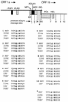
FIG. 1
FIG. 1
MHV gene 1 organization and putative 3CLpro cleavage sites. The diagram shows the organization of the 22-kb gene 1 of the MHV 32-kb RNA. The locations of the PLP-1 and PLP-2 domains, the MP-1 and MP-2 hydrophobic domains, 3CLpro, the GFL domain, RNA-dependent RNA polymerase (POL), and helicase (HEL) are shown as shaded boxes. Locations of predicted MHV 3CLpro cleavage sites are numbered below the diagram. KR, Lys-Arg dipeptide also proposed as a 3CLpro cleavage site (15). The dots denote the confirmed cleavage sites flanking 3CLpro in the polyprotein. The ∗ indicates the Q_N4208 cleavage site identified and described in this paper. The sequences surrounding the confirmed or putative MHV 3CLpro cleavage sites (denoted by MHV) are aligned with the deduced amino acid sequences of HCV 229E (229E) (11), IBV (3), and TGEV (9). Alignments were performed with MacVector version 6.01.
FIG. 2
FIG. 2
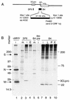
Identification of a 22-kDa protein in MHV-infected DBT cells. (A) The schematic shows the location of the region of gene 1 used to express the protein against which the B4 antibodies were raised. The subclone extends from the XbaI site at nt 12303 to the KpnI site at nt 13906, and the expressed protein extends from aa 4032 to the end of ORF 1a (filled bar). (B) Immunoprecipitation of proteins from MHV-infected DBT cells. Cells were infected (lanes i) or mock infected (lanes m), and proteins were labeled and harvested as described in Materials and Methods. Proteins were immunoprecipitated with antibodies against whole MHV virions (αMHV), the SP9 antisera directed against 3CLpro, or the B4 antiserum. B4-pre indicates preimmune serum from the rabbit used to induce antibodies against the B4 protein. In lanes 4, 8, 9, and 10, proteins were labeled in the presence of the proteinase inhibitors leupeptin (lane L), phenylmethylsulfonyl fluoride (lane P), or E64d (lanes E). Mass markers (in kilodaltons) and the locations of the spike (S), nucleocapsid (N), and matrix proteins (M) are indicated to the left of the gel, and the locations of 3CLpro and other newly identified proteins are shown to the right of the gel.
FIG. 3
FIG. 3
Kinetics of translation and processing of p1a-22. (A) Pulse-label translation. Proteins were radiolabeled with [35S]methionine at 7 h postinfection, and samples were withdrawn from 10 to 120 min and used for immunoprecipitation by B4 antiserum, followed by electrophoresis and fluorography on SDS–5 to 18% gradient polyacrylamide gels. Lysates of mock-infected cells were immunoprecipitated by B4 antiserum (lane mock), and lysates of infected cells were immunoprecipitated with B4 preimmune serum (lane Inf-pre). Molecular mass standards (in kilodaltons) are to the left of the gel, and the location of p1a-22 is shown to the right of the gel. (B) Translation with different durations of labeling and with constant chase. Proteins were labeled in MHV-infected DBT cells for periods from 10 to 120 min and then chased with media containing excess unlabeled methionine and cycloheximide for an additional 90 min. The locations of markers and p1a-22 are shown. (C) Pulse-chase translation. Proteins were radiolabeled for 45 min and then chased in media lacking [35S]Met but containing a 10-fold excess of unlabeled methionine for 10 min (10") to 16 h (16′). All samples were immunoprecipitated and analyzed as described above for panel A. All labeling is as described for panel A. The location of the 200-kDa protein is indicated by the small arrow, and open arrowheads indicate the locations of proteins seen only after prolonged chase.
FIG. 4
FIG. 4
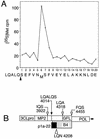
Amino-terminal radiosequencing of p1a-22 from virus-infected cells. (A) p1a-22 was sequenced as described in Materials and Methods. The count of [35S]Met per minute per fraction is shown on the y axis, and the fraction number is shown on the x axis. The presumptive sequence and cleavage site (arrowhead) are shown below the fraction numbers. (B) Schematic of the carboxy-terminal region of the ORF 1a polyprotein shows the confirmed amino terminus of p1a-22 (LQALQ_S4014; filled arrow) and other predicted 3CLpro cleavage sites (open arrows). The extent of the protein used to induce B4 antibodies is indicated by the shaded bar, and the probable extent of the p1a-22 domain is indicated by the black box. The location of the putative LQ_N carboxy-terminal cleavage site of p1a-22 is shown by the open triangle. POL, the RNA-dependent RNA polymerase.
FIG. 5
FIG. 5
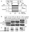
Cloning, expression, and trans cleavage of p1a-22 in vitro. (A) Schematic of the carboxy-terminal region of the ORF 1a polyprotein. The putative p1a-22 domain is shown as filled rectangles. The locations of the sequenced LQ_S cleavage site and the putative IQ_S and LQ_A cleavage sites are shown by filled arrows, and the location of the LQ_N motif is shown by the open arrowhead. The boxed amino acid residues show the amino- and carboxy-terminal residues expressed from the cloned constructs. Residues in parentheses (MQ or MG) were contributed by the primers used to clone the fragments by PCR. The fragment number (1 through 3) and the calculated masses of the individual fragments are shown at the bottom of the diagram, and the constructs are numbered according to the fragments comprising them. (B) The results of the expression of constructs shown in panel A, along with products of cleavage by r3CLpro, are shown. Lane 1 shows p1a-22 immunoprecipitated from MHV-infected DBT cells (DBT RIP), and the location of p1a-22 along with molecular mass standards (in kilodaltons) is indicated to the left. Proteins expressed from construct 2, 1-2, 2-3, or 1-3 were incubated in the presence (+) or absence of r3CLpro and proteinase inhibitor E64 (PI). The masses of expressed proteins and cleavage products are shown to the right of the gel. (C) The products of expression and r3CLpro cleavage were immunoprecipitated with B4 antiserum. All lanes correspond to the proteins seen directly above them in panel B. The levels of contrast of lanes 11 and 12 were equally altered (Photoshop version 4.0) to show the minor 22-kDa cleavage product in lane 12 that was easily seen on the original fluorogram.
FIG. 6
FIG. 6
MHV gene 1 polyprotein processing. This model is based on known and predicted cleavage sites and proteins identified in cells and in vitro. PLP-1 and proteins known to be cleaved in trans by PLP-1 (p28 and p65) are shown as hatched boxes. 3CLpro and proteins known to be cleaved by 3CLpro (3CLpro and p1a-22) are shown as filled boxes. Other predicted proteins are shown as open boxes. Experimentally confirmed cleavage sites are noted by filled arrows, while putative cleavage sites are indicated by open arrowheads. The enlarged region at the bottom of the figure shows the apparent masses of the confirmed 3CLpro and p1a-22 proteins and the location of their cleavage sites (filled boxes and arrows). The calculated masses of other predicted proteins at the end of ORF 1b are shown as open boxes and arrows (–20). POL, RNA-dependent RNA polymerase; HEL, helicase.

Similar articles

Cited by

References

    1. Baker S C, Yokomori K, Dong S, Carlisle R, Gorbalenya A E, Koonin E V, Lai M M. Identification of the catalytic sites of a papain-like cysteine proteinase of murine coronavirus. J Virol. 1993;67:6056–6063. - PMC - PubMed
    1. Bonilla P J, Gorbalenya A E, Weiss S R. Mouse hepatitis virus strain A59 RNA polymerase gene ORF 1a: heterogeneity among MHV strains. Virology. 1994;198:736–740. - PMC - PubMed
    1. Boursnell M F G, Brown T D K, Foulds I J, Green P F, Tomley F M, Binns M M. Completion of the sequence of the genome of the coronavirus avian infectious bronchitis virus. J Gen Virol. 1987;68:57–77. - PubMed
    1. Breedenbeek P J, Pachuk C J, Noten A F H, Charite J, Luytjes W, Weiss S R, Spaan W J M. The primary structure and expression of the second open reading frame of the polymerase gene of the coronavirus MHV-A59; a highly conserved polymerase is expressed by an efficient ribosomal frameshifting mechanism. Nucleic Acids Res. 1990;18:1825–1832. - PMC - PubMed
    1. Brierley I, Boursnell M E G, Binns M M, Billimoria B, Blok V C, Brown T D K, Inglis S C. An efficient ribosomal frame-shifting signal in the polymerase-encoding region of the coronavirus IBV. EMBO J. 1987;6:3779–3785. - PMC - PubMed

Publication types

LinkOut - more resources