. 1998 Mar 9;140(5):1255-63.
doi: 10.1083/jcb.140.5.1255.

Integrin alphavbeta3 requirement for sustained mitogen-activated protein kinase activity during angiogenesis

Affiliations

Integrin alphavbeta3 requirement for sustained mitogen-activated protein kinase activity during angiogenesis

B P Eliceiri et al. J Cell Biol. .

Abstract

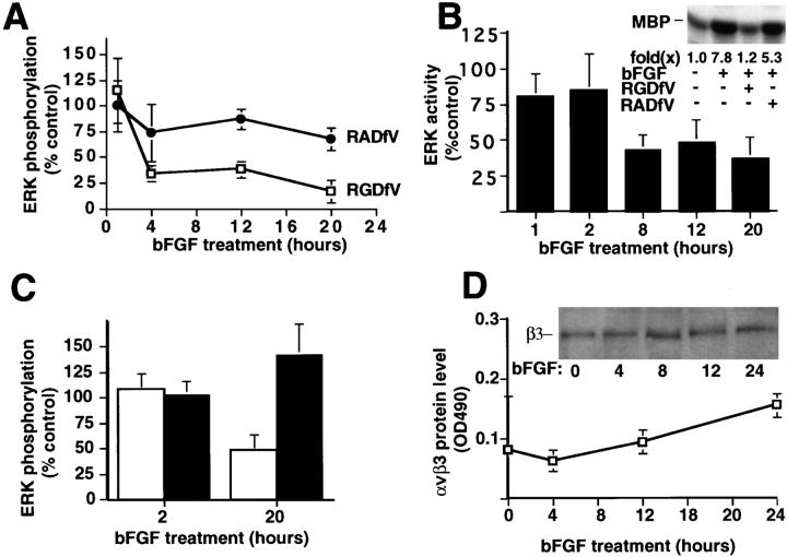
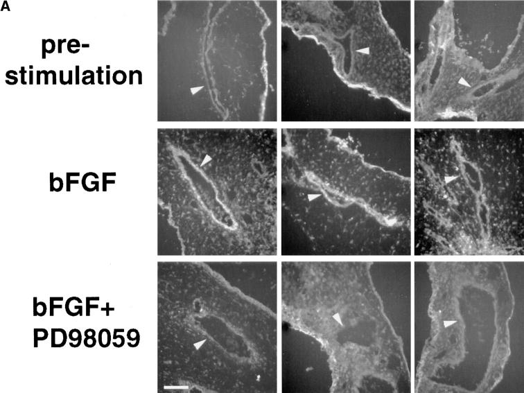
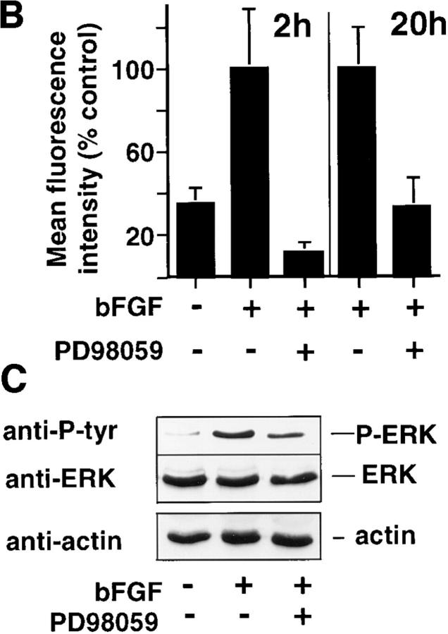
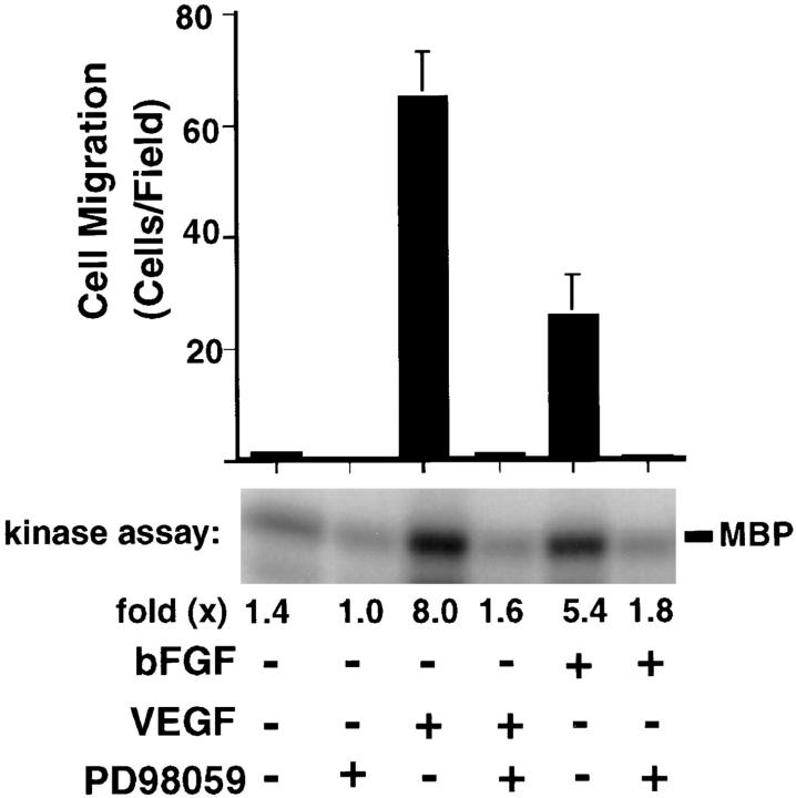
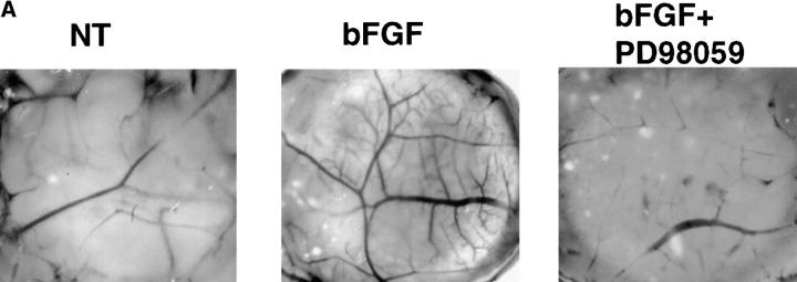
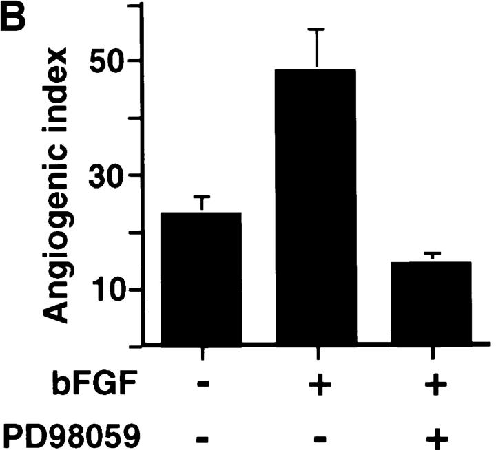
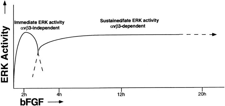
Angiogenesis depends on growth factors and vascular cell adhesion events. Integrins and growth factors are capable of activating the ras/MAP kinase pathway in vitro, yet how these signals influence endothelial cells during angiogenesis is unknown. Upon initiation of angiogenesis with basic fibroblast growth factor (bFGF) on the chick chorioallantoic membrane (CAM), endothelial cell mitogen-activated protein (MAP) kinase (ERK) activity was detected as early as 5 min yet was sustained for at least 20 h. The initial wave of ERK activity (5-120 min) was refractory to integrin antagonists, whereas the sustained activity (4-20 h) depended on integrin alphavbeta3, but not beta1 integrins. Inhibition of MAP kinase kinase (MEK) during this sustained alphavbeta3-dependent ERK signal blocked the formation of new blood vessels while not influencing preexisting blood vessels on the CAM. Inhibition of MEK also blocked growth factor induced migration but not adhesion of endothelial cells in vitro. Therefore, angiogenesis depends on sustained ERK activity regulated by the ligation state of both a growth factor receptor and integrin alphavbeta3.


Figures

Figure 1
Figure 1
Kinetics of bFGF-induced ERK phosphorylation in the CAM. bFGF (+) or PBS (−) saturated filter disks were placed on the CAM of 10-d-old chick embryos. At various times thereafter, CAMs were resected, lysed in detergent, and subjected to immunoprecipitation with an antibody to ERK. The immunoprecipitated material was subjected to SDS-PAGE and immunoblotting with an antiphosphotyrosine antibody (top). Band shifts of phosphorylated ERK in bFGF-treated CAMs was detected by immunoblotting with an anti-ERK antibody of parallel lysates (bottom). The relative increase in kinase activation of endogenous ERK after bFGF treatment compared with CAMs not treated with bFGF was measured by phosphorylation of myelin basic protein in an in vitro kinase assay using anti-ERK immunoprecipitates of CAM tissues as described in the Materials and Methods.
Figure 3
Figure 3
The effect of αvβ3 antagonists on ERK activity during bFGF-induced angiogenesis. (A) ERK phosphorylation was measured in CAMs treated with bFGF-saturated filter disks as described in the Materials and Methods. The αvβ3 antagonist, cyclic peptide RGDfV directed to αvβ3, or a control peptide (RADfV) was added to the filter disks (100 μg/50 μl) 1 h before harvest at the indicated times, after bFGF stimulation. Each point depicts ERK phosphorylation on CAMs treated with bFGF and cyclic peptide compared with CAMs treated with bFGF alone. Data are expressed as the mean percent of control ERK phosphorylation ± SD (n = 3). (B) ERK activity was measured in bFGF-treated CAM lysates in the presence or absence of cyclic peptides (1 h) measured by an in vitro kinase assay (MBP) as described in Materials and Methods. Data are expressed as the mean percent of ERK activity of RGDfV-treated CAMs relative to CAMs treated with control peptide (RADfV). Each bar represents the mean ± SD (n = 3). (Inset) A representative kinase assay from lysates derived from CAMs treated with buffer or bFGF, for a total of 20 h, in the presence or absence of RGDfV or RADfV for 1 h before harvest as described in the Materials and Methods. (C) ERK phosphorylation was detected in bFGF-treated CAMs in the presence of either mAb CSAT (50 μg/50 μl) directed to chick β1 or LM609 directed to αvβ3. Data are expressed as the percentage of ERK phosphorylation relative to control (no antibody). Each bar is the mean ± SD (n = 3). Open bar, LM609; black bar, CSAT. (D) Expression of αvβ3 in CAM tissue after bFGF stimulation in whole tissue lysates was measured for the expression of αvβ3 at each time point indicated by ELISA. Expression of β3 protein was measured by Western blotting of whole tissue extracts (inset) using a primary antibody to the COOH terminus of β3 as described in Materials and Methods. For the ELISA assay, protein extracts from control or bFGF-treated CAM tissue were immobilized on ELISA plates and detected with an anti-αvβ3 primary antibody (LM609) and a horseradish peroxidase–conjugated anti–mouse secondary antibody. Data are expressed as the mean concentration of αvβ3 ± SD (n = 3) and represent arbitrary units of αvβ3 reactivity measured by OD 490.
Figure 2
Figure 2
Localization of bFGF-induced ERK phosphorylation in the CAM. (A) Cryosections (4 μm) of bFGF-treated (2 h) or control CAMs treated without growth factors incubated in the presence or absence of the MEK inhibitor, PD98059 (50 μM), were examined for phosphorylated ERK, by indirect immunofluorescence using a primary antibody directed to the phosphorylated ERK peptide (New England Biolabs) as described in the Materials and Methods. Arrowheads indicate blood vessels. The edge staining of the tissues reflects nonspecific signal, which is routinely observed with secondary antibody alone in these cryopreserved CAM tissues. (B) Quantitation of immunofluorescence staining intensity with antiphosphorylated ERK antibody after bFGF treatment for 2 or 20 h, in the presence or absence of PD98059 by digital image analysis, as described in the Materials and Methods. (C) Phosphorylated ERK (top) and total ERK (middle) in bFGF-treated (20 h) CAM tissue lysates, in the presence or absence of PD98059, was detected by immunoprecipitation with an anti-ERK antibody followed by immunoblotting with either antiphosphotyrosine antibody to detect phosphorylated ERK (top) or anti-ERK antibody (middle) to show levels of ERK protein in the same lysate. Actin content of these CAM tissue extracts was detected by immunoblotting with an antiactin antibody (bottom). Bar, 50 μm.
Figure 2
Figure 2
Localization of bFGF-induced ERK phosphorylation in the CAM. (A) Cryosections (4 μm) of bFGF-treated (2 h) or control CAMs treated without growth factors incubated in the presence or absence of the MEK inhibitor, PD98059 (50 μM), were examined for phosphorylated ERK, by indirect immunofluorescence using a primary antibody directed to the phosphorylated ERK peptide (New England Biolabs) as described in the Materials and Methods. Arrowheads indicate blood vessels. The edge staining of the tissues reflects nonspecific signal, which is routinely observed with secondary antibody alone in these cryopreserved CAM tissues. (B) Quantitation of immunofluorescence staining intensity with antiphosphorylated ERK antibody after bFGF treatment for 2 or 20 h, in the presence or absence of PD98059 by digital image analysis, as described in the Materials and Methods. (C) Phosphorylated ERK (top) and total ERK (middle) in bFGF-treated (20 h) CAM tissue lysates, in the presence or absence of PD98059, was detected by immunoprecipitation with an anti-ERK antibody followed by immunoblotting with either antiphosphotyrosine antibody to detect phosphorylated ERK (top) or anti-ERK antibody (middle) to show levels of ERK protein in the same lysate. Actin content of these CAM tissue extracts was detected by immunoblotting with an antiactin antibody (bottom). Bar, 50 μm.
Figure 4
Figure 4
The role of ERK activity on endothelial cell migration. Endothelial cell migration on vitronectin in modified Boyden chambers of serum-starved HUVECs was induced with bFGF (25 ng/ml) or VEGF (25 ng/ml) in the presence or absence of 50 μM PD98059 for 2 h as described in Materials and Methods. ERK activity as detected by phosphorylation of MBP in an in vitro kinase assay was measured in parallel HUVEC cultures after treatment with bFGF, VEGF, or PD98059 for 30 min as described in the Materials and Methods. Migration data are expressed as the mean number of cells quantitated in at least three 10× microscope fields ± SD (n = 3). The kinase data are from a representative experiment.
Figure 5
Figure 5
The effects of PD98059 on bFGF-induced angiogenesis. (A) 10-d-old chick CAMs were exposed to filter paper disks saturated with bFGF (20 h) or buffer for 20 h and then incubated for an additional 48 h with or without PD98059 (50 μl of 50 μM). Photomicrographs were taken at 4× with a stereomicroscope and are representative of each group of treated CAMs. (B) The level of angiogenesis in bFGF-treated CAMs in the absence or presence of PD98059 was quantified by counting blood vessel branch points double blind as previously described (Friedlander et al., 1995).
Figure 5
Figure 5
The effects of PD98059 on bFGF-induced angiogenesis. (A) 10-d-old chick CAMs were exposed to filter paper disks saturated with bFGF (20 h) or buffer for 20 h and then incubated for an additional 48 h with or without PD98059 (50 μl of 50 μM). Photomicrographs were taken at 4× with a stereomicroscope and are representative of each group of treated CAMs. (B) The level of angiogenesis in bFGF-treated CAMs in the absence or presence of PD98059 was quantified by counting blood vessel branch points double blind as previously described (Friedlander et al., 1995).
Figure 6
Figure 6
Model depicting kinetics of MAP kinase activity during angiogenesis and the distinct requirements for ligation of integrin αvβ3 for immediate versus sustained MAP kinase activity. A rapid increase in bFGF-induced MAP kinase activity that is independent of integrin αvβ3 is followed by a sustained MAP kinase activity which requires αvβ3 ligation.

Similar articles

Cited by

References

    1. Alessi D, Cuenda A, Cohen P, Dudley D, Saltiel A. PD98059 is a specific inhibitor of the activation of mitogen-activated protein kinase kinase in vitro and in vivo. J Biol Chem. 1995;270:27489–27494. - PubMed
    1. Berrou E, Fontenay-Roupie M, Quarck R, McKenzie F, Levy-Toledano S, Tobelem G, Bryckaert M. Transforming growth factor β1 inhibits mitogen-activated protein kinase induced by basic fibroblast growth factor in smooth muscle cells. Biochem J. 1996;316:167–173. - PMC - PubMed
    1. Boudreau N, Andrews C, Srebrow A, Ravanpay A, Cheresh DA. Induction of the angiogenic phenotype by Hox D3. J Cell Biol. 1997;139:257–264. - PMC - PubMed
    1. Boulton TG, Cobb MH. Identification of multiple extracellular signaling kinases (ERKs) with antipeptide antibodies. Cell Regul. 1991;2:357–371. - PMC - PubMed
    1. Boulton T, Nye S, Robbins D, Ip N, Radziejewska E, Morgenbesser S, DePinho R, Panayotatos N, Cobb M, Yancopoulos G. ERKs: a family of protein serine/threonine kinases that are activated and tyrosine phosphorylated in response to insulin and NGF. Cell. 1991;65:663–675. - PubMed

Publication types

Substances