. 1998 Mar 9;140(5):1063-74.
doi: 10.1083/jcb.140.5.1063.

Vac8p, a vacuolar protein with armadillo repeats, functions in both vacuole inheritance and protein targeting from the cytoplasm to vacuole

Affiliations

Vac8p, a vacuolar protein with armadillo repeats, functions in both vacuole inheritance and protein targeting from the cytoplasm to vacuole

Y X Wang et al. J Cell Biol. .

Abstract

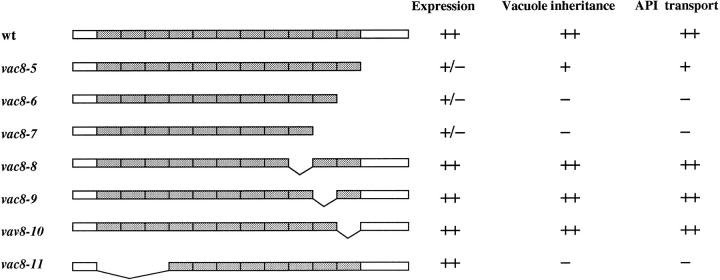
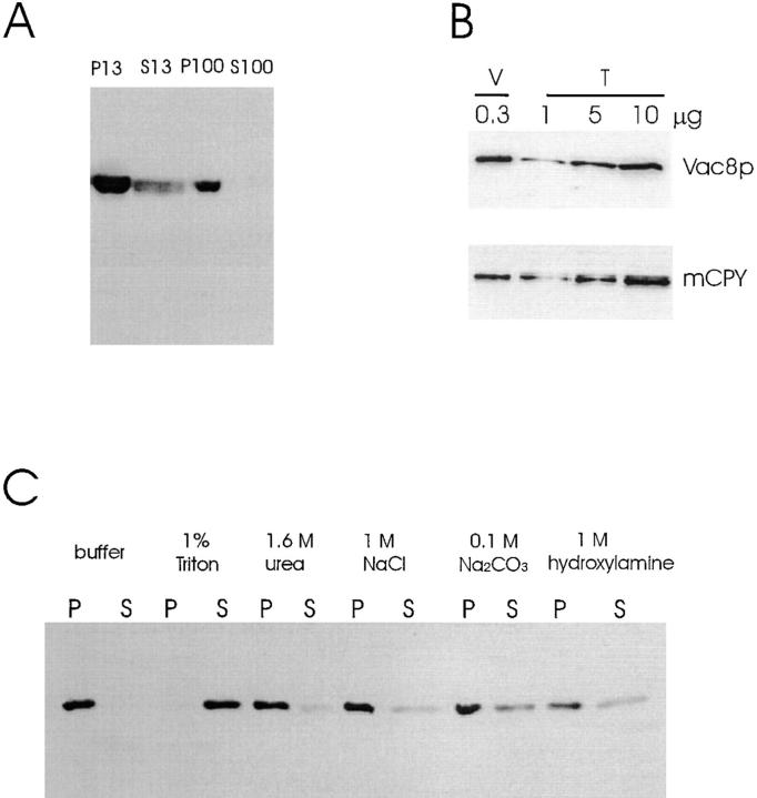
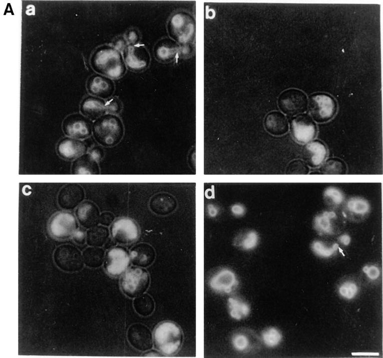
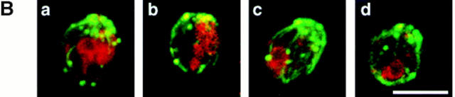
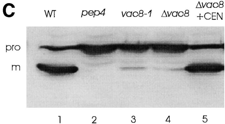
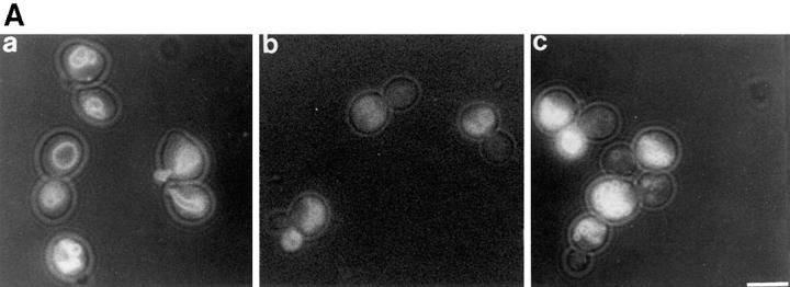
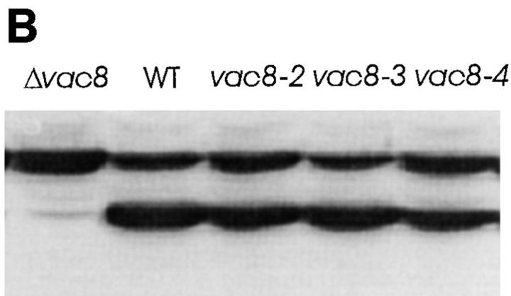
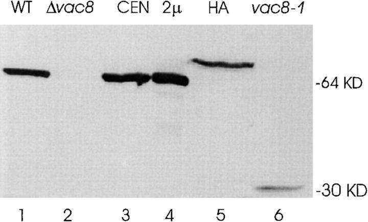
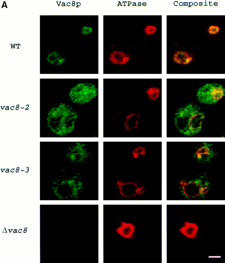
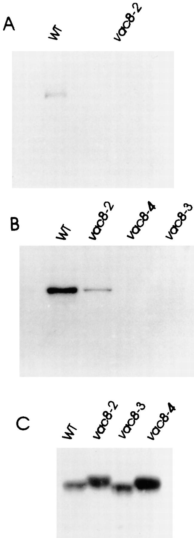
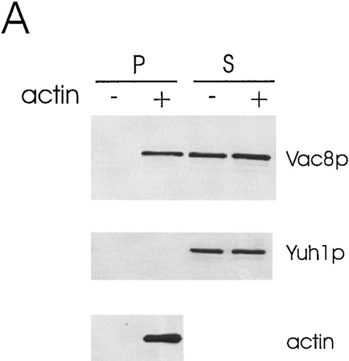
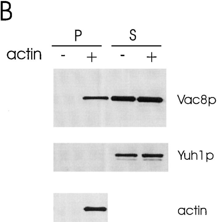
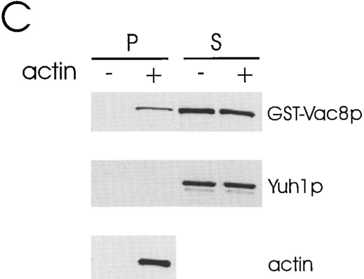
During each cell cycle, the yeast vacuole actively partitions between mother and daughter cells. This process requires actin, profilin, an unconventional myosin (Myo2p), and Vac8p. A mutant yeast strain, vac8, is defective in vacuole inheritance, specifically, in early vacuole migration. Vac8p is a 64-kD protein found on the vacuole membrane, a site consistent with its role in vacuole inheritance. Both myristoylation and palmitoylation are required for complete Vac8p localization. Interestingly, whereas myristoylation of Vac8p is not required for vacuole inheritance, palmitoylation is essential. Thus, palmitoylation appears to play a more direct role in vacuole inheritance. Most of the VAC8 sequence encodes 11 armadillo (Arm) repeats. Arm repeats are thought to mediate protein-protein interactions, and many Arm proteins have multiple functions. This is also true for Vac8p. In addition to its role in early vacuole inheritance, Vac8p is required to target aminopeptidase I from the cytoplasm to the vacuole. Mutant analysis demonstrates that Vac8p functions separately in these two processes. Vac8p cosediments with actin filaments. Vac8p is related to beta-catenin and plakoglobin, which connect a specific region of the plasma membrane to the actin cytoskeleton. In analogy, Vac8p may link the vacuole to actin during vacuole partitioning.


Figures

Figure 1
Figure 1
Sequence analysis of the VAC8 gene. (A) Restriction map of the 2,972-bp SmaI–EcoRI fragment containing VAC8. The Δvac8::HIS3 disruption construct is shown. Ec, EcoRI; Bc, BclI; Xb, XbaI; Sp, SphI; Bg, BglII; Sm, SmaI. (B) Alignment of 11 Arm repeats from Vac8p. Shading indicates that at least 3 of the 11 residues are identical or equivalent (K, R, and H; D and E; L and I were considered equivalent). An asterisk indicates a space. The consensus sequence from the 11 repeats are also shown, where plus indicates K, R, or H; minus indicates D or E; X indicates any amino acids. (C) Acylation motifs in Vac8p and other proteins. Myristoylated glycines and palmitoylated cysteines are highlighted (Milligan et al., 1995).
Figure 1
Figure 1
Sequence analysis of the VAC8 gene. (A) Restriction map of the 2,972-bp SmaI–EcoRI fragment containing VAC8. The Δvac8::HIS3 disruption construct is shown. Ec, EcoRI; Bc, BclI; Xb, XbaI; Sp, SphI; Bg, BglII; Sm, SmaI. (B) Alignment of 11 Arm repeats from Vac8p. Shading indicates that at least 3 of the 11 residues are identical or equivalent (K, R, and H; D and E; L and I were considered equivalent). An asterisk indicates a space. The consensus sequence from the 11 repeats are also shown, where plus indicates K, R, or H; minus indicates D or E; X indicates any amino acids. (C) Acylation motifs in Vac8p and other proteins. Myristoylated glycines and palmitoylated cysteines are highlighted (Milligan et al., 1995).
Figure 8
Figure 8
Deletion analysis of Vac8p. Δvac8 strains harboring CEN plasmids with different VAC8 constructs were analyzed for expression, vacuole inheritance and API transport. ++, normal; +, slightly defective; +/−, greatly reduced expression; −, defective.
Figure 4
Figure 4
Vac8p is associated with vacuolar membranes. (A) Precleared extracts from wild-type cells (LWY7213) were subjected to sequential differential centrifugation as described in Materials and Methods. Equal portions of cell extracts (0.75 OD600 unit equivalents) were separated by SDS-PAGE, transferred to nitrocellulose and probed with Vac8p antiserum. (B) Vacuoles were isolated as described (Haas, 1995). Vacuoles (V, 0.3 μg protein) and precleared total cell extracts (T, 1 μg, 5 μg and 10 μg) were dissolved in SDS-PAGE sample buffer (Laemmli, 1970) and analyzed by immunoblot with Vac8p antiserum and CPY antiserum. (C) The P13 fraction in A was resuspended in lysis buffer. Aliquots of the P13 fraction was treated for 30 min on ice with various reagents indicated. The samples were then respun to generate the second round of P13 and S13 fractions. The presence of Vac8p in the P13 and S13 fractions was determined by Western blot.
Figure 2
Figure 2
Vac8p is required for both vacuole inheritance and API transport. (A) Cells were labeled with FM4-64 for 1 h. Free dye was removed and cells were resuspended in fresh medium and allowed to grow for 3 h at 23°C. The vacuoles were viewed by fluorescence microscopy. Segregation structures are indicated with arrows. a, Wild-type; b, vac8-1; c, Δvac8; d, Δvac8 with CEN VAC8 plasmid. (B) Vac8p is required for polarization of the vacuole to the presumptive bud site. Fixed spheroplasts were double labeled with actin antibody (green) and 60-kD v-ATPase antibody (red), detected with Cy2 conjugated donkey anti–goat IgG and Lissamine rhodamine–conjugated donkey anti–mouse IgG, respectively. (a and b) Wild-type, (c and d) Δvac8. (C) Cell extracts were prepared as described in Materials and Methods. Equal amounts of cell extract (0.3 OD600 equivalents) were subjected to Western analysis with anti-API serum. The positions of precursor (pro) and mature (m) API are shown. Lane 1, wild-type; lane 2, pep4; lane 3, vac8-1; lane 4, Δvac8; lane 5, Δvac8 with CEN VAC8 plasmid. Bars, 5 μm.
Figure 2
Figure 2
Vac8p is required for both vacuole inheritance and API transport. (A) Cells were labeled with FM4-64 for 1 h. Free dye was removed and cells were resuspended in fresh medium and allowed to grow for 3 h at 23°C. The vacuoles were viewed by fluorescence microscopy. Segregation structures are indicated with arrows. a, Wild-type; b, vac8-1; c, Δvac8; d, Δvac8 with CEN VAC8 plasmid. (B) Vac8p is required for polarization of the vacuole to the presumptive bud site. Fixed spheroplasts were double labeled with actin antibody (green) and 60-kD v-ATPase antibody (red), detected with Cy2 conjugated donkey anti–goat IgG and Lissamine rhodamine–conjugated donkey anti–mouse IgG, respectively. (a and b) Wild-type, (c and d) Δvac8. (C) Cell extracts were prepared as described in Materials and Methods. Equal amounts of cell extract (0.3 OD600 equivalents) were subjected to Western analysis with anti-API serum. The positions of precursor (pro) and mature (m) API are shown. Lane 1, wild-type; lane 2, pep4; lane 3, vac8-1; lane 4, Δvac8; lane 5, Δvac8 with CEN VAC8 plasmid. Bars, 5 μm.
Figure 7
Figure 7
Phenotypic analysis of the acylation mutants. (A) Δvac8 strains harboring CEN plasmids with different VAC8 alleles were examined for vacuole inheritance as done in Fig. 2 A. (a) vac8-2; (b) vac8-3; (c) vac8-4. (B) API transport was examined as done in Fig. 2 B. Bar, 5 μm.
Figure 7
Figure 7
Phenotypic analysis of the acylation mutants. (A) Δvac8 strains harboring CEN plasmids with different VAC8 alleles were examined for vacuole inheritance as done in Fig. 2 A. (a) vac8-2; (b) vac8-3; (c) vac8-4. (B) API transport was examined as done in Fig. 2 B. Bar, 5 μm.
Figure 3
Figure 3
Identification of the VAC8 gene product. Cells were grown in SC medium and cell extracts generated by breakage with glass beads. Precleared cell extracts were separated by SDS-PAGE, transferred to nitrocellulose and probed with Vac8p antiserum. Lane 1, 10 μg protein, wild-type; lane 2, 10 μg protein, Δvac8; lane 3, 10 μg protein, Δvac8 with CEN VAC8; lane 4, 5 μg protein, Δvac8 with 2μ VAC8; lane 5, 50 μg protein, Δvac8 with CEN VAC8-HA; lane 6, 50 μg protein, vac8-1.
Figure 6
Figure 6
Mislocalization of Vac8p in the acylation mutants. (A) Cells were processed and incubated with both affinity-purified Vac8p antibody (green) and 60-kD v-ATPase monoclonal antibody (red), detected by Oregon Green 488–conjugated goat anti–rabbit IgG and Lissamine rhodamine–conjugated donkey anti– mouse IgG, respectively. Δvac8 harboring CEN plasmids with different VAC8 alleles was used. (B) Fractionation and determination of Vac8p was performed as in Fig. 4 A. Δvac8 harboring CEN plasmids with different VAC8 alleles was used. Lanes 1–4, WT; lanes 5–8, vac8-2; lanes 9–12, vac8-3; lanes 12–16, vac8-4. Bar, 2 μm.
Figure 6
Figure 6
Mislocalization of Vac8p in the acylation mutants. (A) Cells were processed and incubated with both affinity-purified Vac8p antibody (green) and 60-kD v-ATPase monoclonal antibody (red), detected by Oregon Green 488–conjugated goat anti–rabbit IgG and Lissamine rhodamine–conjugated donkey anti– mouse IgG, respectively. Δvac8 harboring CEN plasmids with different VAC8 alleles was used. (B) Fractionation and determination of Vac8p was performed as in Fig. 4 A. Δvac8 harboring CEN plasmids with different VAC8 alleles was used. Lanes 1–4, WT; lanes 5–8, vac8-2; lanes 9–12, vac8-3; lanes 12–16, vac8-4. Bar, 2 μm.
Figure 5
Figure 5
Vac8p is acylated in vivo. Δvac8 yeast harboring CEN plasmids with different VAC8 alleles were labeled in vivo with [3H]myristic acid (A) or [3H]palmitic acid (B). Vac8p was immunoprecipitated with antiserum as described in Materials and Methods. Antigen was eluted and subjected to SDS-PAGE and fluorography. vac8-2, G2A; vac8-3, C4G, C5T, and C7S; vac8-4, G2A, C4G, C5T, and C7S. (C) Cell extracts prepared from Δvac8 harboring CEN plasmids with different VAC8 alleles were analyzed by immunoblot with anti-Vac8p serum. Note the mobility differences in the VAC8 alleles.
Figure 9
Figure 9
Vac8p cosediments with actin filaments. 20 μl (2 mg/ml final concentration) of a high speed, Triton X-100 solubilized total cell extract (A) or cytosol from the myristoylation-minus mutant vac8-2 (B), or cytosol from Δvac8 (C) was mixed with 20 μl fresh G-actin (0.4 mg/ml final concentration) and G-buffer in a 100 μl reaction. G-buffer without actin was added to adjust the volume in the controls. For the experiments using Δvac8 cytosol, 10 μl purified GST-Vac8 protein (26 μg/ml final concentration) was added. The mixtures were incubated for 1 h on ice and then the polymerized actin was pelleted. 5 μl of the supernatant fraction (200 μl final) and the washed pellet fraction (50 μl final) were separated by SDS-PAGE and transferred to nitrocellulose membranes. The same membrane was cut into two halves. The top half was probed with Vac8p antiserum and the bottom half was probed with the antiserum to Yuh1p. In addition, 0.1 μl of the pellet fraction was run separately and used to detect actin.
Figure 9
Figure 9
Vac8p cosediments with actin filaments. 20 μl (2 mg/ml final concentration) of a high speed, Triton X-100 solubilized total cell extract (A) or cytosol from the myristoylation-minus mutant vac8-2 (B), or cytosol from Δvac8 (C) was mixed with 20 μl fresh G-actin (0.4 mg/ml final concentration) and G-buffer in a 100 μl reaction. G-buffer without actin was added to adjust the volume in the controls. For the experiments using Δvac8 cytosol, 10 μl purified GST-Vac8 protein (26 μg/ml final concentration) was added. The mixtures were incubated for 1 h on ice and then the polymerized actin was pelleted. 5 μl of the supernatant fraction (200 μl final) and the washed pellet fraction (50 μl final) were separated by SDS-PAGE and transferred to nitrocellulose membranes. The same membrane was cut into two halves. The top half was probed with Vac8p antiserum and the bottom half was probed with the antiserum to Yuh1p. In addition, 0.1 μl of the pellet fraction was run separately and used to detect actin.

Similar articles

Cited by

References

    1. Adams J. Cell adhesion-spreading frontiers, intricate insights. Trends Cell Biol. 1997;7:107–110. - PubMed
    1. Berkower C, Loayza D, Michaelis S. Metabolic instability and constitutive endocytosis of STE6, the a-factor transporter of Saccharomyces cerevisiae. . Mol Biol Cell. 1994;5:1185–1198. - PMC - PubMed
    1. Bonangelino CJ, Catlett NL, Weisman LS. Vac7p, a novel vacuolar protein, is required for normal vacuole inheritance and morphology. Mol Cell Biol. 1997;17:6847–6858. - PMC - PubMed
    1. Brodsky JL, Schekman R. A Sec63p-Bip complex from yeast is required for protein translocation in a reconstituted proteoliposome. J Cell Biol. 1993;123:1355–1363. - PMC - PubMed
    1. Casey P. Protein lipidation in cell signaling. Science. 1995;268:221–225. - PubMed

Publication types

MeSH terms