. 1998 Mar;18(3):1266-74.
doi: 10.1128/MCB.18.3.1266.

Functional interference of Sp1 and NF-kappaB through the same DNA binding site

Affiliations

Functional interference of Sp1 and NF-kappaB through the same DNA binding site

F Hirano et al. Mol Cell Biol. 1998 Mar.

Abstract

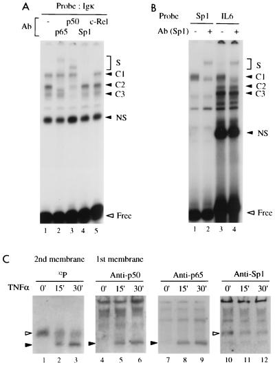
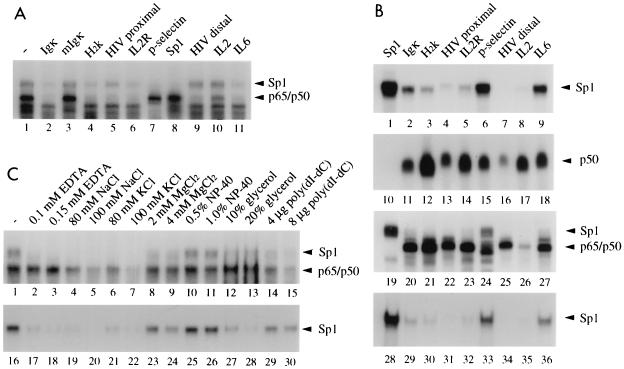
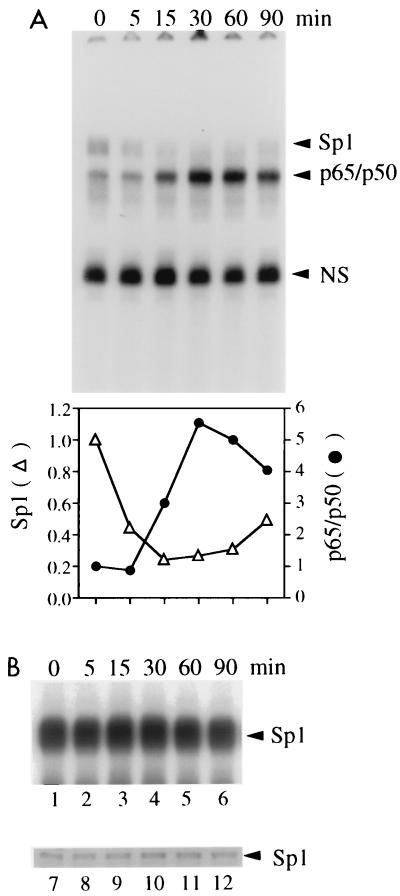
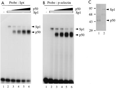
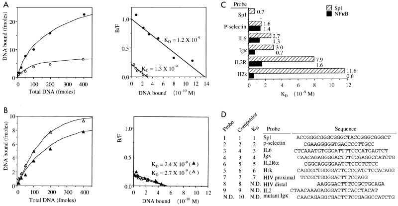
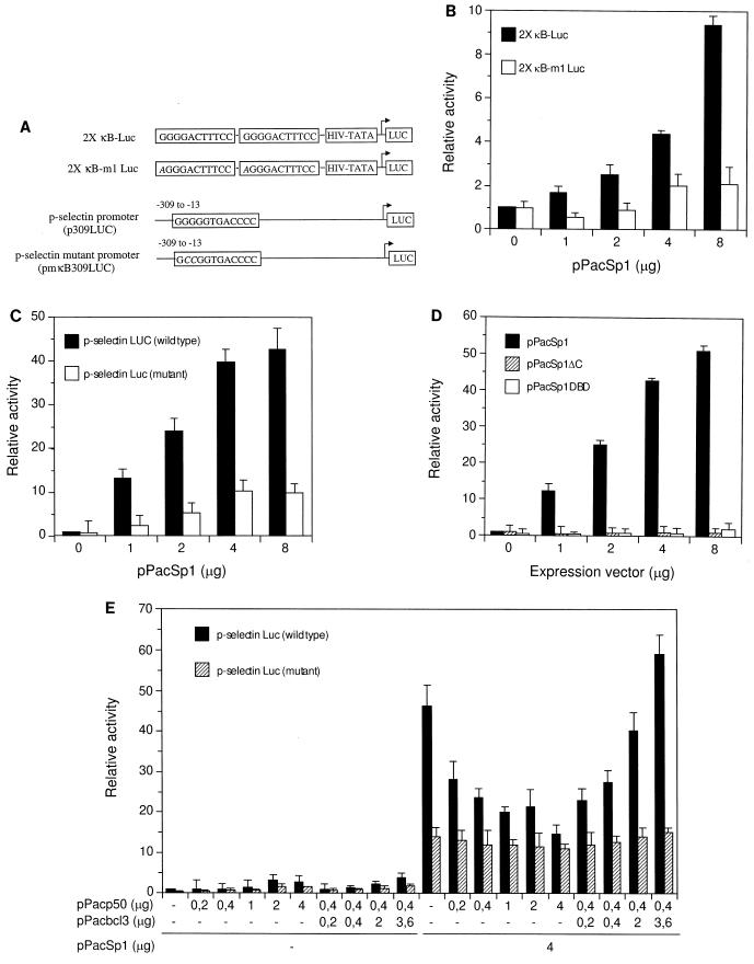
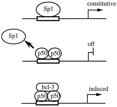
Gene activation by NF-kappaB/Rel transcription factors is modulated by synergistic or antagonistic interactions with other promoter-bound transcription factors. For example, Sp1 sites are often found in NF-kappaB-regulated genes, and Sp1 can activate certain promoters in synergism with NF-kappaB through nonoverlapping binding sites. Here we report that Sp1 acts directly through a subset of NF-kappaB binding sites. The DNA binding affinity of Sp1 to these NF-kappaB sites, as determined by their relative dissociation constants and their relative efficiencies as competitor DNAs or as binding site probes, is in the order of that for a consensus GC box Sp1 site. In contrast, NF-kappaB does not bind to a GC box Sp1 site. Sp1 can activate transcription through immunoglobulin kappa-chain enhancer or P-selectin promoter NF-kappaB sites. p50 homodimers replace Sp1 from the P-selectin promoter by binding site competition and thereby either inhibit basal Sp1-driven expression or, in concert with Bcl-3, stimulate expression. The interaction of Sp1 with NF-kappaB sites thus provides a means to keep an elevated basal expression of NF-kappaB-dependent genes in the absence of activated nuclear NF-kappaB/Rel.


Figures

FIG. 1
FIG. 1
Cellular Sp1 binds to Igκ and IL-6 NF-κB DNA binding sites. (A) Complexes formed between nuclear extract proteins (5 μg) of TNF-α (1 ng/ml, 30 min)-stimulated HeLa cells and an Igκ probe were analyzed by EMSA. The reaction mixtures contained either no (−) antibody (Ab) (lane 1) or antiserum directed against p65, p50, Sp1, or c-Rel (lanes 2 to 5), as indicated. The reactivities with the antibodies identify C1 as Sp1, C2 as p65-p50, and C3 as p50-p50. S, supershifted complex; NS, nonspecific. (B) Complexes formed with an Sp1 (lanes 1 and 2) or IL-6 gene (lanes 3 and 4) NF-κB binding site probe without (lanes 1 and 3) or with (lanes 2 and 4) Sp1 antibody. C1, C2, and C3 contain Sp1, p65-p65, and p50-p65 (not shown), respectively. (C) Shift-Western blotting of complexes formed with the Igκ probe and nuclear extracts of adherent HeLa cells stimulated with TNF-α (10 ng/ml) for the indicated times. EMSA gels were sandwich blotted with a nitrocellulose membrane and then with a PVDF membrane, which were then analyzed by immunoblotting and autoradiography, respectively, to separately visualize bound proteins and labeled DNA of the complexes. The DNAs of two differently migrating complexes (open and solid arrowheads) (lanes 1 to 3) whose intensities were decreased and induced, respectively, upon TNF-α stimulation, were detected. The slower complex (open arrowhead) was detected by an Sp1 antibody (lanes 10 to 12), and the faster one (solid arrowhead) was detected by both p50 and p65 antibodies (lanes 4 to 9). Free DNA is not shown.
FIG. 2
FIG. 2
Differential interaction of Sp1 with various NF-κB binding sites. (A) Complexes formed between the Igκ probe and nuclear extracts (5 μg of protein/lane) of TNF-α (1 ng/ml, 30 min)-stimulated HeLa cells were challenged without (lane 1) or with (lanes 2 to 11) 50-fold molar amounts of competitor oligonucleotides containing the indicated NF-κB DNA binding sites or an Sp1 binding site. The positions of Sp1 and p65-p50 DNA complexes are indicated; free DNA is not shown. (B) Oligonucleotides containing various NF-κB binding sites or an Sp1 binding site, as indicated, were used as radiolabeled probes in gel retardation assays either with affinity-purified Sp1 (1 footprint-producing unit/lane) (lanes 1 to 9 and 28 to 36), with bacterially expressed p50 (20 ng/lane) (lanes 10 to 18), or with nuclear extract proteins (5 μg/lane) of TNF-α (10 ng/ml, 30 min)-stimulated HeLa cells (lanes 19 to 27). Either NF-κB buffer conditions [20 mM HEPES (pH 8.4), 60 mM KCl, 5 mM DTT, 1 μg of BSA, 2 μg of poly(dI-dC), 4% Ficoll] (lanes 1 to 27) or Sp1 buffer conditions [10 mM Tris-HCl (pH 8.0), 50 mM NaCl, 0.1 mM ZnCl2, 0.2 μg of poly(dA-dT), 10% glycerol] (lanes 28 to 36) were used. The positions of complexes are indicated. Free DNA is not shown. (C) Effect of buffer components on Sp1 or NF-κB complex formation with the IL-6 gene NF-κB site. Either nuclear extracts of TNF-α-stimulated HeLa cells (lanes 1 to 15) or purified Sp1 (lanes 16 to 30) were used in EMSA with the indicated components included in the standard NF-κB binding buffer. NP-40, Nonidet P-40.
FIG. 3
FIG. 3
Mutually exclusive interaction of Sp1 or NF-κB with the same site. (A) Gel retardation assay with the IL-6 probe and nuclear extracts of adherent HeLa cells stimulated with TNF-α (10 ng/ml) for the indicated times. The increase of NF-κB complex formation after induction and the concomitant decrease of Sp1 complex formation are also visualized by quantitation of the counts/minute of each complex with a BAS2000 phosphorimaging analyzer in arbitrary units (bottom). NS, nonspecific. (B) The nuclear extracts used for panel A were assayed for Sp1 complex formation with an Sp1 DNA probe in a gel retardation assay (lanes 1 to 6; only complexes are shown) and for Sp1 protein amounts in a Western blot (lanes 7 to 12).
FIG. 4
FIG. 4
Competition of Sp1 by p50 homodimers on Igκ (A) or P-selectin (B) NF-κB binding sites, determined by EMSA using purified Sp1 protein from HeLa cells (1 footprint-producing unit (fpu)/lane) (A and B, lanes 2 to 6) and increasing amounts of recombinant p50 (0, 5, 10, 20, and 40 ng/lane) (lanes 2 to 6, respectively). Lanes 1 in panels A and B, no protein added. (C) Silver-stained SDS-polyacrylamide gel of 2 fpu of Sp1 and 20 ng of p50. Sizes are indicated in kilodaltons.
FIG. 5
FIG. 5
Determination of the dissociation constants for the interaction of Sp1 and NF-κB with Sp1 and NF-κB binding sites by quantitative EMSA and Scatchard analysis. (A and B) Binding curves (left) and Scatchard plots (right) for complex formation of p65-p50 (A) or Sp1 (B) with the IL-6 probe in nuclear extracts of nonstimulated and TNF-α (10 ng/ml, 30 min)-stimulated HeLa cells (open and closed symbols, respectively). B/F, bound/free. (C) Summary of the dissociation constants determined in HeLa cell nuclear extracts for the interaction of Sp1 or NF-κB with various NF-κB and Sp1 sites, as indicated. (D) Alignment of the Sp1 and NF-κB binding sites used in this study in order of relative affinity to Sp1. The left columns indicate similar orders of affinity as determined when we used them as probes, as competitors, or for KD determination. ND, not determined.
FIG. 6
FIG. 6
Sp1 activates transcription through Igκ and P-selectin NF-κB binding sites. Functional interference with p50 and Bcl-3 was analyzed. (A) Schematic presentation of heterologous HIV-TATA luciferase reporter genes containing two tandem wild-type or mutant Igκ NF-κB binding sites and of the P-selectin luciferase promoter constructs with either the wild-type or mutated NF-κB binding site in the context of the complete promoter sequence (42). (B) Sp1 activates through Igκ NF-κB binding sites. Drosophila SL2 cells were transfected with reporter constructs containing wild-type (2× κB-Luc, 8 μg) or mutant (2× κB-m1 Luc, 8 μg) Igκ NF-κB binding sites with increasing amounts of pPacSp1 expression vector, as indicated. (C) Sp1 activates transcription through a P-selectin promoter NF-κB binding site. SL2 cells were transfected with the P-selectin promoter luciferase construct (8 μg) harboring a wild-type or mutated NF-κB binding site along with increasing amounts of Sp1 expression vector. (D) Activation through the P-selectin NF-κB site by Sp1 requires both DNA binding and transactivation domains of Sp1. Cell were transfected with P-selectin constructs as in panel C along with expression vectors encoding Sp1 (pPacSp1, amino acids 83 to 778) or Sp1 lacking the zinc finger region (pPacSp1ΔC, amino acids 83 to 611) or containing only the DNA binding domain of Sp1 (pPacSp1DBD, amino acids 612 to 778), as indicated. (E) Functional interference between Sp1, p50, and Bcl-3 at the P-selectin NF-κB binding site. The P-selectin wild-type or mutant promoter was transfected into SL2 cells with increasing amounts of p50 or constant amounts of p50 and increasing amounts of Bcl-3. In addition, cells were transfected without or with Sp1 expression vector, as indicated. Relative luciferase activity is shown in panels B to E as mean values with standard deviations of three to four independent experiments.
FIG. 7
FIG. 7
Hypothetical scheme for the functional interaction of Sp1 with NF-κB sites. Sp1 occupies an NF-κB site and constitutively activates transcription in the absence of NF-κB (top). The exchange of Sp1 by NF-κB/Rel at certain sites (e.g., P-selectin) allows a switch from constitutive activation by Sp1 to repression by transcriptionally inactive p50 homodimers (center). The repressed gene can be induced depending on the availability of the Bcl-3 coactivator, which forms a ternary complex (bottom).

Similar articles

Cited by

References

    1. Augustin L B, Felsheim R F, Min B H, Fuchs S M, Fuchs J A, Loh H H. Genomic structure of the mouse delta opioid receptor gene. Biochem Biophys Res Commun. 1995;207:111–119. - PubMed
    1. Baeuerle P A, Baltimore D. NF-κB: ten years after. Cell. 1996;87:13–20. - PubMed
    1. Ballard D W, Bohnlein E, Lowenthal J W, Wano Y, Franza B R, Greene W C. HTLV-I tax induces cellular proteins that activate the κB element in the IL-2 receptor α gene. Science. 1988;241:1652–1655. - PubMed
    1. Beg A A, Baldwin A S., Jr The IκB proteins: multifunctional regulators of Rel/NF-κB transcription factors. Genes Dev. 1993;7:2064–2070. - PubMed
    1. Beg A A, Sha W C, Bronson R T, Ghosh S, Baltimore D. Embryonic lethality and liver degeneration in mice lacking the RelA component of NF-κB. Nature. 1995;376:167–170. - PubMed

Publication types

LinkOut - more resources