. 1998 Mar 3;95(5):2642-7.
doi: 10.1073/pnas.95.5.2642.

Chronic lithium treatment robustly protects neurons in the central nervous system against excitotoxicity by inhibiting N-methyl-D-aspartate receptor-mediated calcium influx

Affiliations

Chronic lithium treatment robustly protects neurons in the central nervous system against excitotoxicity by inhibiting N-methyl-D-aspartate receptor-mediated calcium influx

S Nonaka et al. Proc Natl Acad Sci U S A. .

Abstract

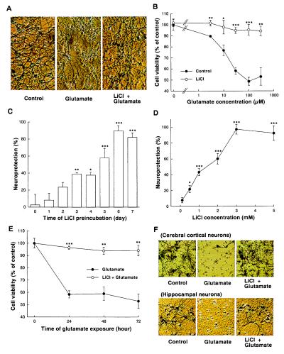
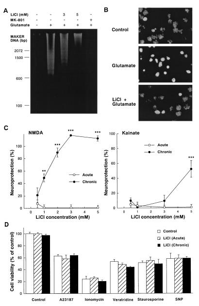
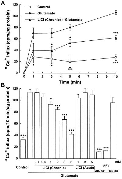
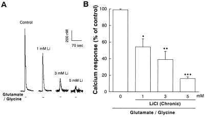
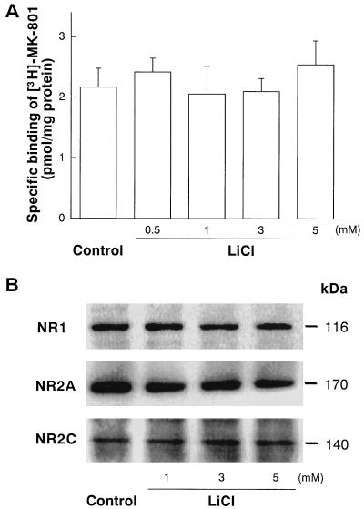
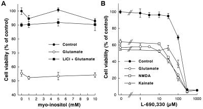
Lithium is the most commonly used drug for the treatment of manic depressive illness. The precise mechanisms underlying its clinical efficacy remain unknown. We found that long-term exposure to lithium chloride dramatically protects cultured rat cerebellar, cerebral cortical, and hippocampal neurons against glutamate-induced excitotoxicity, which involves apoptosis mediated by N-methyl-D-aspartate (NMDA) receptors. This neuroprotection is long-lasting, occurs at therapeutically relevant concentrations of lithium with an EC50 of approximately 1.3 mM, and requires treatment for 6-7 days for complete protection to occur. In contrast, a 24-h treatment with lithium is ineffective. The protection in cerebellar neurons is specific for glutamate-induced excitotoxicity and can be attributed to inhibition of NMDA receptor-mediated calcium influx measured by 45Ca2+ uptake studies and fura-2 fluorescence microphotometry. The long-term effects of lithium are not caused by down-regulation of NMDA receptor subunit proteins and are unlikely related to its known ability to block inositol monophosphatase activity. Our results suggest that modulation of glutamate receptor hyperactivity represents at least part of the molecular mechanisms by which lithium alters brain function and exerts its clinical efficacy in the treatment for manic depressive illness. These actions of lithium also suggest that abnormality of glutamatergic neurotransmission as a pathogenic mechanism underlying bipolar illness warrants future investigation.


Figures

Figure 1
Figure 1
Chronic lithium pretreatment protects neurons against glutamate toxicity in cultured cerebellar granule, cerebral cortical, and hippocampal cells. (A) Lithium-induced excitoprotection revealed by cell morphology. Cultured cerebellar granule cells were pretreated with LiCl (2 mM) for 7 days. Cultures were then exposed to glutamate (100 μM) and live cells were examined for morphology 24 h later after MTT staining. Cells were photographed with a phase-contrast microscope at a magnification of ×200. Note that the glutamate exposure resulted in the typical appearance of dead cells, which were round, smaller, translucent, and unable to metabolize MTT to blue formazan product. Chronic lithium prevented this aspect of glutamate neurotoxicity. (B) The extent of lithium-induced excitoprotection in cerebellar granule neurons at various glutamate concentrations. Cultured cells were pretreated with LiCl (3 mM) for 7 days. Cultures were then exposed to indicated concentrations of glutamate, and neuronal survival was determined 24 h after addition of glutamate by using the MTT colorimetric assay. (C) Preincubation time-dependent excitoprotection elicited by lithium. LiCl (2 mM) was added to cells at various times (0–7 days) before addition of glutamate (100 μM) on the 7th day in cultures to vary the time of lithium exposure. Neuronal survival was determined 24 h after addition of glutamate by using the MTT colorimetric assay. The results are expressed as percent of neuroprotection. Note that maximal protection was achieved by pretreatment with lithium for 6 days or longer. (D) Concentration-dependent induction of the excitoprotective state. Cells were pretreated with various concentrations of LiCl (0.1–5 mM) for 6 days and then exposed to 100 μM glutamate for 24 h before measurement of neuronal survival by MTT assays. The results are expressed as percent of neuroprotection. (E) Time course of glutamate-induced neurotoxicity. Cultured cerebellar granule cells were pretreated with LiCl (3 mM) for 7 days and then exposed to glutamate (100 μM) as indicated. Neuronal survival was determined 24–72 h after addition of glutamate by using the MTT colorimetric assay. (F) Morphology of cultured cerebral cortical and hippocampal neurons exposed to glutamate in the absence and presence of lithium pretreatment. Cultured cortical or hippocampal neurons were pretreated with 1 mM LiCl for 7 days, starting from the second day in culture. Cultures were then exposed to glutamate (100 μM), and live cells were examined for morphology 24 h later by MTT staining. Cells were photographed with a phase-contrast microscope at a magnification of ×200. Chronic lithium prevented glutamate-induced neurotoxicity. Data are the mean ± SEM of viability measurements from four cultures. ∗, P < 0.05; ∗∗, P < 0.01; ∗∗∗, P < 0.001, compared with the group treated with glutamate alone (one-way ANOVA with Bonferroni–Dunn test).
Figure 2
Figure 2
Effects of chronic lithium pretreatment on neurotoxicity of cerebellar granule cells. (A) Chronic lithium prevents glutamate-induced internucleosomal DNA fragmentation of cerebellar granule cell. Cells were pretreated with various concentrations of LiCl (3–5 mM) for 7 days and then exposed to glutamate (100 μM) as described in Fig. 1. Soluble DNA was extracted from cells 24 h after the addition of glutamate, subjected to agarose gel electrophoresis, stained with ethidium bromide, and photographed. (B) Chronic lithium inhibits glutamate-induced chromatin condensation of cerebellar granule cell. Cells were pretreated with LiCl (3 mM) for 7 days and then exposed to glutamate (100 μM). Chromatin condensation was detected by nucleus staining with Hoechst 33258. Nuclei were photographed with a Zeiss Axiophot fluorescence microscope at a magnification of ×1,000. (C) Chronic lithium pretreatment protects cells against NMDA and kainate toxicity. Lithium chloride (0.5–5 mM) was added to cultured neurons 1 h (acute) or 7 days (chronic) before NMDA (1 mM) or kainate (100 μM). Additionally MK-801 (1 μM) was added to the kainate-treated groups. After 24 h of stimulation, neuronal viability was measured with the MTT colorimetric assay. The results are expressed as percent of neuroprotection. (D) Effects of lithium on A23187, ionomycin, veratridine, staurosporine, and sodium nitroprusside (SNP)-induced neurotoxicity. Lithium chloride (3 mM) was added to cultured cerebellar granule neurons 1 h (acute) or 7 days (chronic) prior to their exposure to A23187 (300 nM), ionomycin (3 μM), veratridine (1 μM), staurosporine (30 nM), and SNP (100 μM). Neuronal survival was determined 24 h after addition by using the MTT colorimetric assay. Data are the mean ± SEM of viability measurements from four or five cultures. ∗∗, P < 0.01; ∗∗∗, P < 0.001, compared with the group of control (one-way ANOVA with Bonferroni–Dunn test).
Figure 3
Figure 3
Chronic lithium exposure inhibits glutamate-induced 45Ca2+ influx into cerebellar granule cells. (A) Time course of basal and glutamate-induced 45Ca2+ influx. Cells were treated with LiCl (3 mM) for 7 days before adding 100 μM glutamate and 45CaCl2 (1 μCi/ml) to the culture medium. At indicated times, the influx was terminated by rapid washing of the culture dishes three times with ice-cold buffer (154 mM choline chloride/2 mM EGTA/10 mM Hepes, pH 7.4) and then solubilized in 0.6 ml of 0.5 M NaOH. An aliquot of 0.5 ml was used for measuring 45Ca2+ radioactivity and the remainder was used for protein determination. (B) Concentration dependence of inhibition by lithium of the 45Ca2+ influx. Cells were pretreated chronically (7 days) or acutely (1 h) with various concentrations of LiCl (0.1–5 mM) and then exposed to glutamate (100 μM) in the presence of 45Ca2+. The 45Ca2+ influx was determined 10 min after glutamate addition. When used, the concentrations of MK-801, 2-amino-5-phosphonopentanoate (APV), and 6-cyano-7-nitroquinoxaline-2,3-dione (CNQX) were 1 μM, 200 μM, and 20 μM, respectively. Data are the mean ± SEM of 45Ca2+ uptake measurements from three cultures. ∗, P < 0.05; ∗∗, P < 0.01; ∗∗∗, P < 0.001, compared with the group treated with glutamate alone (one-way ANOVA with Bonferroni–Dunn test).
Figure 4
Figure 4
Chronic lithium inhibits the peak elevation in [Ca2+]i after exposure to glutamate in cerebellar granule neurons. Cerebellar granule neurons were pretreated with LiCl (1–5 mM) for 7 days and [Ca2+]i was then measured by using microfluorimetry in cells prelabeled with the fluorescent Ca2+ indicator fura-2. Glutamate-induced [Ca2+]i increase was elicited by a 10-sec pulse with glutamate (100 μM)/glycine (10 μM) followed by a 60-sec wash. (A) Graphic tracing of glutamate/glycine-induced [Ca2+]i in untreated and lithium-treated cerebellar granule cells. (B) Averaged peak of [Ca2+]i from three cultures of cells pretreated for 7 days with various concentrations of LiCl (1–5 mM). Data are the mean ± SEM of percent of glutamate/glycine response in untreated cells. ∗, P < 0.05; ∗∗, P < 0.01; ∗∗∗, P < 0.001, compared with the group treated with glutamate/glycine alone without lithium pretreatment (one-way ANOVA with Bonferroni–Dunn test). The 100% values are 645 ± 20 nM.
Figure 5
Figure 5
Chronic lithium treatment does not affect the levels of NMDA receptor binding and immunoreactive NMDA receptor subunit protein in cerebellar granule cells. (A) Specific binding of [3H]MK-801 to NMDA receptors in intact cells was performed essentially as described (27). Briefly, cells grown on 24-well plates were incubated with indicated concentration of LiCl for 7 days, washed, and incubated with 1 nM [3H]MK-801 (24 Ci/mmol, New England Nuclear) in Na+-free PBS containing 100 μM glutamate, 10 μM glycine, and 30 μM MgSO4, at 2°C for 2 h. Nonspecific binding was determined in the presence of 100 μM unlabeled MK-801. Data are the amount of [3H]MK-801 bound (pmol/mg of protein) from three experiments (mean ± SEM). Note that level of [3H]MK-801 specific binding to NMDA receptors was unaffected by pretreatment with 0.5–5 mM LiCl. (B) Cells were pretreated with 1–5 mM for 7 days and Western immunoblotting for NR1, NR2A, and NR2C proteins was performed by using their specific antibodies. Note that the levels of NR1 (116 kDa), NR2A (170 kDa), and NR2C (140 kDa) immunoreactive proteins were not changed by lithium pretreatment. The levels of NR2B protein was not detected in untreated and lithium-treated cells. The immunoblots shown are representative of three experiments.
Figure 6
Figure 6
myo-Inositol does not affect and L-690,330 does not mimic lithium protection. (A) Cerebellar granule cells were pretreated with various concentrations of myo-inositol (1–10 mM) in the absence or presence of LiCl (3 mM) for 7 days and then exposed to 100 μM glutamate. (B) Cerebellar granule cells were pretreated with various concentrations of L-690,330 (a potent inositol monophosphatase inhibitor; 1–1,000 μM) for 7 days and then exposed to glutamate (100 μM), NMDA (1 mM), or kainate (100 μM). Neuronal survival was determined at 24 h after addition of drugs by using the MTT colorimetric assay. Data are the mean ± SEM of viability measurements from three cultures and are expressed as percent of untreated control. Note that myo-inositol (1–10 mM) did not affect and L-690,330 (1–30 μM) did not mimic LiCl-induced neuroprotection. At concentrations in the range of 100–1,000 μM, L-690,330 induced a dose-dependent neurotoxicity.

Similar articles

Cited by

References

    1. Goodwin F K, Jamison K R. Manic-Depressive Illness. New York: Oxford Univ. Press; 1990.
    1. Birch N J. Lithium and the Cell: Pharmacology and Biochemistry. San Diego: Academic; 1991.
    1. Post R M, Weiss S R B, Chuang D-M. J Clin Psychopharmacol. 1992;12:23S–35S. - PubMed
    1. Collingridge G L, Watkins J C. The NMDA Receptor. New York: Oxford Univ. Press; 1994.
    1. Collingridge G L, Bliss T V. Trends Neurosci. 1995;18:54–56. - PubMed

Publication types

MeSH terms

LinkOut - more resources