. 1998 Feb 23;140(4):779-93.
doi: 10.1083/jcb.140.4.779.

A novel dynamin-like protein associates with cytoplasmic vesicles and tubules of the endoplasmic reticulum in mammalian cells

Affiliations

A novel dynamin-like protein associates with cytoplasmic vesicles and tubules of the endoplasmic reticulum in mammalian cells

Y Yoon et al. J Cell Biol. .

Abstract

Dynamins are 100-kilodalton guanosine triphosphatases that participate in the formation of nascent vesicles during endocytosis. Here, we have tested if novel dynamin-like proteins are expressed in mammalian cells to support vesicle trafficking processes at cytoplasmic sites distinct from the plasma membrane. Immunological and molecular biological methods were used to isolate a cDNA clone encoding an 80-kilodalton novel dynamin-like protein, DLP1, that shares up to 42% homology with other dynamin-related proteins. DLP1 is expressed in all tissues examined and contains two alternatively spliced regions that are differentially expressed in a tissue-specific manner. DLP1 is enriched in subcellular membrane fractions of cytoplasmic vesicles and endoplasmic reticulum. Morphological studies of DLP1 in cultured cells using either a specific antibody or an expressed green fluorescent protein (GFP)- DLP1 fusion protein revealed that DLP1 associates with punctate cytoplasmic vesicles that do not colocalize with conventional dynamin, clathrin, or endocytic ligands. Remarkably, DLP1-positive structures coalign with microtubules and, most strikingly, with endoplasmic reticulum tubules as verified by double labeling with antibodies to calnexin and Rab1 as well as by immunoelectron microscopy. These observations provide the first evidence that a novel dynamin-like protein is expressed in mammalian cells where it associates with a secretory, rather than endocytic membrane compartment.


Figures

Figure 1
Figure 1
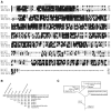
Amino acid sequencing of an 80-kD protein isolated with a pan dynamin antibody revealed strong sequence similarity to the dynamin family. An 80-kD protein recognized by the pan dynamin antibody MC12 was isolated by large scale immunoprecipitation from rat liver homogenate. The immunoprecipitate was run on a 5–15% polyacrylamide gradient gel, and the 80-kD protein band was excised and eluted from the gel. Amino acid sequencing of six tryptic peptides was conducted and compared with rat dynamins I, II, and III as well as Vps1p and Dnm1p. Peptides A to E aligned with the NH2-terminal region of the dynamins, Vps1p and Dnm1p, and peptide F aligned with the COOH terminus of these proteins. Sequence information obtained from these peptides indicated that this protein was strongly related to the dynamins, yet different enough to represent a novel and distinct gene product. Boxes represent conserved residues.
Figure 2
Figure 2
A full-length clone of a novel dynamin-like protein (DLP1) shares homology with dynamin and other dynamin-related proteins. (A) Amino acid sequence comparison of rat DLP1 with rat dynamin I, yeast Dnm1p, and Vps1p was performed by the Clustal method using the DNASTAR sequence analysis program. Conserved amino acid residues are boxed in black. The three regions with asterisks are GTP-binding elements. There is a high degree of homology within the NH2-terminal domains, especially in the tripartite GTP-binding motif (asterisks), but all four sequences are completely divergent at the region aligned with the PH domain of dynamin I (aa 510–633 of Dyn1). Homology is partially restored at the extreme COOH-terminal end of DLP1. Proline-rich tails are not present in DLP1, Dnm1p, or Vps1p. (B) Table showing percent homology among dynamin-related proteins. Rat DLP1 shares up to 42% homology with other dynamin-related proteins. (C) Phylogenetic tree of the large GTPase superfamily showing that DLP1 is subgrouped with the yeast dynamin-related proteins.
Figure 3
Figure 3
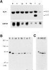
Rat DLP1 is expressed in all tissues examined. (A) Northern blot analysis showing ubiquitous expression of rat DLP1. Total RNA (15 μg/lane) from various rat tissues was hybridized with a DLP1-specific probe. An unknown lower band was detected in testis. The same blot was also hybridized with a GAPDH probe for internal control. (B and C) Western blot analyses showing that DLP1 is present ubiquitously in rat tissues (B) and cultured mouse and human cells (C). Total protein from various rat tissues and tissue cultured cells was run on SDS–polyacrylamide gels, and DLP1 was detected by an affinity-purified anti-DLP1 antibody (DLP-MID). Each lane was loaded with 20 μg total protein, except the brain lane (10 μg). Note that DLP1 in brain, mouse hepatocytes, and human fibroblasts runs differently in SDS-PAGE from DLP1 in other tissues. B, brain; H, heart; Lg, lung; K, kidney; Sp, spleen; T, testis; Lv, liver; P, pancreas; MH, mouse hepatocyte cell line; HF, cultured human fibroblasts.
Figure 4
Figure 4
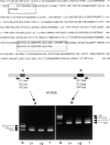
DLP1 has two short alternatively spliced regions that are differentially expressed in tissues. (Top) Amino acid sequence of rat DLP1 showing two alternatively spliced regions. Each region has at least three differentially spliced forms. (Bottom) Brain expresses longer forms of DLP1 in both alternative splicing regions, and other tissues predominantly express the shorter forms. Two sets of specific PCR primers flanking two splicing regions, A and B, were used for RT-PCR with RNA from four different rat tissues (brain, liver, lung, and testis). Details in text.
Figure 5
Figure 5
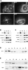
DLP1 is associated with numerous punctate vesicular structures in cultured mammalian cells. Immunofluorescence microscopy of two distinct cultured cell lines, human fibroblasts (A) and a rat hepatocyte cell line, clone 9 (B), stained with affinity-purified antibodies to DLP1 (DLP-N). DLP1 is associated with numerous vesicular structures that are distributed throughout the cytoplasm and appear aligned along cytoskeletal filaments that extend outward from perinuclear foci (arrows). Higher magnification images of boxed regions in A and B are shown in A′ and B′, respectively, and strongly suggest a vesicular association. N, nucleus. Bar, 10 μm.
Figure 6
Figure 6
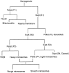
DLP1-associated organelles do not colocalize with multiple endocytic organelle markers but are fractionated into light membrane and ER fractions. (A) Cultured mouse hepatocytes (top two panels of A) or human cholangiocytes (bottom two panels of A) were double stained with antibodies to DLP1 (DLP1-N) and FITC-conjugated dextran (Dx) to label endocytic compartments and lysosomes, and FITC-conjugated transferrin (Tf) to label the recycling endosomal compartment. Little, if any, colocalization between DLP1 and the labeled endocytic compartments is observed. N, nucleus. (B) Immunoblots of rat liver subcellular fractions with DLP1 and various endocytic marker antibodies. Rat liver membranes were fractionated by differential centrifugation and sucrose gradient centrifugation. Each fraction was separated by SDS-PAGE and immunoblotted with anti-DLP1 antibody (DLP-MID) and endocytic marker antibodies including: MC63 for conventional dynamin, anti–α-adaptin, anti-Rab5, and anti–β-galactosidase. While DLP1 is abundant in cytosol and a light vesicle fraction (LM *), conventional dynamin is enriched in the plasma membrane (PM) with little seen in the LM fraction. α-adaptin and Rab5 are enriched in the plasma membrane fraction, and β-galactosidase is in multiple fractions. (C) Same fractions (as prepared in B) were immunoblotted with anti-DLP1 antibody (DLP-MID) and marker antibodies for secretory vesicles including anti–γ-adaptin for Golgi-associated clathrin-coated vesicles, anti-Sec23p for COPII-coated vesicles, anti-Rab8 for post-Golgi secretory vesicles, and anti-μ3 for vesicles associated with the newly identified nonendocytic adaptor complex AP-3. There is substantial coenrichment of DLP1 with the secretory vesicle marker proteins in the LM fraction. Cyt, cytosol; PM, plasma membranes; LM, light microsomes; G, Golgi fraction. (D) Rat liver total microsomes were fractionated by sucrose step gradient centrifugation and fractions were immunoblotted with anti-DLP1 (DLP-MID). DLP1 was enriched in the ER and 1.22 M sucrose fraction, in addition to GL. LS, low speed spin supernatant; LP, low speed spin pellet, TM, total microsomes; GL, light Golgi fraction; GH, heavy Golgi fraction; 1.15, 1.15 M sucrose fraction; 1.22, 1.22 M sucrose fraction; ER, endoplasmic reticulum fraction. (E) Extended fractionation for the liver microsomes (as prepared in B and C). Fractions were immunoblotted with anti-DLP1 and anti-Rab1, showing DLP1 enrichment in ER. HM, heavy microsomes; SM, smooth microsomes; RM, rough microsomes. 40 μg protein from each fraction was loaded onto the gels in B–E. Bars, 20 μm.
Figure 7
Figure 7
DLP1 does not localize with dynamin- or clathrin-associated organelles. Clone 9 cells were labeled with anti-DLP1 (DLP-N) and anti-dynamin (hudy-1) antibodies (A), or with anti-DLP1 (DLP-N) and anti-clathrin heavy chain antibodies (B). In both images, the DLP1 staining (red fluorescence) does not colocalize with dynamin or clathrin (green fluorescence). Insets show higher magnification images of boxed regions N, nucleus. Bars, 10 μm.
Figure 8
Figure 8
Distribution of DLP1-vesicles are coincident with microtubule arrays in cultured cells. Double immunofluorescence microscopy of cultured human fibroblasts (A and B) and clone 9 cells (C) with antibodies to DLP1 and α-tubulin. There is high degree of overlap (arrows) between the distribution of DLP1-stained vesicles (A) and the microtubule array (B). (C) A clone 9 cell stained for DLP1 (red) and microtubules (green) showing DLP1-vesicles aligned along the microtubule network. Inset is a higher magnification image of the boxed region revealing the intimate association between the DLP1 vesicles and microtubules. N, nucleus. Bars: (B) 20 μm; (C and inset) 10 μm.
Figure 9
Figure 9
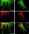
DLP1 localizes to tubules of the endoplasmic reticulum. Double immunofluorescence microscopy of cultured human fibroblasts with antibodies to DLP1 (A and D) and calnexin (B and E). DLP1 spots are organized into long tracks that extend out into the peripheral cytoplasm and coalign perfectly with ER tubules (arrows). Overlap images are seen in C and F. Note that the linear extension of DLP1 is only observed when associated with the ER tubule. N, nucleus. Bars, 10 μm.
Figure 10
Figure 10
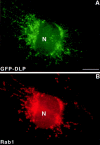
DLP1 is associated with Rab1-positive ER tubules. Clone 9 cells expressing GFP-DLP1 were prepermeabilized with digitonin before fixation and immunostaining with anti-Rab1 antibodies. GFP-DLP1 distributes along the linear structures (A). These linear structures are ER tubules to which Rab1 is associated (B). (C) Overlaying image of GFP-DLP1 and Rab1 staining showing DLP1 and Rab1 are localized on the same ER tubules. (D) Enlarged image of boxed region in C. Note that most GFP-DLP1 spots alternate with Rab1 along the ER tubules. N, nucleus. Bars, 10 μm.
Figure 10
Figure 10
DLP1 is associated with Rab1-positive ER tubules. Clone 9 cells expressing GFP-DLP1 were prepermeabilized with digitonin before fixation and immunostaining with anti-Rab1 antibodies. GFP-DLP1 distributes along the linear structures (A). These linear structures are ER tubules to which Rab1 is associated (B). (C) Overlaying image of GFP-DLP1 and Rab1 staining showing DLP1 and Rab1 are localized on the same ER tubules. (D) Enlarged image of boxed region in C. Note that most GFP-DLP1 spots alternate with Rab1 along the ER tubules. N, nucleus. Bars, 10 μm.
Figure 11
Figure 11
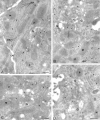
Immunogold labeling of DLP1 showing DLP1 on ER cisternae. Ultrathin cryosections of rat liver were immunolabeled with rabbit anti-DLP1 (DLP-N) followed by goat anti–rabbit IgG 10 nm gold. DLP1 labeling is observed over parallel ER cisternae (rER and arrowheads), and tubular reticular network profiles (small arrows), some of which surround the Golgi apparatus (G). Golgi stacks are largely devoid of DLP1 immunoreactivity as are peroxisomes (P), mitochondria (M), and lysosomes (L). LAT, lateral plasma membrane; bc, bile canalicular plasma membrane. Bars, 400 nm.
Figure
Figure
Diagram 1.

Similar articles

Cited by

References

    1. Adams, M., A. Kerlavage, R. Fleischmann, R. Fuldner, C. Bult, N. Lee, E. Kirkness, K. Weinstock, J. Gocayne, O. White, et al. 1995. Initial assessment of human gene diversity and expression patterns based upon 83 million nucleotides of cDNA sequence. Nature. 377(Suppl.):3–174. - PubMed
    1. Ahle S, Mann A, Eichelsbacher U, Ungewickell E. Structural relationships between clathrin assembly proteins from the Golgi and the plasma membrane. EMBO (Eur Mol Biol Organ) J. 1988;7:919–929. - PMC - PubMed
    1. Barlowe C, Orci L, Yeung T, Hosobuchi M, Hamamoto S, Salama N, Rexach MF, Ravazzola M, Amherdt M, Schekman R. COPII: a membrane coat formed by Sec proteins that drive vesicle budding from the endoplasmic reticulum. Cell. 1994;77:895–907. - PubMed
    1. Chomczynski P, Sacchi N. Single-step method of RNA isolation by acid guanidium thiocyanate-phenol-chloroform extraction. Anal Biochem. 1987;162:156–159. - PubMed
    1. Clark J, Moore L, Krasinskas A, Way J, Battey J, Tamkun J, Kahn RA. Selective amplification of additional members of the ADP-ribosylation factor (ARF) family: cloning of additional human and DrosophilaARF-like genes. Proc Natl Acad Sci USA. 1993;90:8952–8956. - PMC - PubMed

Publication types

MeSH terms