

Cell cycle-dependent subcellular localization of the TSG101 protein and mitotic and nuclear abnormalities associated with TSG101 deficiency
- PMID: 9465061
- PMCID: PMC19109
- DOI: 10.1073/pnas.95.4.1595
Cell cycle-dependent subcellular localization of the TSG101 protein and mitotic and nuclear abnormalities associated with TSG101 deficiency
Abstract
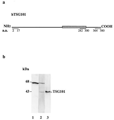
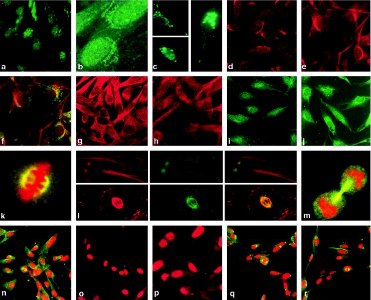
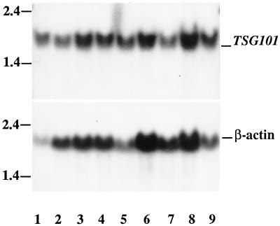
TSG101 is a recently discovered tumor susceptibility gene whose functional inactivation in mouse fibroblasts results in cell transformation and the ability to form metastatic tumors in nude mice. Although restoration of TSG101 activity reverses tumorigenesis, neoplasia is irreversible in some cells, suggesting that permanent genetic alteration can occur during TSG101 inactivation. Here we describe studies that support this notion. We find that localization of TSG101 is cell cycle-dependent, occurring in the nucleus and Golgi complex during interphase, and in mitotic spindles and centrosomes during mitosis; cells made neoplastic by a deficiency in TSG101 expression show a series of mitosis-related abnormalities, including multiple microtubule organizing centers, aberrant mitotic spindles, abnormal distribution of metaphase chromatin, aneuploidy, and nuclear anomalies. Our findings suggest that TSG101 deficiency may lead to genome instability in addition to previously reported reversible neoplastic transformation.
Figures

Similar articles
-
Perturbation of TSG101 protein affects cell cycle progression.Cancer Res. 1998 Jul 1;58(13):2699-702. Cancer Res. 1998. PMID: 9661875
-
TSG101 protein steady-state level is regulated posttranslationally by an evolutionarily conserved COOH-terminal sequence.Cancer Res. 2000 Mar 15;60(6):1736-41. Cancer Res. 2000. PMID: 10749147
-
Aberrant expression of TSG101 in Taiwan Chinese breast cancer.Breast Cancer Res Treat. 2000 Apr;60(3):259-66. doi: 10.1023/a:1006426400524. Breast Cancer Res Treat. 2000. PMID: 10930114
-
Centrosomes and the art of mitotic spindle maintenance.Int Rev Cell Mol Biol. 2014;313:179-217. doi: 10.1016/B978-0-12-800177-6.00006-2. Int Rev Cell Mol Biol. 2014. PMID: 25376493 Review.
-
The role of mitotic kinases in coupling the centrosome cycle with the assembly of the mitotic spindle.J Cell Sci. 2014 Oct 1;127(Pt 19):4111-22. doi: 10.1242/jcs.151753. Epub 2014 Aug 15. J Cell Sci. 2014. PMID: 25128564 Review.
Cited by
-
Re-splicing of mature mRNA in cancer cells promotes activation of distant weak alternative splice sites.Nucleic Acids Res. 2012 Sep;40(16):7896-906. doi: 10.1093/nar/gks520. Epub 2012 Jun 6. Nucleic Acids Res. 2012. PMID: 22675076 Free PMC article.
-
Identification of TSG101 functional domains and p21 loci required for TSG101-mediated p21 gene regulation.PLoS One. 2013 Nov 11;8(11):e79674. doi: 10.1371/journal.pone.0079674. eCollection 2013. PLoS One. 2013. PMID: 24244542 Free PMC article.
-
The ESCRT machinery: from the plasma membrane to endosomes and back again.Crit Rev Biochem Mol Biol. 2014 May-Jun;49(3):242-61. doi: 10.3109/10409238.2014.881777. Epub 2014 Jan 24. Crit Rev Biochem Mol Biol. 2014. PMID: 24456136 Free PMC article. Review.
-
Direct isolation and characterization of circulating exosomes from biological samples using magnetic nanowires.J Nanobiotechnology. 2019 Jan 7;17(1):1. doi: 10.1186/s12951-018-0433-3. J Nanobiotechnology. 2019. PMID: 30612562 Free PMC article.
-
Cell cycle arrest and cell death are controlled by p53-dependent and p53-independent mechanisms in Tsg101-deficient cells.J Biol Chem. 2004 Aug 20;279(34):35984-94. doi: 10.1074/jbc.M400408200. Epub 2004 Jun 21. J Biol Chem. 2004. PMID: 15210712 Free PMC article.
References
Publication types
MeSH terms
Substances
LinkOut - more resources
Full Text Sources
Other Literature Sources
Molecular Biology Databases
