. 1998 Feb 1;12(3):435-46.
doi: 10.1101/gad.12.3.435.

The Homothorax homeoprotein activates the nuclear localization of another homeoprotein, extradenticle, and suppresses eye development in Drosophila

Affiliations

The Homothorax homeoprotein activates the nuclear localization of another homeoprotein, extradenticle, and suppresses eye development in Drosophila

C Y Pai et al. Genes Dev. .

Abstract

The Extradenticle (Exd) protein in Drosophila acts as a cofactor to homeotic proteins. Its nuclear localization is regulated. We report the cloning of the Drosophila homothorax (hth) gene, a homolog of the mouse Meis1 proto-oncogene that has a homeobox related to that of exd. Comparison with Meis1 finds two regions of high homology: a novel MH domain and the homeodomain. In imaginal discs, hth expression coincides with nuclear Exd. hth and exd also have virtually identical, mutant clonal phenotypes in adults. These results suggest that hth and exd function in the same pathway. We show that hth acts upstream of exd and is required and sufficient for Exd protein nuclear localization. We also show that hth and exd are both negative regulators of eye development; their mutant clones caused ectopic eye formation. Targeted expression of hth, but not of exd, in the eye disc abolished eye development completely. We suggest that hth acts with exd to delimit the eye field and prevent inappropriate eye development.


Figures

Figure 1
Figure 1
The hth locus and cDNAs. (A) Map of the hth locus. The mapped hth genomic region and the clone λSY46 is shown with horizontal lines. The region deleted in P1-K1–8 is shown as a hatched box. Sites of P[lacW] insertion are indicated. Restriction fragments hybridizing to the cDNA clones 7, 5, or 1 are shown below as thick lines with arrows indicating the transcription direction. The arrow indicates the position of an exon-intron junction. Restriction sites are: (B) BamHI; (E) EcoRI, and (H) HindIII. (B) Alignment of the group II (hth) cDNA clones. The different regions are marked differently to indicate their relationship. The extra 45 bp is indicated as a dark gray box in clones 8, 7, 3, 9, 10, and 4. Six vertical bars on top of clone 5 indicate the positions of the first six in-frame ATGs. (An) Poly(A) stretches. The ORF and the two regions (the MH and HD domains), which are highly homologous to Meis1, are indicated. The arrow indicates the position of an exon–intron junction (same as in A). Arrowheads indicate potential splice junctions.
Figure 2
Figure 2
hth encodes a HD protein related to Meis1. (A) The peptide sequence of Hth translated from cDNA. The Meis1–Hth homology domain (MH domain) is boxed. The HD is underlined. The 15 residues present in some clones are double underlined. (B) The relative location of MH and HD in the Meis1 and Hth proteins. (C) Comparison of the HD domain of Hth to that of the Mies1, Pbx, Knotted, and MaTα2 subfamilies. Representatives from different phylogenetic groups are shown. Members in the Meis1 family include three genes in mouse, Meis1 (U33629, U33630), Meis2 (four alternatively spliced isoform that do not affect the HD: 2a, U57343, AJ000504; 2b, U68383, AJ000505; 2c, AJ000506; 2d, U68384, AJ000507), and Meis3 (U57344); three genes in human, MEIS1 (U85707), PKNOX1 (U69727), and several ESTs defining the MEIS3 (F05816, H25643, R35310; two other short ESTs probably have unspliced intron, D31072, T10795); three genes in Xenopus laevis: meisl-1 (U68386), meisl-2 (U68387), and meisl-3 (U68388). More distantly related members (not shown) include human TGIF (X89750), chicken AKR (U25353), Arabidopsis ATH1 (X80126), BELL1 (U39944), and an EST (Z35398), a C. elegans EST (M88963), yeast CUP9 (L36815), and a gene identified in the yeast genomic sequence (1723892). Residues identical to the Hth sequence (top) are indicated by dots. The consensus sequence of classic HDs is shown at the bottom. The position of three extra residues in the TALE HD family is shown as dashes in the classic HD. Residues that are highly conserved among all HDs are indicated by an asterisk. (D). Comparison of the MH domain of Hth to that of other members of the Meis–hth family. The arrowhead indicates the position of a 6-amino-acid (DGASAG) insertion in the mouse Meis3 protein.
Figure 3
Figure 3
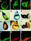
Expression pattern of hth in imaginal discs are similar to those of nuclear Exd. Expression pattern of hth in imaginal discs (leg disc, A,D,G; wing disc, B,E,H; eye–antennea disc, C,F,I) was detected by anti-Hth immunostaining in wild-type (A–C), lacZ activity staining in the hth1422-4 enhancer trap line (D–F), and in situ hybridization in wild type (G–I). Anti-ELAV staining (red in C) was used to show the position of the photoreceptor neurons. Double staining of Hth (J) and Exd (K) in the eye disc showed Hth colocalize with nuclear Exd (both channels merged in L). Outside of the Hth domain, there is very weak cytoplasmic staining of Exd. The image is of the ventral–anterior margin of eye disc.
Figure 4
Figure 4
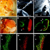
Clonal mutant phenotype of hth. hth mutant clones on the head cuticle were detected as y and non-Sb bristles. Those in the compound eyes were detected as w. (A) A clone on the third antennal segment transformed the entire segment into distal leg structures with a claw (arrowhead). (B) Scanning electron microscope (SEM) picture of a similar clone showing antenna to leg transformation. (C) A clone ventral to the normal eye produced a small ectopic eye (arrowhead) at the tip of a tubular outgrowth. (D) A ventral clone caused ectopic eye development (arrowhead). The clone is entirely white, suggesting that the effect of hth mutation is cell-autonomous. However, clones within the compound eye did not have abnormal phenotypes (arrow). (E) Eye–antennal discs were double stained with phalloidin (green) and anti-ELAV (red) to reveal the location of the morphogenic furrow (arrow) and the photoreceptors, respectively. Ectopic photoreceptors (arrowhead) were detected in the ventral margin. (F) Eye–antennal discs were double stained with anti-Hth (green) and anti-ELAV (red) to reveal the position of hth mutant clones relative to the photoreceptors. A clone at the ventral margin of eye disc (arrow) caused ectopic photoreceptor development and local outgrowth. Two clones at the dorsal margin of eye disc (arrowheads) caused no ectopic photoreceptor formation. (G–I)Eye–antennal discs were double stained with anti-Hth (G) and anti-Exd (H). An hth mutant clone (arrowhead) has no Hth staining (G) and no Exd staining (H). (I) A merged image of the Hth and Exd signals.
Figure 5
Figure 5
Hth ectopic expression induced the accumulation and nuclear transport of Exd. (A–F) Ectopically expressed Exd is located in the cytoplasm. Eye–antennal discs from UAS–exd/+; GMR–gal4/+ larvae were triple stained by anti-Hth, anti-Exd, and the DNA dye SYTOX. Ectopic Exd is induced in the GMR expression domain, posterior to the morphogenetic furrow (B). The merged image (C) showed that the ectopically induced Exd is not accompanied by Hth. The endogenous Hth and Exd were colocalized (confirmed at higher magnification, not shown), although they were not expressed at the same relative level in all cells. (D–F) A region (boxed in C) of the GMR domain at higher magnification. (D) SYTOX labeling revealed the position of the nucleus. (E) Ectopically expressed Exd is primarily located in the cytoplasm. This is more clearly demonstrated in the merged image (F). (G–L) When Exd and Hth were coexpressed ectopically, both were located in the nucleus. Eye–antennal discs from UAS–exd/+; GMR–gal4/+; UAS–hth/+ larvae were triple stained by SYTOX, anti-Hth, and anti-Exd. Ectopic Hth (G), as well as Exd (H), was induced in the GMR domain, posterior to the furrow. (I–L) A region (boxed in H) of the GMR domain at higher magnification. The ectopic Hth and Exd were located in the nucleus. (L) Merged image of SYTOX (green) and anti-Exd (red) showed that Exd and DNA colocalize (yellow), except in a few dividing cells, where Exd is in the cytoplasm. Hth and Exd staining is stronger in alternating oblique rows of cells. This is probably attributable to the GMR specificity, as GMR–gal4-induced Ubx expression gave a similar pattern (not shown). (M–R) Ectopic expression of Hth resulted in an elevated level of Exd. The eye–antennal discs from GMR–gal4/+; UAS–hth/+; larvae were double stained for anti-Hth and anti-Exd. Ectopic induction of Hth also induced elevated levels of Exd in the GMR domain. A merged view (O) showed that the Exd appears several rows of cells posterior to the anterior border of Hth expression. This is clearer in a higher magnification (P–R).
Figure 6
Figure 6
Ectopic expression of hth suppresses furrow progression/initiation and eye development. Clonal hth ectopic expression were induced. Typical phenotypes in the adult compound eye includes a posterior–anterior scar (A), sometimes accompanied by local outgrowth (B). (C) In one case the scar is fused in the anterior end. (D) In two cases, the eye is split into a dorsal and a ventral small eye. All heads are oriented with anterior to the left and dorsal to the top. (E) The eye–antennal discs were triple stained with anti-Hth (green), anti-ELAV (red), and Phalloidin (blue). An ectopic hth-expressing clone (arrowhead) had inhibited MF progression and the subsequent photoreceptor differentiation. (F) dpp–gal4-induced hth expression completely abolished eye development.

Similar articles

Cited by

References

    1. Arkhipova IR. Promoter elements in Drosophila melanogaster revealed by sequence analysis. Genetics. 1995;139:1359–1369. - PMC - PubMed
    1. Aspland SE, White RA. Nucleocytoplasmic localisation of extradenticle protein is spatially regulated throughout development in Drosophila. Development. 1997;124:741–747. - PubMed
    1. Bennassayag C, Seroude L, Boube M, Erard M, Cribbs DL. A homeodomain point mutation of the Drosophila proboscipedia protein provokes eye loss independently of homeotic function. Mech Dev. 1997;63:187–198. - PubMed
    1. Bier E, Vaessin H, Shepherd S, Lee K, McCall K, Barbel S, Ackerman L, Carretto R, Uemura T, Grell E, et al. Searching for pattern and mutation in the Drosophila genome with a P-lacZ vector. Genes & Dev. 1989;3:1273–1287. - PubMed
    1. Bonini NM, Choi K-W. Early decisions in Drosophila eye morphogenesis. Curr Opin Genet Dev. 1995;5:507–515. - PubMed

Publication types

MeSH terms

Associated data