. 1998 Feb;18(2):790-8.
doi: 10.1128/MCB.18.2.790.

Activation of the mitogen-activated protein kinase/extracellular signal-regulated kinase pathway by conventional, novel, and atypical protein kinase C isotypes

Affiliations

Activation of the mitogen-activated protein kinase/extracellular signal-regulated kinase pathway by conventional, novel, and atypical protein kinase C isotypes

D C Schönwasser et al. Mol Cell Biol. 1998 Feb.

Abstract

Phorbol ester treatment of quiescent Swiss 3T3 cells leads to cell proliferation, a response thought to be mediated by protein kinase C (PKC), the major cellular receptor for this class of agents. We demonstrate here that this proliferation is dependent on the activation of the extracellular signal-regulated kinase/mitogen-activated protein kinase (ERK/MAPK) cascade. It is shown that dominant-negative PKC-alpha inhibits stimulation of the ERK/MAPK pathway by phorbol esters in Cos-7 cells, demonstrating a role for PKC in this activation. To assess the potential specificity of PKC isotypes mediating this process, constitutively active mutants of six PKC isotypes (alpha, beta, delta, epsilon, eta, and zeta) were employed. Transient transfection of these PKC mutants into Cos-7 cells showed that members of all three groups of PKC (conventional, novel, and atypical) are able to activate p42 MAPK as well as its immediate upstream activator, the MAPK/ERK kinase MEK-1. At the level of Raf, the kinase that phosphorylates MEK-1, the activation cascade diverges; while conventional and novel PKCs (isotypes alpha and eta) are potent activators of c-Raf1, atypical PKC-zeta cannot increase c-Raf1 activity, stimulating MEK by an independent mechanism. Stimulation of c-Raf1 by PKC-alpha and PKC-eta was abrogated for RafCAAX, which is a membrane-localized, partially active form of c-Raf1. We further established that activation of Raf is independent of phosphorylation at serine residues 259 and 499. In addition to activation, we describe a novel Raf desensitization induced by PKC-alpha, which acts to prevent further Raf stimulation by growth factors. The results thus demonstrate a necessary role for PKC and p42 MAPK activation in 12-O-tetradecanoylphorbol-13-acetate induced mitogenesis and provide evidence for multiple PKC controls acting on this MAPK cascade.


Figures
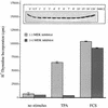
FIG. 1
FIG. 1
Effects of phorbol ester on Swiss 3T3 cells. Phorbol ester treatment induces DNA synthesis via the ERK/MAPK pathway. Quiescent Swiss 3T3 cells were treated for 40 h with 400 nM TPA or 20% fetal calf serum or left without stimulus in the presence (+) or absence (−) of the MEK-1 inhibitor PD 098059 (30 μM). DNA synthesis was assessed by measuring [3H]thymidine incorporation. Each value is the mean ± the standard error of the mean for three dishes representative of two independent experiments. (Inset) Phorbol ester treatment activates p42 MAPK. Quiescent Swiss 3T3 cells were treated with 400 nM TPA for different time intervals, and p42 MAPK activation was measured by mobility shift analysis of the p42 MAPK protein in a 10% polyacrylamide gel as a readout for kinase activity. Phorbol ester treatment of Swiss 3T3 cells causes a rapid activation of p42 MAPK (1 min) which is sustained for up to 2 h.
FIG. 2
FIG. 2
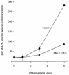
Dominant-negative PKC-α inhibits p42 MAPK activation after TPA treatment. Serum-starved Cos-7 cells were cotransfected with myc-p42 MAPK and dominant-negative PKC-α(T/A)3 or with empty vector as a control. After transfection (48 h), cells were stimulated for 0, 1, 3, and 6 min with TPA (250 nM) and myc-p42 MAPK activity was determined in immune complex kinase assays with MBP as a substrate. Activities were normalized to myc-p42 MAPK protein levels in the immune complex. For each time point, triplicate samples were used, and the standard error is indicated (error bars) when it is >8% of the corresponding mean value. By the t test, the 6-min time points result in a P value of less than 0.001.
FIG. 3
FIG. 3
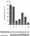
p42 MAPK is activated by various PKC isotypes in vivo. Six different constitutively active PKC isotypes representing the conventional (α, and β1), novel (δ, ɛ, and η), and atypical (ζ) subclasses of this family and empty vector were cotransfected with myc-p42 MAPK into Cos-7 cells. Duplicate dishes were harvested 48 h after transfection, myc-p42 MAPK was immunoprecipitated, and its activity was determined. myc-p42 MAPK activity is presented in arbitrary units as a function of protein expression. The panels below the graph show the amounts of substrate phosphorylation expressed as units of the PhosphorImager scanner (Molecular Dynamics) (upper panel) and the amounts of protein which was present in the reactions (duplicates; lower panel). Results from one of three similar experiments are shown. The stimulation of a control sample, transfected with empty vector and myc-p42 MAPK, by a mixture of TPA (400 nM) and FCS (20%) 20 min before harvesting of the cells resulted in myc-p42 MAPK activation of between 14- and 83-fold depending on the individual experiment (data not shown). The asterisk indicates a longer exposure of the Western blot showing myc-p42 MAPK protein in the PKC-η-transfected cells than for the other blots.
FIG. 4
FIG. 4
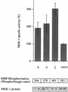
PKC-α, -η, and -ζ activate MEK-1 in vivo. Constitutively active forms of PKC-α, -η, and -ζ and empty vector were coexpressed with a myc-tagged MEK-1 construct in Cos-7 cells. Cells were cultured for 48 h before they were harvested and myc-MEK-1 was immunoprecipitated. Myc-MEK-1 activity was determined in a coupled in vitro kinase assay with recombinant p42 MAPK protein and MBP as a substrate. The panels below the graph represent 32P incorporation into MBP (upper panel) and the amount of myc-MEK-1 enzyme present (duplicates; lower panel) in each reaction. Each value is the average for duplicate samples. Error bars indicate standard error. Stimulation of a control sample, which was transfected with empty vector and myc-MEK-1, by a mixture of TPA (400 nM) and FCS (20%) for 20 min before harvesting resulted in 8- to 10-fold myc-MEK-1 activation (data not shown). The data shown are from one of two similar experiments.
FIG. 5
FIG. 5
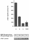
c-Raf 1 is activated by PKC-α and -η but not by PKC-ζ. Constitutively active constructs of PKC-α, -η, and -ζ, and empty vector were cotransfected with a myc-tagged c-Raf1 construct into Cos-7 cells. Forty-five hours after transfection, the myc-Raf protein was immunoprecipitated and its activity was measured in a coupled in vitro kinase assay with recombinant MEK and p42 MAPK proteins and MBP as a substrate. The amount of substrate phosphorylation (upper panel) and the quantity of myc-Raf enzyme in each reaction (duplicates; lower panel) are shown in the pairs of panels below the graph. Stimulation of a control sample, which was transfected with empty vector and myc-Raf, by a mixture of TPA (400 nM) and FCS (20%) for 10 min before harvesting resulted in 6- to 22-fold myc-Raf activation (data not shown). The data shown are from one of three similar experiments.
FIG. 6
FIG. 6
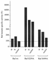
PKC-α and -η are able to activate Raf S259A and Raf S499A. Cos-7 cells were cotransfected with constructs of constitutively active PKC-α and -η or empty vector in combination with a myc-tagged wild-type (wt) c-Raf1 or mutant Raf construct c-Raf S259A or c-Raf S499A. After transfection (40 h), the cells were harvested, the wild-type or mutant Raf proteins were immunoprecipitated, and their activities were measured in a coupled in vitro kinase assay. Substrate phosphorylation was normalized to the amount of Raf protein in each kinase reaction; Raf activation is presented as a percentage of the activity of the empty vector control. Stimulation of control samples, which were transfected with empty vector and either myc-Rafwt, myc-Raf S259A, or myc-Raf S499A, by a mixture of TPA (400 nM) and FCS (20%) for 10 min before harvesting resulted in 6-fold, 4- to 10-fold, and 7- to 9-fold activation, respectively (data not shown). This experiment was carried out in duplicate and is representative of three independent experiments.
FIG. 7
FIG. 7
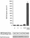
A membrane-localized mutant of Raf cannot be activated by constitutively active PKCs. Cos-7 cells were cotransfected with myc-RafCAAX and either constitutively active PKC-α, -η, or -ζ empty vector. Forty-five hours after transfection, control cells were either left untreated or stimulated for 10 min with 20% serum and 400 nM TPA as indicated and then harvested. A fraction of the total cell lysate was used to immunoprecipitate myc-RafCAAX and to determine its activity in a coupled assay that depends on the addition of recombinant MEK and p42 MAPK and MBP. Substrate phosphorylation (upper panel) was normalized to the amount of protein in each sample (duplicates; lower panel). Stimulation of a control sample, which was transfected with empty vector and myc-RafCAAX, by a mixture of TPA (400 nM) and FCS (20%) for 10 min before harvesting resulted in 3- to 8-fold myc-RafCAAX activation (data not shown). This experiment was carried out in duplicate and is representative of three independent assays.
FIG. 8
FIG. 8
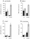
PKC-α exerts a desensitization effect at different levels of the MAPK cascade. Cos-7 cells were cotransfected with constitutively active PKC-α (α) or empty vector in combination with a myc epitope-tagged p42 MAPK (A), myc-MEK-1 (B), myc-c-Raf1 (C), or myc-RafCAAX (D). After 40 h of expression, half of the samples were stimulated with 20% serum and 400 nM TPA for 20 min in the case of p42 MAPK and MEK-1 or 10 min in the case of c-Raf 1 and RafCAAX; unstimulated cells are presented in dark gray, and serum-TPA-stimulated cells are presented in light gray. After immunoprecipitation of the myc-tagged proteins, the activities of p42 MAPK, MEK-1, c-Raf1, and RafCAAX were determined. All data shown have been normalized to the amount of reporter construct expressed in each sample. Each assay was done in duplicate.
FIG. 9
FIG. 9
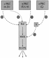
Model illustrating how different PKC isotypes activate the ERK/MAPK cascade. cPKCs (α and β1) have the potential to activate c-Raf1 in vivo while at the same time blocking further activation by growth factors and other PKC isotypes. PKC-δ, -ɛ, and -η, representing isotypes of the novel subclass of the PKC superfamily, activate the MAPK cascade but show no desensitization effect. aPKC-ζ differs from the other PKC isotypes with regard to MEK activation. While PKC-α and -η signal to MEK via Raf, PKC-ζ works through a Raf-independent pathway.

Similar articles

Cited by

References

    1. Adams P D, Parker P J. TPA-induced activation of MAP kinase. FEBS Lett. 1991;290:77–82. - PubMed
    1. Ahmed S, Kozma R, Monfries C, Hall C, Lim H H, Smith P, Lim L. Human brain n-chimaerin cDNA encodes a novel phorbol ester receptor. Biochem J. 1990;272:767–773. - PMC - PubMed
    1. Alessi D R, Cuenda A, Cohen P, Dudley D T, Saltiel A R. PD 098059 is a specific inhibitor of the activation of mitogen-activated protein kinase kinase in vitro and in vivo. J Biol Chem. 1995;270:27489–27494. - PubMed
    1. Alessi D R, Saito Y, Campbell D G, Cohen P, Sithanandam G, Rapp U, Ashworth A, Marshall C J, Cowley S. Identification of the sites in MAP kinase kinase-1 phosphorylated by p74raf-1. EMBO J. 1994;13:1610–1619. - PMC - PubMed
    1. Avruch J, Zhang X F, Kyriakis J M. Raf meets Ras: completing the framework of a signal transduction pathway. Trends Biochem Sci. 1994;19:279–283. - PubMed

MeSH terms

Substances