. 1998 Jan 15;12(2):186-97.
doi: 10.1101/gad.12.2.186.

Smad6 inhibits BMP/Smad1 signaling by specifically competing with the Smad4 tumor suppressor

Affiliations

Smad6 inhibits BMP/Smad1 signaling by specifically competing with the Smad4 tumor suppressor

A Hata et al. Genes Dev. .

Abstract

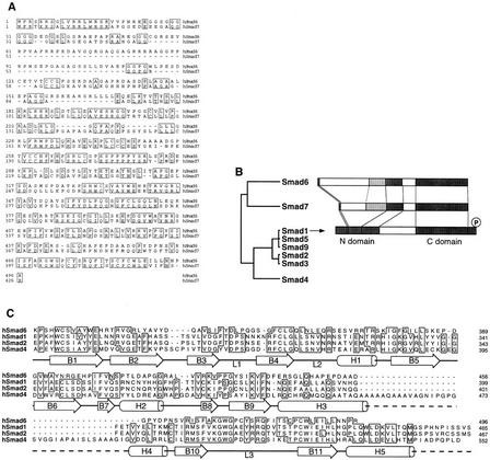
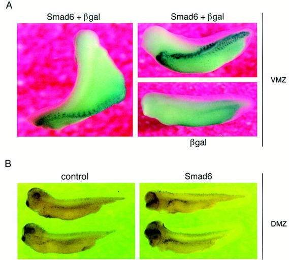
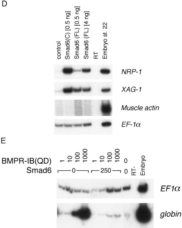
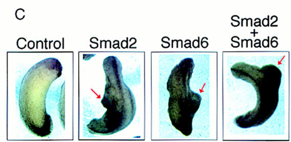
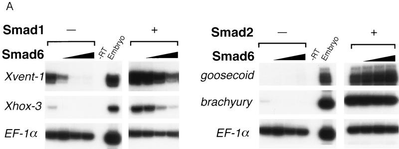
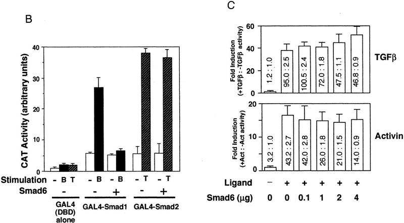
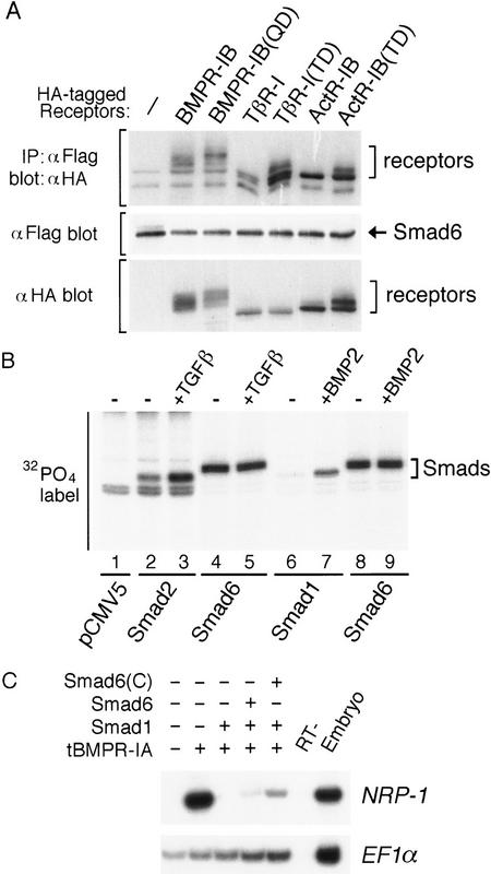
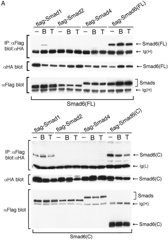
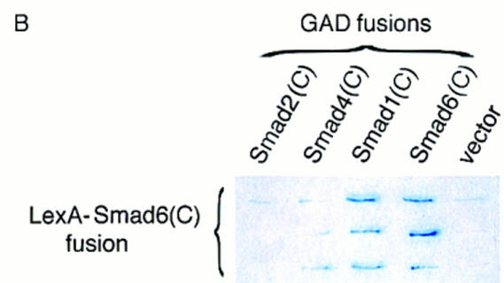
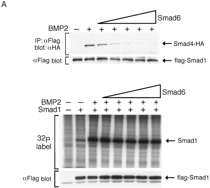
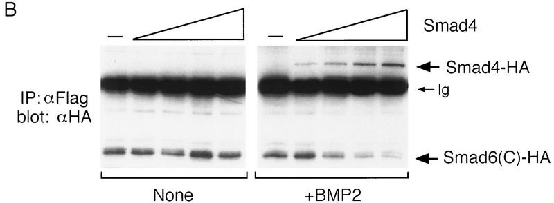
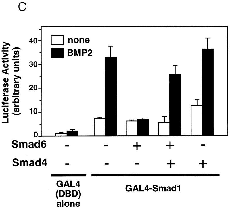
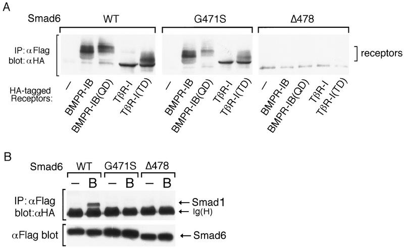
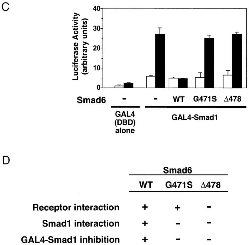
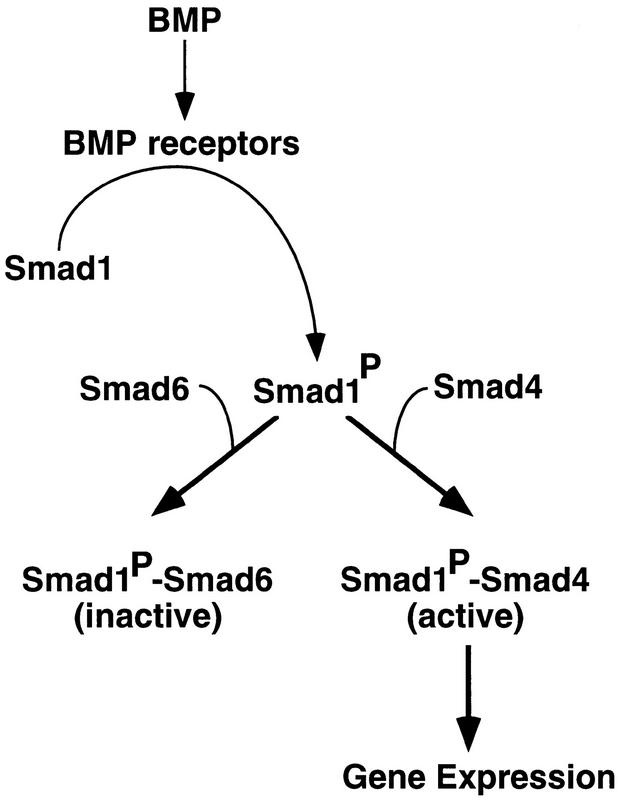
Bone morphogenetic protein (BMP) receptors signal by phosphorylating Smad1, which then associates with Smad4; this complex moves into the nucleus and activates transcription. Here we report the existence of a natural inhibitor of this process, Smad6, a longer version of the previously reported JV15-1. In Xenopus embryos and in mammalian cells, Smad6 specifically blocks signaling by the BMP/Smad1 pathway. Smad6 inhibits BMP/Smad1 signaling without interfering with receptor-mediated phosphorylation of Smad1. Smad6 specifically competes with Smad4 for binding to receptor-activated Smad1, yielding an apparently inactive Smad1-Smad6 complex. Therefore, Smad6 selectively antagonizes BMP-activated Smad1 by acting as a Smad4 decoy.


Figures

Figure 1
Figure 1
Structure of Smad6. (A) Alignment of the predicted amino acid sequence of human Smad6 and Smad7. Identical residues are boxed. (B) Smad homology tree and schematic comparison of the structures of Smad6, Smad7, and Smad1. The amino and carboxy-terminal homologous regions (N- and C-domain) are darkly shaded. A region conserved only between Smad6 and Smad7 is lightly shaded. (C) Alignment of C-domain sequences of Smad6 and other Smads, indicating the secondary structure elements of the Smad4 C-domain (Shi et al. 1997). In the crystal structure of Smad4 this region forms an α-helix (α-helix 5) that associates with α-helices 3 and 4 in a three-helix bundle. This structure contributes the formation of a Smad4 homotrimer by interacting with loops 1 and 2 of the adjacent monomer. The region corresponding to loops 1 and 2 in Smad6 is also very divergent from Smad4 and the receptor-regulated Smads. However, most of the components that constitute the β-sandwich core structure of the Smad4 C-domain are conserved in Smad6.
Figure 2
Figure 2
Smad6 induces secondary axes and neuralizes ectoderm in Xenopus. (A) Smad6 overexpression induces the formation of a secondary axis. Smad6 RNA (0.5–4 ng) was coinjected with nuclear β-galactosidase (β-gal)RNA (100 pg) into the ventral vegetal blastomeres of eight cell stage embryos. Unlike control embryos injected with β-gal alone (bottom right), Smad6 coinjected embryos develop an ectopic dorsal axis (left and top right). β-Galactosidase staining of the injected embryos reveals that the progeny of the injected blastomere directly contribute to the ectopic axis. Dorsal at left; anterior at top. Lateral at right; anterior at left. (B) Smad6 does not affect formation of the primary dorsal axis. Smad6 RNA (1–4 ng) was injected into the dorsal marginal zone of four cell stage embryos. Compared to control embryos (left panel), Smad6-injected tadpoles display enlarged heads and cyclopia, but have normal body axes (right). (C) Secondary axis formation by the activin signaling molecule Smad2 is enhanced by coinjection of Smad6. Smad2 RNA (1 ng), Smad6 RNA (1 ng), or both RNAs (1 ng each) were injected into ventral vegetal blastomeres of eight cell stage embryos. Coinjection of Smad2 and Smad6 produces secondary axes (red arrows) that are more complete than those obtained by injection of either Smad alone. Anterior is at top. (D) Smad6 neuralizes ectodermal explants. Smad6 and Smad6 C-domain RNAs were injected at the indicated amount in the animal pole of two-cell stage embryos. At the blastula stage, animal caps were dissected and cultured in saline solution. At the tailbud stages (stage 22), total RNA was harvested and analyzed by RT–PCR for the presence of the indicated transcripts. Full-length human Smad6 (0.5 and 4 ng) induces NRP-1, a pan–neural marker, and XAG-1, a marker of cement gland. Cement gland is induced efficiently even at the lower dose; induction of neural tissue requires a higher dose of Smad6. The isolated C-domain RNA is a more potent inducer of both XAG-1 and NRP-1 than full-length Smad6. Neither construct induced muscle actin, a marker of dorsal (paraxial) mesoderm. EF-1α, ubiquitously expressed, is a loading control. RNA from whole embryos (Embryo) provides the positive control. The RT lane is identical to the embryo lane, except that reverse transcriptase was omitted. (E) Smad6 interferes with blood induction by a constitutively active BMP receptor. BMPR-IB(QD) RNA (1–1000 pg) was injected either alone or together with Smad6 RNA (250 pg) in the animal pole. BMPR-IB(QD)-injected ectodermal explants show induction of globin at stage 30, and this response is blocked by coexpressed Smad6.
Figure 2
Figure 2
Smad6 induces secondary axes and neuralizes ectoderm in Xenopus. (A) Smad6 overexpression induces the formation of a secondary axis. Smad6 RNA (0.5–4 ng) was coinjected with nuclear β-galactosidase (β-gal)RNA (100 pg) into the ventral vegetal blastomeres of eight cell stage embryos. Unlike control embryos injected with β-gal alone (bottom right), Smad6 coinjected embryos develop an ectopic dorsal axis (left and top right). β-Galactosidase staining of the injected embryos reveals that the progeny of the injected blastomere directly contribute to the ectopic axis. Dorsal at left; anterior at top. Lateral at right; anterior at left. (B) Smad6 does not affect formation of the primary dorsal axis. Smad6 RNA (1–4 ng) was injected into the dorsal marginal zone of four cell stage embryos. Compared to control embryos (left panel), Smad6-injected tadpoles display enlarged heads and cyclopia, but have normal body axes (right). (C) Secondary axis formation by the activin signaling molecule Smad2 is enhanced by coinjection of Smad6. Smad2 RNA (1 ng), Smad6 RNA (1 ng), or both RNAs (1 ng each) were injected into ventral vegetal blastomeres of eight cell stage embryos. Coinjection of Smad2 and Smad6 produces secondary axes (red arrows) that are more complete than those obtained by injection of either Smad alone. Anterior is at top. (D) Smad6 neuralizes ectodermal explants. Smad6 and Smad6 C-domain RNAs were injected at the indicated amount in the animal pole of two-cell stage embryos. At the blastula stage, animal caps were dissected and cultured in saline solution. At the tailbud stages (stage 22), total RNA was harvested and analyzed by RT–PCR for the presence of the indicated transcripts. Full-length human Smad6 (0.5 and 4 ng) induces NRP-1, a pan–neural marker, and XAG-1, a marker of cement gland. Cement gland is induced efficiently even at the lower dose; induction of neural tissue requires a higher dose of Smad6. The isolated C-domain RNA is a more potent inducer of both XAG-1 and NRP-1 than full-length Smad6. Neither construct induced muscle actin, a marker of dorsal (paraxial) mesoderm. EF-1α, ubiquitously expressed, is a loading control. RNA from whole embryos (Embryo) provides the positive control. The RT lane is identical to the embryo lane, except that reverse transcriptase was omitted. (E) Smad6 interferes with blood induction by a constitutively active BMP receptor. BMPR-IB(QD) RNA (1–1000 pg) was injected either alone or together with Smad6 RNA (250 pg) in the animal pole. BMPR-IB(QD)-injected ectodermal explants show induction of globin at stage 30, and this response is blocked by coexpressed Smad6.
Figure 3
Figure 3
Smad6 specifically inhibits the BMP/Smad1 signaling pathway. (A) Smad6 blocks induction of ventral mesoderm by Smad1 but not induction of dorsal mesoderm by Smad2. An increasing amount of Smad6 RNA (0.5–4 ng) was coinjected with or without a fixed amount of Smad1 or Smad2 RNA (2 ng). Animal caps from injected embryos were collected at gastrula stage (stage 11.5) and subjected to RT–PCR. Xvent-1 and Xhox-3 are markers of ventral mesoderm; goosecoid is a dorsal mesoderm marker and brachyury is a pan–mesodermal marker. (B) BMP-dependent transcriptional activation of GAL4–Smad1 fusion protein is blocked by Smad6. R-1B/L17 cells were transfected with the reporter gene (G1E1BCAT, 1 μg), appropriate receptors (TβR-I for TGFβ or BMPR–IB and BMPR–II for BMP2), and either a vector containing the GAL4 DNA-binding domain (DBD) alone or fusion constructs of DBD with Smad1 or Smad2 in the presence or absence of Smad6 (2 μg). Cells were incubated with or without 5 nm BMP2 (B) or 100 pm TGFβ (T) for 18 hr. CAT activity is expressed as the mean ± s.d. of three independent experiments. (C) TGFβ or activin-induced transcriptional activation of the 3TP promoter is not inhibited by Smad6. R-1B/L17 cells were transfected with the reporter gene (p3TP–lux) and type I receptors for TGFβ (TβR-l) or activin (ActR-IB) and were treated with or without 100 pm TGFβ (top) or 2 nm activin (bottom) for 18 hr. The ratios of stimulated to unstimulated levels of luciferase activity are indicated numerically, and their quotients plotted in the bar graph. Data are the mean ± s.d. of triplicate values. Notice that although Smad6 transfection decreased the basal as well as the agonist-induced levels of luciferase activity, it did not decrease the relative induction by TGFβ or activin.
Figure 3
Figure 3
Smad6 specifically inhibits the BMP/Smad1 signaling pathway. (A) Smad6 blocks induction of ventral mesoderm by Smad1 but not induction of dorsal mesoderm by Smad2. An increasing amount of Smad6 RNA (0.5–4 ng) was coinjected with or without a fixed amount of Smad1 or Smad2 RNA (2 ng). Animal caps from injected embryos were collected at gastrula stage (stage 11.5) and subjected to RT–PCR. Xvent-1 and Xhox-3 are markers of ventral mesoderm; goosecoid is a dorsal mesoderm marker and brachyury is a pan–mesodermal marker. (B) BMP-dependent transcriptional activation of GAL4–Smad1 fusion protein is blocked by Smad6. R-1B/L17 cells were transfected with the reporter gene (G1E1BCAT, 1 μg), appropriate receptors (TβR-I for TGFβ or BMPR–IB and BMPR–II for BMP2), and either a vector containing the GAL4 DNA-binding domain (DBD) alone or fusion constructs of DBD with Smad1 or Smad2 in the presence or absence of Smad6 (2 μg). Cells were incubated with or without 5 nm BMP2 (B) or 100 pm TGFβ (T) for 18 hr. CAT activity is expressed as the mean ± s.d. of three independent experiments. (C) TGFβ or activin-induced transcriptional activation of the 3TP promoter is not inhibited by Smad6. R-1B/L17 cells were transfected with the reporter gene (p3TP–lux) and type I receptors for TGFβ (TβR-l) or activin (ActR-IB) and were treated with or without 100 pm TGFβ (top) or 2 nm activin (bottom) for 18 hr. The ratios of stimulated to unstimulated levels of luciferase activity are indicated numerically, and their quotients plotted in the bar graph. Data are the mean ± s.d. of triplicate values. Notice that although Smad6 transfection decreased the basal as well as the agonist-induced levels of luciferase activity, it did not decrease the relative induction by TGFβ or activin.
Figure 4
Figure 4
Smad6 is a phosphoprotein which binds to type I receptors nonspecifically and inhibits downstream of BMP receptors. (A) Smad6 nonspecifically interacts with type I receptors of the TGFβ superfamily. COS cells were transfected with Flag-tagged Smad6 and HA-tagged wild-type or constitutively active type I receptor (QD and TD) for BMPs (BMPR-IB), TGFβ (TβR-I), or activins (ActR–IB). Cell lysates were subjected to anti-Flag immunoprecipitation using a monoclonal antibody followed by immunoblotting using anti-HA polyclonal antibody (toppanel). Immunoprecipitates of cells transfected with receptor alone did not contain specific proteins (data not shown). Similar levels of Smad6 and receptor expression were confirmed by analyzing aliquots of total cell lysate by SDS-PAGE followed by immunoblotting (middle and bottom panels). (B) The level of phosphorylation of Smad6 is unchanged by BMP2 or TGFβ stimulation. R-1B/L17 cells were transiently transfected with an empty vector (pCMV5) or with the Flag-tagged Smads indicated at the bottom. Smad2- or Smad6-transfected cells (lanes 2–5) were cotransfected with TβR-I; Smad1- or Smad6-transfected cells (lanes 6–9) were cotransfected with BMPR-IB and BMPR-II. Cells were labeled with [32P]phosphate, stimulated with (+) or without (−) 100 pm TGFβ or 5 nm BMP2 for 20 min. Flag-tagged Smads were purified by immunoprecipitation with anti-Flag M2 antibody and analyzed by SDS-PAGE and autoradiography. (C) Smad6 inhibits receptor-independent Smad1 signaling. Expression of dominant-negative BMP type I receptor (tBMPR-IA, 1 ng of RNA) induces neural tissue (NRP-1 marker) in Xenopus animal caps. Smad1 (1 ng of RNA) prevents neuralization when coexpressed with tBMPR-IA, but its activity is inhibited by Smad6 and Smad6(C) (2 ng of RNA). EF1α is the loading control.
Figure 5
Figure 5
Smad6 interact with itself and Smad1, but not with Smad2 or Smad4. (A) Specific interaction between Smad1 and Smad6. COS cells were transiently transfected with Flag-tagged Smads as indicated on the top, and HA-tagged Smad6 (toppanel) or Smad6 C-domain (bottom panel). Cells were treated with 5 nm BMP2 or 100 pm TGFβ for 1 hr before harvest. Cell lysates were subjected to immunoprecipitation with anti-Flag antibody and then immunoblotting using the anti-HA monoclonal antibody 12CA5. Expression of Smads was measured by anti-Flag or anti-HA immunoprecipitation of aliquots of cell lysates, followed by anti-Flag or anti-HA immunoblotting (bottompanel). [Ig(H) and Ig(L)] Immunoglobulin heavy and light chain bands, respectively. (B) The interaction between Smad6 C-domain and Smad1 C-domain is specific and direct. The C-domains of Smad1, Smad2, Smad4, and Smad6 fused to the GAL4 activation domain (GAD) were tested for interaction with Smad6 C-domain fused to the LexA DNA-binding domain in yeast. Interaction was monitored by the β-galactosidase assay, which allowed us to score for association by the presence of blue color.
Figure 5
Figure 5
Smad6 interact with itself and Smad1, but not with Smad2 or Smad4. (A) Specific interaction between Smad1 and Smad6. COS cells were transiently transfected with Flag-tagged Smads as indicated on the top, and HA-tagged Smad6 (toppanel) or Smad6 C-domain (bottom panel). Cells were treated with 5 nm BMP2 or 100 pm TGFβ for 1 hr before harvest. Cell lysates were subjected to immunoprecipitation with anti-Flag antibody and then immunoblotting using the anti-HA monoclonal antibody 12CA5. Expression of Smads was measured by anti-Flag or anti-HA immunoprecipitation of aliquots of cell lysates, followed by anti-Flag or anti-HA immunoblotting (bottompanel). [Ig(H) and Ig(L)] Immunoglobulin heavy and light chain bands, respectively. (B) The interaction between Smad6 C-domain and Smad1 C-domain is specific and direct. The C-domains of Smad1, Smad2, Smad4, and Smad6 fused to the GAL4 activation domain (GAD) were tested for interaction with Smad6 C-domain fused to the LexA DNA-binding domain in yeast. Interaction was monitored by the β-galactosidase assay, which allowed us to score for association by the presence of blue color.
Figure 6
Figure 6
Smad6 inhibits the formation of the Smad1–Smad4 complex. (A) Smad6 inhibits the BMP-dependent complex formation between Smad1 and Smad4 but does not inhibit the BMP-dependent phosphorylation of Smad1. COS cells were transiently transfected with BMPR-IB/BMPR-II (0.1 μg) (BMP2 + lanes), Flag-tagged Smad1 (1 μg), and increasing amount of Smad6 (1, 2.5, 5, 7.5, and 10 μg). One-half of the cells was treated with 5 nm BMP2 for 30 min and harvested. Cell lysates were subjected to immunoprecipitation with anti-Flag M2 antibody followed by immunoblotting with anti-HA polyclonal antibody (Y-11) (top panel, IP; αFlag; blot, αHA). Expression of Smad1 was monitored by analyzing aliquots of total cell lysate by SDS-PAGE followed by immunoblotting with anti-Flag antibody (top panel, αFlag blot). The remaining half of the cells was labeled with [32P]phosphate, stimulated with 5 nm BMP2 for 20 min, and harvested. Cell lysates were subjected to immunoprecipitation with anti-Flag M2 antibody and analyzed by SDS-PAGE and autoradiography (bottom panel, 32P label). Expression of Smad1 was monitored by immunoprecipitation followed by immunoblotting using anti-Flag M2 antibody (bottom panel, αFlag blot). (B) Smad4 disrupts the formation of a complex between Smad1 and Smad6(C). COS cells were transiently transfected with Flag-tagged Smad1 (2 μg), HA-tagged Smad6 C-domain (2 μg), and increasing amounts of HA-tagged Smad4 (1, 2, 4, 8 μg). Cells in the right panel were treated with 5 nm BMP2 for 1 hr before harvest. Cell lysates were subjected to immunoprecipitation with anti-Flag M2 antibody followed by immunoblotting using anti-HA monoclonal antibody (12CA5). The migration of Smad6 C-domain and Smad4 proteins is indicated at right. Increasing doses of Smad4 do not affect Smad1–Smad6(C) complex formation in the absence of BMP2 stimulation; however, in the presence of BMP2, formation of the Smad1–Smad4 complex inhibits Smad1–Smad6(C) complex formation. (C) Smad4 rescues the Smad6-induced inhibition of BMP-dependent Smad1 activation. R-1B/L17 cells were transfected with the reporter gene (GAL4–lux, 1 μg), BMPR-IB (1 μg), BMPR-II (0.1 μg), and either a vector containing the GAL4 DNA binding domain (DBD) alone or a GAL4(DBD)–Smad1 fusion construct. Smad6 (2 μg) and/or Smad4 (4 μg) were cotransfected where indicated. Cells were incubated with (solid bars) or without (open bars) 5 nmr for 18 hr. Luciferase activity is expressed as the mean ± s.d. of two independent experiments.
Figure 6
Figure 6
Smad6 inhibits the formation of the Smad1–Smad4 complex. (A) Smad6 inhibits the BMP-dependent complex formation between Smad1 and Smad4 but does not inhibit the BMP-dependent phosphorylation of Smad1. COS cells were transiently transfected with BMPR-IB/BMPR-II (0.1 μg) (BMP2 + lanes), Flag-tagged Smad1 (1 μg), and increasing amount of Smad6 (1, 2.5, 5, 7.5, and 10 μg). One-half of the cells was treated with 5 nm BMP2 for 30 min and harvested. Cell lysates were subjected to immunoprecipitation with anti-Flag M2 antibody followed by immunoblotting with anti-HA polyclonal antibody (Y-11) (top panel, IP; αFlag; blot, αHA). Expression of Smad1 was monitored by analyzing aliquots of total cell lysate by SDS-PAGE followed by immunoblotting with anti-Flag antibody (top panel, αFlag blot). The remaining half of the cells was labeled with [32P]phosphate, stimulated with 5 nm BMP2 for 20 min, and harvested. Cell lysates were subjected to immunoprecipitation with anti-Flag M2 antibody and analyzed by SDS-PAGE and autoradiography (bottom panel, 32P label). Expression of Smad1 was monitored by immunoprecipitation followed by immunoblotting using anti-Flag M2 antibody (bottom panel, αFlag blot). (B) Smad4 disrupts the formation of a complex between Smad1 and Smad6(C). COS cells were transiently transfected with Flag-tagged Smad1 (2 μg), HA-tagged Smad6 C-domain (2 μg), and increasing amounts of HA-tagged Smad4 (1, 2, 4, 8 μg). Cells in the right panel were treated with 5 nm BMP2 for 1 hr before harvest. Cell lysates were subjected to immunoprecipitation with anti-Flag M2 antibody followed by immunoblotting using anti-HA monoclonal antibody (12CA5). The migration of Smad6 C-domain and Smad4 proteins is indicated at right. Increasing doses of Smad4 do not affect Smad1–Smad6(C) complex formation in the absence of BMP2 stimulation; however, in the presence of BMP2, formation of the Smad1–Smad4 complex inhibits Smad1–Smad6(C) complex formation. (C) Smad4 rescues the Smad6-induced inhibition of BMP-dependent Smad1 activation. R-1B/L17 cells were transfected with the reporter gene (GAL4–lux, 1 μg), BMPR-IB (1 μg), BMPR-II (0.1 μg), and either a vector containing the GAL4 DNA binding domain (DBD) alone or a GAL4(DBD)–Smad1 fusion construct. Smad6 (2 μg) and/or Smad4 (4 μg) were cotransfected where indicated. Cells were incubated with (solid bars) or without (open bars) 5 nmr for 18 hr. Luciferase activity is expressed as the mean ± s.d. of two independent experiments.
Figure 7
Figure 7
The inhibitory activity of Smad 6 segregates with its ability to interact with Smad1. (A) Smad6(G471S) interacts with the type I receptors of TGFβ family. Flag-tagged wild-type Smad6 (WT) or two different mutants (G471S and Δ478) were cotransfected into COS cells with HA-tagged wild-type or constitutively active type I receptor (QD and TD) for BMPs (BMPR-IB) or TGFβ (TβR-I). Cell lysates were subjected to anti-Flag immunoprecipitation using a monoclonal antibody followed by immunoblotting using anti-HA polyclonal antibody. Similar levels of receptor and Smad6 expression were confirmed (data not shown). (B) Smad6 mutants fail to interact with Smad1 on BMP stimulation. Flag-tagged wild-type Smad6(WT) or mutants (G471S and Δ478) were cotransfected into COS cells with HA-tagged Smad1. Cell lysates were subjected to anti-Flag immunoprecipitation using a monoclonal antibody followed by immunoblotting using anti-HA polyclonal antibody (top panel). Similar levels of receptor and Smad6 expression were confirmed by anti-Flag Western blot (bottom panel). (C) Smad4 prevents Smad6 from inhibiting Smad1. R1B/L17 cells were transfected with the reporter gene (GAL4–lux, 1 μg), BMPR-IB (1 μg), BMPR-II (0.1 μg) and either a vector containing the GAL4 DNA-binding domain (DBD) alone or a GAL4(DBD)–Smad1 fusion construct. Smad6 (2 μg) and/or Smad4 (4 μg) were cotransfected where indicated. Cells were incubated with (solid bar) or without (open bar) 5 nm BMP2 for 18 hr. Luciferase activity is expressed as the mean ± s.d. of two independent experiments. (D) Summary table of the activities of Smad6 mutants.
Figure 7
Figure 7
The inhibitory activity of Smad 6 segregates with its ability to interact with Smad1. (A) Smad6(G471S) interacts with the type I receptors of TGFβ family. Flag-tagged wild-type Smad6 (WT) or two different mutants (G471S and Δ478) were cotransfected into COS cells with HA-tagged wild-type or constitutively active type I receptor (QD and TD) for BMPs (BMPR-IB) or TGFβ (TβR-I). Cell lysates were subjected to anti-Flag immunoprecipitation using a monoclonal antibody followed by immunoblotting using anti-HA polyclonal antibody. Similar levels of receptor and Smad6 expression were confirmed (data not shown). (B) Smad6 mutants fail to interact with Smad1 on BMP stimulation. Flag-tagged wild-type Smad6(WT) or mutants (G471S and Δ478) were cotransfected into COS cells with HA-tagged Smad1. Cell lysates were subjected to anti-Flag immunoprecipitation using a monoclonal antibody followed by immunoblotting using anti-HA polyclonal antibody (top panel). Similar levels of receptor and Smad6 expression were confirmed by anti-Flag Western blot (bottom panel). (C) Smad4 prevents Smad6 from inhibiting Smad1. R1B/L17 cells were transfected with the reporter gene (GAL4–lux, 1 μg), BMPR-IB (1 μg), BMPR-II (0.1 μg) and either a vector containing the GAL4 DNA-binding domain (DBD) alone or a GAL4(DBD)–Smad1 fusion construct. Smad6 (2 μg) and/or Smad4 (4 μg) were cotransfected where indicated. Cells were incubated with (solid bar) or without (open bar) 5 nm BMP2 for 18 hr. Luciferase activity is expressed as the mean ± s.d. of two independent experiments. (D) Summary table of the activities of Smad6 mutants.
Figure 8
Figure 8
Proposed mechanism of action of Smad6. Upon phosphorylation by the BMP type I receptor, Smad1 can interact with either Smad4 or Smad6. Although the Smad1–Smad6 complex is inactive, the Smad1–Smad4 complex triggers the expression of BMP responsive genes. The ratio between Smad4 and Smad6 in the cell can modulate the strength of the BMP signal.

Similar articles

Cited by

References

    1. Asashima M, Nakano H, Shimada K, Kinoshita K, Ishii K, Shibai H, Ueno N. Mesodermal induction in early amphibian embryos by activin A (erythroid differentiation factor) Wilhelm Roux’s Arch Dev Biol. 1990;198:330–335. - PubMed
    1. Attisano L, Wrana JL, Montalvo E, Massagué J. Activation of signaling by the activin receptor complex. Mol Cell Biol. 1996;16:1066–1073. - PMC - PubMed
    1. Baker J, Harland RM. A novel mesoderm inducer, mMadr-2, functions in the activin signal transduction pathway. Genes & Dev. 1996;10:1880–1889. - PubMed
    1. Chen X, Rubock MJ, Whitman M. A transcriptional partner of MAD proteins in TGF-β signalling. Nature. 1996;383:691–696. - PubMed
    1. Eppert K, Scherer SW, Ozcelik H, Pirone R, Hoodless P, Kim H, Tsui L-C, Bapat B, Gallinger S, Andrulis IL, Thomsen GH, Wrana JL, Attisano L. MADR2 maps to 18q21 and encodes a TGFβ-regulated MAD-related protein that is functionally mutated in colorectal carcinoma. Cell. 1996;86:543–552. - PubMed

Publication types

MeSH terms