. 1998 Jan;18(1):260-8.
doi: 10.1128/MCB.18.1.260.

A novel mre11 mutation impairs processing of double-strand breaks of DNA during both mitosis and meiosis

Affiliations

A novel mre11 mutation impairs processing of double-strand breaks of DNA during both mitosis and meiosis

H Tsubouchi et al. Mol Cell Biol. 1998 Jan.

Abstract

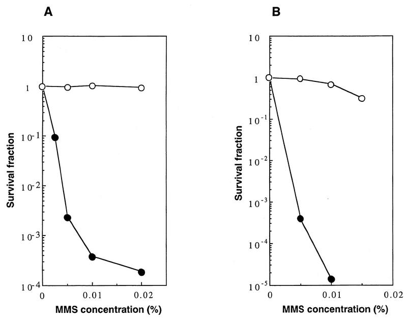
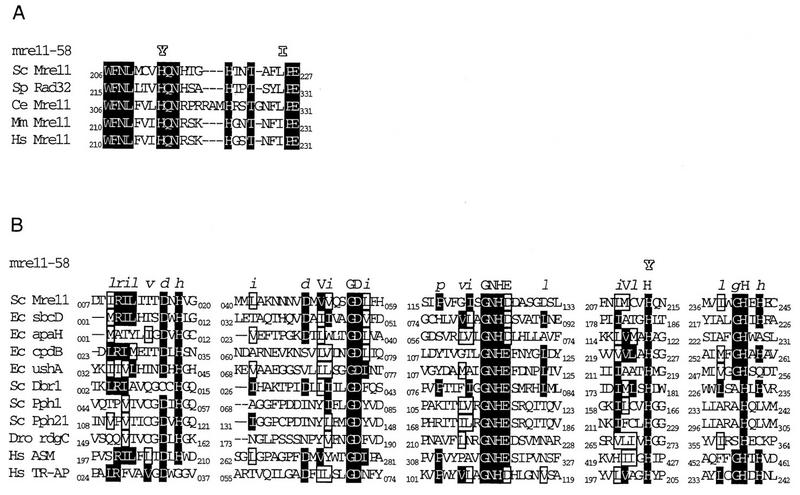
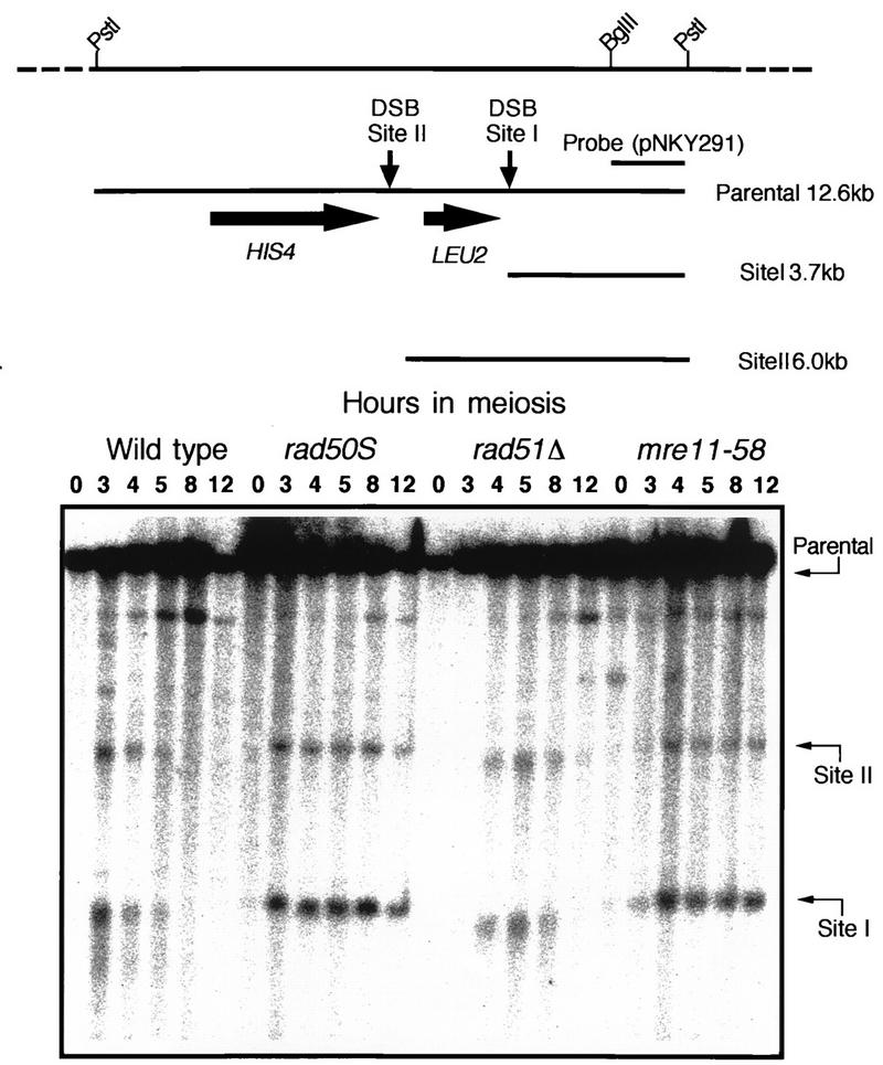
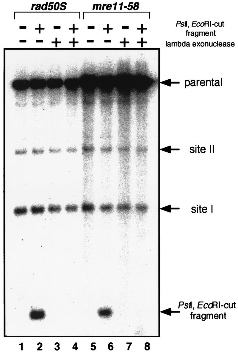
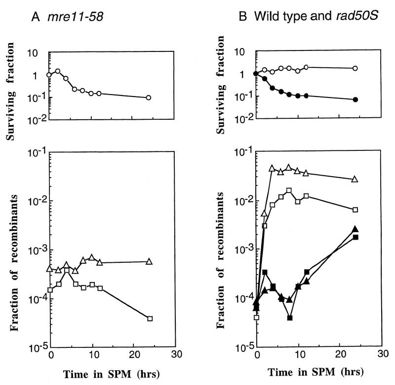
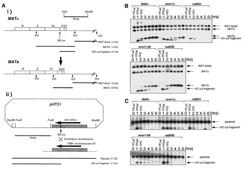
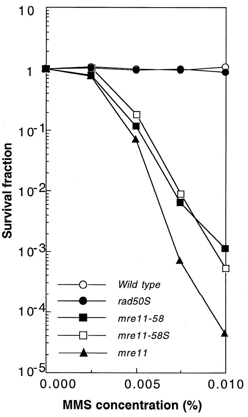
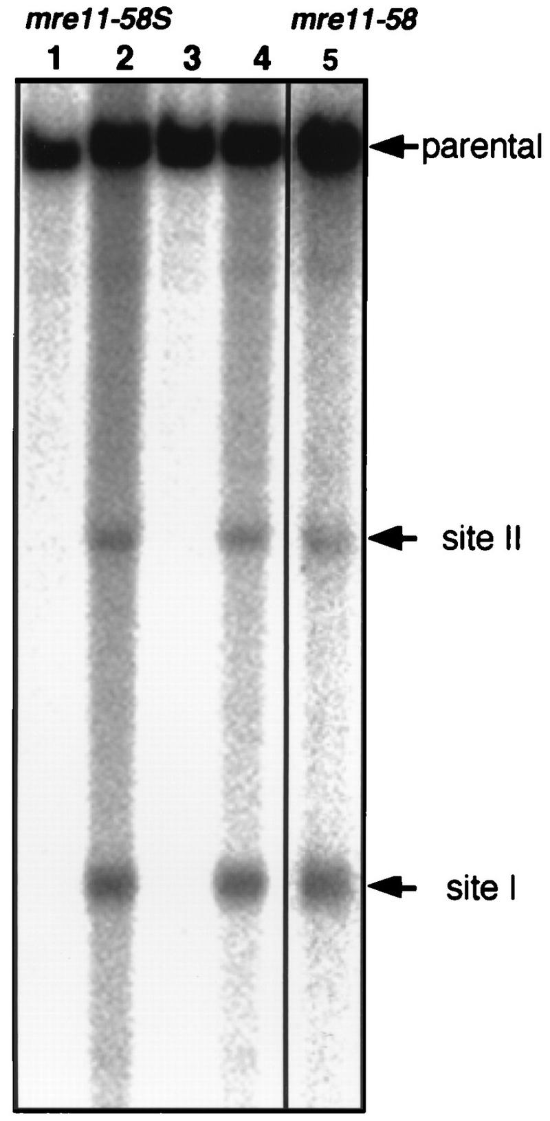
Using complementation tests and nucleotide sequencing, we showed that the rad58-4 mutation was an allele of the MRE11 gene and have renamed the mutation mre11-58. Two amino acid changes from the wild-type sequence were identified; one is located at a conserved site of a phosphodiesterase motif, and the other is a homologous amino acid change at a nonconserved site. Unlike mre11 null mutations, the mre11-58 mutation allowed meiosis-specific double-strand DNA breaks (DSBs) to form at recombination hot spots but failed to process those breaks. DSB ends of this mutant were resistant to lambda exonuclease treatment. These phenotypes are similar to those of rad50S mutants. In contrast to rad50S, however, mre11-58 was highly sensitive to methyl methanesulfonate treatment. DSB end processing induced by HO endonuclease was suppressed in both mre11-58 and the mre11 disruption mutant. We constructed a new mre11 mutant that contains only the phosphodiesterase motif mutation of the Mre11-58 protein and named it mre11-58S. This mutant showed the same phenotypes observed in mre11-58, suggesting that the phosphodiesterase consensus sequence is important for nucleolytic processing of DSB ends during both mitosis and meiosis.


Figures

FIG. 1
FIG. 1
(A) Lack of complementation for MMS sensitivity between the rad58-4 mutant and the mre11 disruptant. Diploids obtained by mating rad58-4 (20B-D3142) with an mre11 (HTY231) (solid circles) strain and with a wild-type strain (CG379) (open circles) were grown in yeast-peptone-dextrose (YPD) liquid medium, diluted appropriately, and spread on YPD plates containing various concentrations of MMS, and the plates were incubated at 30°C for 3 days. Colonies growing on each plate were counted. (B) Complementation of the repair defect of the rad58-4 mutant with the cloned MRE11 gene. The rad58-4 mutant (HTY553) carrying the MRE11 gene on a single-copy vector, pHT62 (open circles), or the vector alone, YCp50 (solid circles), was tested for MMS sensitivity. Each transformant was grown in SD-URA, diluted appropriately, and spread on SD-URA plates containing various concentrations of MMS.
FIG. 2
FIG. 2
(A) Comparison of amino acid sequences of Mre11 homologs around the mutant sites of mre11-58; (B) comparison of amino acid sequences of various phosphoesterases. The motif sequence is shown at the top (from reference 3a). Shading, identical amino acids; boxes, similar amino acids. Sc, S. cerevisiae; Sp, Schizosaccharomyces pombe; Ec, E. coli; Mm, Mus musculus; Hs, Homo sapiens; Dro, Drosophila; Ce, Caenorhabditis elegans. Sc Mre11, 1513065; Ec sbcD, 1586770; Ec apaH, 1003022; Ec cpdB, 67263; Ec ushA, 137173; Sc Dbr1, 171382; Sc Pph1, 319859; Sc Pph21, 4203; Dro rdcC, 158238; Hs ASM, 179095; and Hs TR-AP, 130722. Numbers indicate NCBI sequence identity.
FIG. 3
FIG. 3
DSB formation at the HIS4LEU2 locus in mre11-58. The physical map of the HIS4LEU2 locus is shown in the upper panel. Horizontal arrows indicate the major two sites of DSBs, called sites I and II. Genomic DNAs were prepared from cells collected at various times after entering meiosis and cut with PstI, and the fragments corresponding to the parent (12.6 kb), site I (3.7 kb), and site II (6.0 kb) were detected by Southern blotting with the 1.5-kb PstI-EcoRI fragment of pNKY291, which contains the PstI-BglII fragment shown in the upper panel, as a probe. The lower panel represents the images of DSBs at the HIS4LEU2 locus, and the numbers above the images indicate the times (in hours) after entry into meiosis. Strains: HTY525 (wild type), HTY603 (rad50S), HTY533 (rad51Δ), and HTY703 (mre11-58).
FIG. 4
FIG. 4
Lambda exonuclease digestion of mre11-58 meiotic DNA. Genomic DNAs from meiotic cells were prepared when DSBs were fully formed and were treated with lambda exonuclease. After inactivating exonuclease, genomic DNAs were cut with PstI and subjected to Southern hybridization after agarose gel electrophoresis as shown in Fig. 3. A DNA fragment cut produced by PstI EcoRI double digestion was added before exonuclease treatment as an internal control. Lanes 1 to 4, rad50S DNA; lanes 5 to 8, mre11-58 DNA.
FIG. 5
FIG. 5
Meiotic recombination deficiency of mre11-58. Diploid strains were introduced synchronously into meiosis, as described in Materials and Methods. Surviving fractions were obtained by dividing the numbers of CFU on complete medium (MYPD) after each incubation time by those at 0 h. Recombinant fractions at two sets of heteroalleles, his4X/his4B and arg4-nsp/arg4-bgl, were obtained. Each value plotted is the ratio of the number of HIS+ or ARG+ CFU to the total number of CFU at each time point. (A) Open circles, mre11-58 (HTY703) surviving fractions; open triangles, ARG+ fractions; open squares, HIS+ fractions; (B) Open symbols, wild type (HTY525); solid symbols, rad50S (HTY603); circles, surviving fractions; squares, HIS+ fractions; triangles, ARG+ fractions. SPM, sporulation medium.
FIG. 6
FIG. 6
Kinetics of repair of HO endonuclease-induced DSBs. Haploid strains were transformed with pHT51 carrying the HO gene under the control of the galactose promoter. HO endonuclease expression was induced by suspending the cells in a galactose-containing medium for 1 h and was then repressed by suspending the cells in a glucose-containing medium, and the DSB repair kinetics were observed as described in Materials and Methods. (A) Physical maps used for DSB repair detection. (i) The MATα locus. The indicated probe (a 1.0-kb NdeI-HindIII fragment) was used to detect the 2.2-kb MAT distal, 1.8-kb MATα, and 0.7-kb HO-cut fragments after StyI digestion. HO endonuclease produces a 0.7-kb HO-cut fragment from the 1.8-kb MATα fragment, and this 0.7-kb fragment is replaced by a 0.9-kb MATa fragment when the mating type switches from MATα to MATa. (ii) pHT51. The indicated probe (a 2.4-kb PvuII-PvuII fragment obtained from pJH283) was used to detect 5.7-kb parental and 3.3-kb HO-cut fragments after HindIII digestion. Stippled and solid rectangles, the homologous regions of pHT51 and the genome and the 35-mer HO recognition sequence, respectively. (B) Kinetics of DSB repair at the MATα locus. (C) Kinetics of DSB repair on pHT51. Strains: HTY925 (rad+), HTY927 (mre11Δ), HTY929 (rad52Δ), HTY931 (mre11-58), and HTY933 (rad50S).
FIG. 7
FIG. 7
The mre11-58S mutant is as sensitive to MMS as mre11-58. Haploid strains HTY1114 (mre11-58S), HTY693 (mre11-58), HTY1075 (mre11 null), and NKY1003 (rad50S), and a wild-type strain (HTY464) were grown in YPD liquid medium, diluted appropriately, spread on MYPD plates containing various concentrations of MMS, the plates were incubated at 30°C for 5 days, and the numbers of colonies growing on each plate were counted.
FIG. 8
FIG. 8
DSB formation at the HIS4LEU2 locus in mre11-58S. Horizontal arrows, major two sites of DSBs, called sites I and II. Genomic DNAs from mre11-58S (HTY1115) and mre11-58 (HTY703) were prepared from cells collected at 0 and 8 h (only 8 h for mre11-58) after entering meiosis, and Southern blotting was performed to detect fragments corresponding to the parent (12.6 kb), site I (3.7 kb), and site II (6.0 kb) as described in the legend to Fig. 3. Lanes 1 and 3, 0-h sample; lanes 2 and 4, 8-h sample. Lanes 1 and 2 and 3 and 4 are the results of independent clones of HTY1115. Lane 5, mre11-58 (HTY703).
FIG. 9
FIG. 9
Lambda exonuclease digestion of mre11-58S meiotic DNA. Genomic DNA of the mre11-58S strain was isolated from meiotic cells at 8 h after entering meiosis when DSBs were fully formed, and that of the wild type was isolated from mitotic cells and from cells at 3 h after entering meiosis. The genomic DNAs were treated with lambda exonuclease. After inactivation of exonuclease, genomic DNAs were digested with PstI and subjected to Southern hybridization after agarose gel electrophoresis as shown in Fig. 3. DNA fragments cut with PstI and EcoRI double digestion were added before exonuclease treatment as an internal control. Lanes 1 to 4, wild-type DNA (0 h); lanes 5 to 8, wild-type DNA (3 h); lanes 9 to 12, mre11-58S DNA (8 h). Strains used were HTY525 (wild type), and HTY1115 (mre11-58S).

Similar articles

Cited by

References

    1. Ajimura M, Leem S-H, Ogawa H. Identification of new genes required for meiotic recombination in Saccharomyces cerevisiae. Genetics. 1993;133:51–66. - PMC - PubMed
    1. Alani E, Cao L, Kleckner N. A method for gene disruption that allows repeated use of URA3 selection in the construction of multiply disrupted yeast strains. Genetics. 1987;116:541–545. - PMC - PubMed
    1. Alani E, Padmore R, Kleckner N. Analysis of wild-type and rad50 mutants of yeast suggests an intimate relationship between meiotic chromosome synapsis and recombination. Cell. 1990;61:419–436. - PubMed
    1. Baum B. Mre11 and S. pombe Rad32 are phosphoesterases. Yeast Update Newsl. 1995;3.7:1.
    1. Bergerat A, de Massy B, Gadelle D, Varoutas P-C, Nicolas A, Forterre P. An atypical topoisomerase II from archaea with implications for meioric recombination. Nature. 1997;386:414–417. - PubMed

Publication types

LinkOut - more resources