. 1997 Dec 29;139(7):1635-43.
doi: 10.1083/jcb.139.7.1635.

Extracellular ATP activates transcription factor NF-kappaB through the P2Z purinoreceptor by selectively targeting NF-kappaB p65

Affiliations

Extracellular ATP activates transcription factor NF-kappaB through the P2Z purinoreceptor by selectively targeting NF-kappaB p65

D Ferrari et al. J Cell Biol. .

Abstract

Cells of the macrophage lineage express a peculiar surface receptor for extracellular ATP, designated P2Z/P2X7 purinergic receptor, that induces pore formation and collapse of the plasma membrane potential. Although the function of the P2Z receptor is largely unknown, accumulating evidence implicates its role in cell signaling and immune reactions. Here, we investigated the effect of P2Z receptor ligation on the activation of NF-kappaB, a transcription factor controlling cytokine expression and apoptosis. Exposure of microglial cells to ATP but not other nucleotides resulted in potent NF-kappaB activation. This effect was specifically mediated by the P2Z receptor, because selective receptor antagonists prevented NF-kappaB activation. NF-kappaB activation required reactive oxygen intermediates and proteases of the caspase family, because it was abolished by antioxidants and specific protease inhibitors. The subunit composition of the ATP-induced NF- kappaB-DNA complex was rather unusual. Whereas exposure to LPS-induced prototypical NF-kappaB p50 homo- and p65 (RelA)/p50 heterodimers, ATP stimulation resulted in the sole appearance of a p65 homodimer. This is the first demonstration that a certain stimulus activates a particular NF-kappaB subunit. Because different NF-kappaB complexes exhibit distinct transcriptional and DNA-binding activities, ATP may control the expression of a subset of NF-kappaB target genes distinct from those activated by classical proinflammatory mediators.


Figures
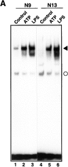
Figure 1
Figure 1
Induction of NF-κB activation by ATP. (A) The effect of ATP on NF-κB activation in N9 and N13 microglial cells. N9 (lanes 1–3) or N13 cells (lanes 4–6) were either left untreated or treated with 3 mM ATP or 100 ng/ml LPS. After 3 h total cell extracts were prepared and analyzed by EMSA using a 32P-labeled oligonucleotide containing a high affinity κB-binding motif. The NF-κB–DNA complex is indicated by a filled arrowhead. A faster migrating non-specific complex is marked by a circle. (B) Dose dependency of NF-κB activation in response to ATP or LPS. N9 cells were treated for 3 h with the indicated concentrations of ATP or LPS and analyzed for NF-κB activation. (C) The effect of ATP and LPS on the DNA-binding activity of AP-1. The same cellular extracts and protein amounts as in Fig. 1 A were analyzed with an AP-1 specific oligonucleotide in an EMSA.
Figure 1
Figure 1
Induction of NF-κB activation by ATP. (A) The effect of ATP on NF-κB activation in N9 and N13 microglial cells. N9 (lanes 1–3) or N13 cells (lanes 4–6) were either left untreated or treated with 3 mM ATP or 100 ng/ml LPS. After 3 h total cell extracts were prepared and analyzed by EMSA using a 32P-labeled oligonucleotide containing a high affinity κB-binding motif. The NF-κB–DNA complex is indicated by a filled arrowhead. A faster migrating non-specific complex is marked by a circle. (B) Dose dependency of NF-κB activation in response to ATP or LPS. N9 cells were treated for 3 h with the indicated concentrations of ATP or LPS and analyzed for NF-κB activation. (C) The effect of ATP and LPS on the DNA-binding activity of AP-1. The same cellular extracts and protein amounts as in Fig. 1 A were analyzed with an AP-1 specific oligonucleotide in an EMSA.
Figure 2
Figure 2
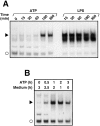
Time dependence of NF-κB activation in response to ATP and LPS. (A) Kinetics of NF-κB activation: total extracts of cells treated for the indicated periods of time with 3 mM ATP or 100 ng/ml LPS were prepared and analyzed by EMSA. Only a section of the autoradiogram is shown. The filled arrowhead indicates the position of the inducible NF-κB–DNA complex; the open circle denotes a non-specific complex. (B) Pulse-chase experiment: N9 cells were stimulated for different times (0.5–3 h) with 3 mM ATP and further incubated for the indicated time periods in culture medium without ATP. After a total incubation time of 3 h, cells were harvested and analyzed for NF-κB DNA-binding activity.
Figure 3
Figure 3
Subunit composition of the induced NF-κB–DNA complexes after treatment with LPS (A) or ATP (B). Total cell extracts of non-stimulated (lanes 1) or stimulated (lanes 2) N9 cells were prepared and either left untreated (lanes 1 and 2) or incubated with specific antibodies against the NF-κB subunits p50 (lanes 3), p65 (lanes 4), c-Rel (lanes 5) and RelB (lanes 6). Lanes 7 denote an extract of ATP-treated cells incubated with recombinant IκB-α that releases NF-κB–specific complexes from bound DNA. Only sections of the autoradiograms are shown.
Figure 4
Figure 4
Specificity of ATP-induced NF-κB activation. (A) The effect of different nucleotides on NF-κB activation. N9 cells were treated with 3 mM ATP, ADP, UTP, CTP, or GTP. After 3 h, cell extracts were prepared and analyzed by EMSA. (B) Induction of NF-κB activation by ATP or its pharmacological analogues ATPγS (3 mM) and benzoylbenzoyl-ATP (BzATP, 1 mM). (C) Inhibition of ATP-induced NF-κB activation by oxidized ATP (oATP). Cells were either left untreated or stimulated with ATP following a 2 h pretreatment with the indicated concentrations of oxidized ATP. (D) Lack of ATP-induced NF-κB activation in the P2Z receptor-deficient N9 derivative clones N9R14 and N9R17. Parental N9 cells and the ATP-resistant N9R14 and N9R17 were either left untreated or incubated for 3 h in the presence of 3 mM ATP or LPS (100 ng/ml).
Figure 5
Figure 5
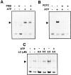
The effect of the LPS inhibitor polymyxin B (A), the antioxidant PDTC (B), and the proteasome inhibitor lactacystin (C) on NF-κB activation in response to ATP. N9 cells were pretreated for 30 min with polymyxin B (PMB, 10 μg/ml; A) or 50 μM PDTC (B), or for 90 min with the indicated concentrations of lactacystin (LC; C), followed by the activation with 3 mM ATP for 3 h. Total cell extracts were then prepared and analyzed by EMSA for NF-κB DNA-binding activity. Only sections of the autoradiograms are shown.
Figure 6
Figure 6
Involvement of ICE proteases in ATP-induced NF-κB activation. (A) The effect of the ICE tetrapeptide inhibitor YVAD-CHO (YVAD) on NF-κB activation. Cells were pretreated for 30 min with the indicated concentrations of YVAD and then stimulated with 3 mM ATP for 3 h. (B) Proteolytic processing of ICE by ATP treatment. N9 cells were incubated for the indicated times with 3 mM ATP. Total cell extracts were then prepared and analyzed by SDS-PAGE and immunoblotting. Proteolytic cleavage of the ICE precursor to the active protease was monitored using an anti-ICE specific antiserum that detects pro-ICE and the p20 proteolytic cleavage product. (C) Effect of ATP on cell viability. N9 cells were treated with 3 mM ATP. After the indicated time points, LDH released from dead cells was measured in the supernatants and calculated as the percentage of total cellular LDH activity from an untreated sample.
Figure 7
Figure 7
Lack of involvement of IL-1β, TNF-α, or soluble factors in ATP-induced NF-κB activation. (A) Effect of IL-1RA. N9 cells were either left untreated or incubated with 3 mM ATP for 3 h in the presence of the indicated concentrations of IL-1RA. (B) Neutralizing anti-TNF-α does not interfere with ATP-induced NF-κB activation. N9 cells were either left untreated or incubated with 3 mM ATP for 3 h in the presence of the indicated concentrations of rabbit anti–mouse TNF antibodies. (C) Secretion of soluble proteins is not required for NF-κB activation. In lanes 1–6, cells were incubated for the indicated times with 3 mM ATP. In lanes 10–13, cells were treated for 0.5–3 h with ATP. Supernatants (SN) of these cultures were then transferred to untreated N9 cells which were analyzed for NF-κB activation after the indicated incubation periods. In lane 9, N9 cells were incubated for 4 h with a culture supernatant from control cells.
Figure 8
Figure 8
Effect of ATP on NF-κB target gene expression. N9 cells were transfected by electroporation with an NF-κB-controlled luciferase construct. 24 h after transfection, cells were either left untreated (Control) or stimulated with the indicated amounts of ATP. In some samples, cells were pretreated with 600 μM oATP before ATP stimulation. Cells were harvested after an additional 4 h and assayed for luciferase activity. Mean values ±SD of NF-κB activity given as relative light units from triplicate experiments are shown.

Similar articles

Cited by

References

    1. Baeuerle PA, Henkel T. Function and activiation of NF-κB in the immune system. Annu Rev Immunol. 1994;12:141–179. - PubMed
    1. Baeuerle PA, Baltimore D. NF-κB: ten years after. Cell. 1996;87:13–20. - PubMed
    1. Baichwal VR, Baeuerle PA. Activate NF-κB or die? . Curr Biol. 1997;7:R94–R96. - PubMed
    1. Baldwin AS. The NF-κB and IκB proteins: new discoveries and insights. Annu Rev Immunol. 1996;242:540–546. - PubMed
    1. Bauer MKA, Lieb K, Schulze-Osthoff K, Bauer J, Gebicke-Haerter PJ, Fiebich BI. Expression and regulation of cyclooxygenase-2 in rat microglia. Eur J Biochem. 1997;243:726–731. - PubMed

Publication types

MeSH terms