

Accumulation of major histocompatibility complex class II molecules in mast cell secretory granules and their release upon degranulation
- PMID: 9398681
- PMCID: PMC25733
- DOI: 10.1091/mbc.8.12.2631
Accumulation of major histocompatibility complex class II molecules in mast cell secretory granules and their release upon degranulation
Abstract
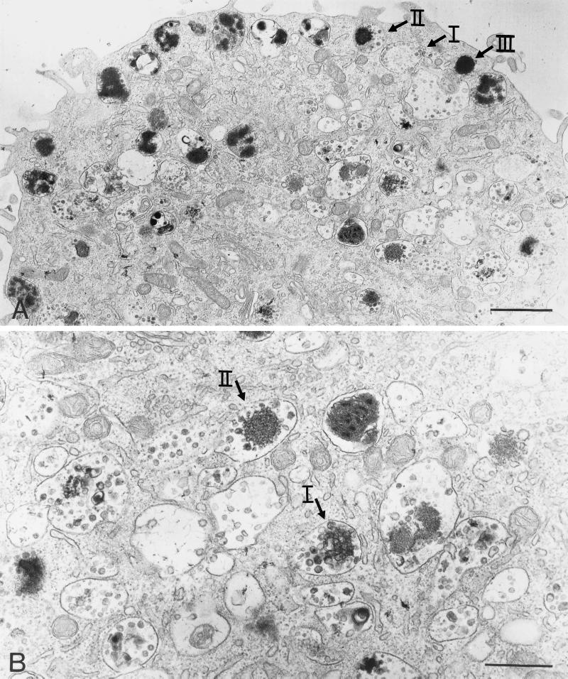
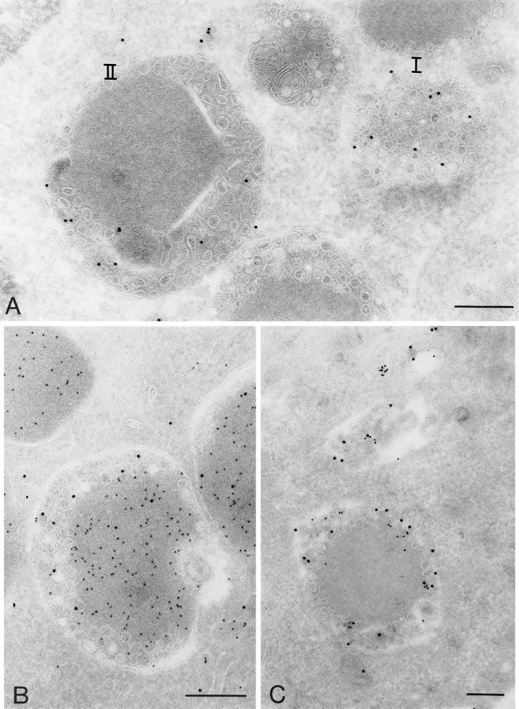
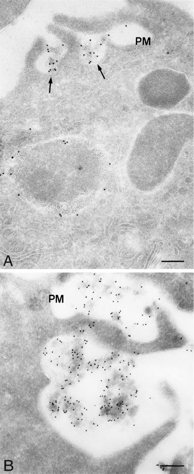
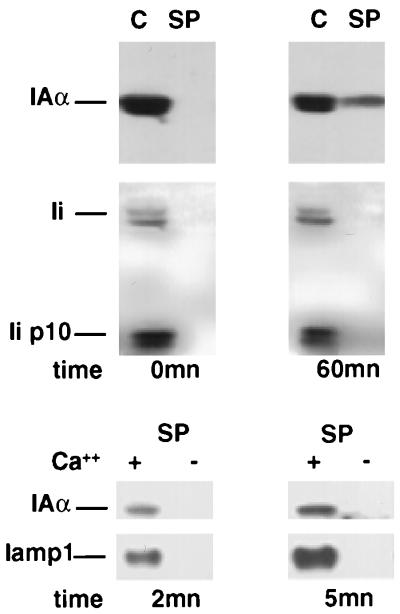
To investigate the relationship between major histocompatibility complex (MHC) class II compartments, secretory granules, and secretory lysosomes, we analyzed the localization and fate of MHC class II molecules in mast cells. In bone marrow-derived mast cells, the bulk of MHC class II molecules is contained in two distinct compartments, with features of both lysosomal compartments and secretory granules defined by their protein content and their accessibility to endocytic tracers. Type I granules display internal membrane vesicles and are accessed by exogenous molecules after a time lag of 20 min; type II granules are reached by the endocytic tracer later and possess a serotonin-rich electron-dense core surrounded by a multivesicular domain. In these type I and type II granules, MHC class II molecules, mannose-6-phosphate receptors and lysosomal membrane proteins (lamp1 and lamp2) localize to small intralumenal vesicles. These 60-80-nm vesicles are released along with inflammatory mediators during mast cell degranulation triggered by IgE-antigen complexes. These observations emphasize the intimate connection between the endocytic and secretory pathways in cells of the hematopoietic lineage which allows regulated secretion of the contents of secretory lysosomes, including membrane proteins associated with small vesicles.
Figures


Similar articles
-
Secretory granules of mast cells accumulate mature and immature MHC class II molecules.J Cell Sci. 2001 Jan;114(Pt 2):323-34. doi: 10.1242/jcs.114.2.323. J Cell Sci. 2001. PMID: 11148134
-
The lysosomal protease cathepsin D is efficiently sorted to and secreted from regulated secretory compartments in the rat basophilic/mast cell line RBL.J Cell Sci. 2000 Sep;113 ( Pt 18):3289-98. doi: 10.1242/jcs.113.18.3289. J Cell Sci. 2000. PMID: 10954426
-
Major histocompatibility complex class II compartments in human B lymphoblastoid cells are distinct from early endosomes.J Exp Med. 1995 Aug 1;182(2):325-34. doi: 10.1084/jem.182.2.325. J Exp Med. 1995. PMID: 7629497 Free PMC article.
-
Mast cell secretory granules: armed for battle.Nat Rev Immunol. 2014 Jul;14(7):478-94. doi: 10.1038/nri3690. Epub 2014 Jun 6. Nat Rev Immunol. 2014. PMID: 24903914 Review.
-
Biogenesis of secretory granules. Implications arising from the immature secretory granule in the regulated pathway of secretion.FEBS Lett. 1991 Jul 22;285(2):220-4. doi: 10.1016/0014-5793(91)80805-d. FEBS Lett. 1991. PMID: 1906810 Review.
Cited by
-
Conformational Alterations of the Cell Surface of Monomeric and Dimeric β2m-Free HLA-I (Proto-HLA) May Enable Novel Immune Functions in Health and Disease.Curr Issues Mol Biol. 2024 Jul 4;46(7):6961-6985. doi: 10.3390/cimb46070416. Curr Issues Mol Biol. 2024. PMID: 39057057 Free PMC article. Review.
-
Myeloma as a model for the process of metastasis: implications for therapy.Blood. 2012 Jul 5;120(1):20-30. doi: 10.1182/blood-2012-01-379024. Epub 2012 Apr 24. Blood. 2012. PMID: 22535658 Free PMC article.
-
The roles of tumor-derived exosomes in cancer pathogenesis.Clin Dev Immunol. 2011;2011:842849. doi: 10.1155/2011/842849. Epub 2011 Nov 30. Clin Dev Immunol. 2011. PMID: 22190973 Free PMC article. Review.
-
Exosome-Encapsulated miR-31, miR-192, and miR-375 Serve as Clinical Biomarkers of Gastric Cancer.J Oncol. 2023 Feb 16;2023:7335456. doi: 10.1155/2023/7335456. eCollection 2023. J Oncol. 2023. PMID: 36844871 Free PMC article.
-
Myrrh and Chamomile Flower Extract Inhibit Mediator Release from IgE-stimulated Mast-Cell-Like RBL-2H3 Cells.Plants (Basel). 2022 Dec 8;11(24):3422. doi: 10.3390/plants11243422. Plants (Basel). 2022. PMID: 36559534 Free PMC article.
References
-
- Amigorena S, Drake JR, Webster P, Mellman I. Transient accumulation of new class II MHC molecules in a novel endocytic compartment in B lymphocytes. Nature. 1994;369:113–120. - PubMed
-
- Bentfeld-Barker ME, Bainton DF. Cytochemical localization of arylsulfatase B in rat basophils and mast cells. J Histochem Cytochem. 1980;28:1055–1061. - PubMed
-
- Bhattacharya A, Dorf ME, Springer TA. A shared alloantigenic determinant on Ia antigens encoded by the I-A and I-E subregions: evidence for I region gene duplication. J Immunol. 1981;127:2488–2495. - PubMed
Publication types
MeSH terms
Substances
LinkOut - more resources
Full Text Sources
Other Literature Sources
Research Materials
Miscellaneous
