. 1997 Dec 15;186(12):2013-21.
doi: 10.1084/jem.186.12.2013.

Syk tyrosine kinase is required for the positive selection of immature B cells into the recirculating B cell pool

Affiliations

Syk tyrosine kinase is required for the positive selection of immature B cells into the recirculating B cell pool

M Turner et al. J Exp Med. .

Abstract

The tyrosine kinase Syk has been implicated as a key signal transducer from the B cell antigen receptor (BCR). We show here that mutation of the Syk gene completely blocks the maturation of immature B cells into recirculating cells and stops their entry into B cell follicles. Furthermore, using radiation chimeras we demonstrate that this developmental block is due to the absence of Syk in the B cells themselves. Syk-deficient B cells are shown to have the life span of normal immature B cells. If this is extended by over-expression of Bcl-2, they accumulate in the T zone and red pulp of the spleen in increased numbers, but still fail to mature to become recirculating follicular B cells. Despite this defect in maturation, Syk-deficient B cells were seen to give rise to switched as well as nonswitched splenic plasma cells. Normally only a proportion of immature B cells is recruited into the recirculating pool. Our results suggest that Syk transduces a BCR signal that is absolutely required for the positive selection of immature B cells into the recirculating B cell pool.


Figures

Figure 1
Figure 1
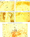
Immunohistochemistry of splenic B cells in an adult Syk −/− mouse. Sections were stained with antibodies to IgD or IgM; these were revealed with peroxidase (brown) or with antibodies to CD3 or B220 revealed with alkaline phosphatase (blue) as indicated. Sections labeled “none” were stained only with the peroxidase-coupled secondary antibody to reveal endogenous peroxidase activity. (a–c) Serial sections from the spleen of an exceptional Syk −/− mouse that survived to adulthood; small numbers of B220+ cells are found in the outer T zone (T) and a proportion of these are IgM+ (arrows in b), but none are IgD+ nor have they entered the follicles. The B220+IgM cells in this spleen were shown to be CD3 (data not shown). Note in c the brown staining of eosinophils which contain endogenous peroxidase activity; the same eosinophils are seen staining brown in a and b. (d–f ) Serial sections from a heterozygous littermate showing the development of normal follicles (F), which contain IgM+IgD+ recirculating B cells; IgM+IgD plasma cells are present in extra-follicular foci (E) in this spleen. Scale: black bar represents 100 μm. Analysis of a second Syk −/− mouse gave similar results.
Figure 2
Figure 2
Immature Syk −/− B cells migrate to the spleen but do not mature into follicular B cells. Immunohistochemistry of peripheral B cells in radiation chimeras whose nature is shown to the left above each micrograph; i.e., whether the reconstituting livers were Syk +/− or Syk −/− and if they contained the 3-83 or Bcl-2 transgenes (29, 33). All fetal liver donors were of the IgHb immunoglobulin allotype; all hosts were IgHa. 20 Syk −/− nontransgenic chimeras and at least three chimeras from each group of transgenic donors were analyzed. The specificity of the antibodies used for staining the sections is shown to the right above each micrograph; the molecule stained gold by immunoperoxidase is shown first, the one stained blue by immunoalkaline phosphatase is shown second and, where used, BrdU staining is in red. Cells double positive for gold and blue appear dark brown. Scale: black bar represents 100 μm. (a) Donor IgMb+ Syk −/− immature B cells (small arrows) are present in the T zone (T) and red pulp (R) of the spleen, but not in the partially filled follicles (F), which contain mature IgD+ (gold) cells of host origin. Donor IgMb+ Syk −/− plasma cells in the red pulp are indicated by large arrows. (b) Section through a lymph node from a similar chimera to that in a. Well-developed primary follicles are present but no donor IgMb+ Syk −/− B cells were detected in this or in any lymph node from similar chimeras. (c) Spleen section from a Syk +/−/3-83 chimera that, like the ones shown in d and e, had been given BrdU for 12 h, 8 d before the spleen was taken. The follicular mantles are filled with mature 3-83+IgD+ (dark brown) donor B cells; occasional cells (arrowed) have a red-stained nucleus, indicating that they had taken up BrdU during the 12-h pulse 8 d previously. Some 3-83+IgD (blue) donor B cells can be seen in the outer T zone and red pulp; some of these may be immature B cells, but none are BrdU+. The germinal center (G) is filled with donor-derived 3-83IgHb+ B cells (data not shown) that arise from occasional rearrangement of endogenous Ig genes in the 3-83 transgenics. (d) Spleen section from a Syk −/−/3-83 chimera. Small numbers of 3-83+IgD donor B cells (blue and arrowed) can be seen in the outer T zone and red pulp; none of these are BrdU+. As in a the follicles exclusively contain host IgD+ cells (gold) but no donor B cells. (e) Spleen section from a Syk −/−/3-83/Bcl-2Tg chimera. More donor B cells are present in the T zone and red pulp than in the chimera in d not carrying Bcl-2Tg. Nevertheless, there were no donor B cells in follicles. Some of the donor 3-83+ B cells had BrdU in their nuclei (arrowed and red). These must have been in cell cycle as pre-B cells 8 d previously, indicating an extended life span for these immature B cells.
Figure 3
Figure 3
Positive selection of immature B cells. Flow cytometric analysis of cells from radiation chimeras reconstituted with liver cells from Syk +/−/3-83 (Syk+/−), Syk −/−/3-83 (Syk−/−), and Syk −/−/3-83/Bcl-2Tg (Syk−/− + Bcl-2) fetuses or from an adult Syk +/−/3-83/Bcl-2Tg (Syk+/− + Bcl-2) mouse. In separate experiments chimeras made with Syk +/−/3-83/Bcl-2Tg fetal liver gave essentially the same results as presented in this figure (data not shown). Chimeras made with Syk +/+/3-83 fetal liver gave results indistinguishable from Syk +/−/3-83 chimeras (data not shown). (a) The first column from the left shows the expression of B220 and the transgenic BCR (3-83) on bone marrow cells; B220+3-83+ cells are boxed and their fraction as a percentage of all lymphoid cells is shown. The second column shows the expression of IgMa and IgDa by B220+3-83+ cells with gating to show immature (IgM+IgD), transitional (IgM+IgDlow), and mature (IgM+IgDhigh) B cells; the percentage for each gated population is expressed as a fraction of all lymphocytes. (b) The first column shows the gating on immature (B220low3-83low) cells that was used to generate the histograms of CD43 expression on B220+3-83+ bone marrow cells shown in the second column; percentages represent the fraction of gated immature B220low3-83low cells that are CD43. (c) Expression of B220 and 3-83 in the spleen; B220+3-83+ cells are boxed and their fraction as a percentage of all lymphoid cells is shown. (d) Mean numbers ± SEM of B220+3-83+ cells in the bone marrow (per femur) and spleen are shown. In the marrow, these have been subdivided into immature (IgM+IgD), transitional (IgM+IgDlow) and mature (IgM+IgDhigh) B cells using the gating shown in a. Data in (a–c) are representative of at least six mice of each genotype. Absolute cell numbers in the marrow were determined using eight Syk +/−/3-83 mice, six Syk −/−/3-83 mice, three Syk +/−/3-83/Bcl-2Tg mice, and four Syk −/−/3-83/Bcl-2Tg mice; data for the spleen cell numbers was determined using four Syk +/−/3-83 mice, four Syk −/−/3-83 mice, three Syk +/−/3-83/Bcl-2Tg mice, and two Syk −/−/3-83/Bcl-2Tg mice.
Figure 3
Figure 3
Positive selection of immature B cells. Flow cytometric analysis of cells from radiation chimeras reconstituted with liver cells from Syk +/−/3-83 (Syk+/−), Syk −/−/3-83 (Syk−/−), and Syk −/−/3-83/Bcl-2Tg (Syk−/− + Bcl-2) fetuses or from an adult Syk +/−/3-83/Bcl-2Tg (Syk+/− + Bcl-2) mouse. In separate experiments chimeras made with Syk +/−/3-83/Bcl-2Tg fetal liver gave essentially the same results as presented in this figure (data not shown). Chimeras made with Syk +/+/3-83 fetal liver gave results indistinguishable from Syk +/−/3-83 chimeras (data not shown). (a) The first column from the left shows the expression of B220 and the transgenic BCR (3-83) on bone marrow cells; B220+3-83+ cells are boxed and their fraction as a percentage of all lymphoid cells is shown. The second column shows the expression of IgMa and IgDa by B220+3-83+ cells with gating to show immature (IgM+IgD), transitional (IgM+IgDlow), and mature (IgM+IgDhigh) B cells; the percentage for each gated population is expressed as a fraction of all lymphocytes. (b) The first column shows the gating on immature (B220low3-83low) cells that was used to generate the histograms of CD43 expression on B220+3-83+ bone marrow cells shown in the second column; percentages represent the fraction of gated immature B220low3-83low cells that are CD43. (c) Expression of B220 and 3-83 in the spleen; B220+3-83+ cells are boxed and their fraction as a percentage of all lymphoid cells is shown. (d) Mean numbers ± SEM of B220+3-83+ cells in the bone marrow (per femur) and spleen are shown. In the marrow, these have been subdivided into immature (IgM+IgD), transitional (IgM+IgDlow) and mature (IgM+IgDhigh) B cells using the gating shown in a. Data in (a–c) are representative of at least six mice of each genotype. Absolute cell numbers in the marrow were determined using eight Syk +/−/3-83 mice, six Syk −/−/3-83 mice, three Syk +/−/3-83/Bcl-2Tg mice, and four Syk −/−/3-83/Bcl-2Tg mice; data for the spleen cell numbers was determined using four Syk +/−/3-83 mice, four Syk −/−/3-83 mice, three Syk +/−/3-83/Bcl-2Tg mice, and two Syk −/−/3-83/Bcl-2Tg mice.
Figure 4
Figure 4
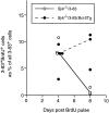
Life span of Syk −/− immature B cells is increased by expression of Bcl-2. Chimeric mice reconstituted with Syk −/−/3-83 fetal livers with and without Bcl-2Tg were given a 12-h pulse of BrdU. Graph shows the percentage of splenic 3-83+ B cells that were also labeled with BrdU either 4 or 8 d after the BrdU pulse. At each time point, spleens from three or four mice were analyzed by quantitative immunohistology as previously described (4). The lines are drawn between median values. The number of cells enumerated was 168–263 cells/spleen in the Syk −/−/ 3-83 chimeras and 673–994 cells/spleen in the Syk −/−/3-83/Bcl-2Tg chimeras.

Similar articles

Cited by

References

    1. Rajewsky K. Clonal selection and learning in the antibody system. Nature. 1996;381:751–758. - PubMed
    1. Bazin H, Platteau B, MacLennan ICM, Johnson GD. B-cell production and differentiation in adult rats. Immunology. 1985;54:79–88. - PMC - PubMed
    1. Lortan JE, Roobottom CA, Oldfield S, MacLennan ICM. Newly produced virgin B-cells migrate to secondary lymphoid organs but their capacity to enter follicles is restricted. Eur J Immunol. 1987;17:1311–1316. - PubMed
    1. Chan EY-T, MacLennan ICM. Only a small proportion of splenic B cells in adults are short-lived virgin cells. Eur J Immunol. 1993;23:357–363. - PubMed
    1. Gray D. Population kinetics of rat peripheral B cells. J Exp Med. 1988;167:805–816. - PMC - PubMed

Publication types

MeSH terms