Comparative Study
. 1997 Dec 1;139(5):1157-68.
doi: 10.1083/jcb.139.5.1157.

The mammalian protein (rbet1) homologous to yeast Bet1p is primarily associated with the pre-Golgi intermediate compartment and is involved in vesicular transport from the endoplasmic reticulum to the Golgi apparatus

Affiliations
Comparative Study

The mammalian protein (rbet1) homologous to yeast Bet1p is primarily associated with the pre-Golgi intermediate compartment and is involved in vesicular transport from the endoplasmic reticulum to the Golgi apparatus

T Zhang et al. J Cell Biol. .

Abstract

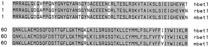
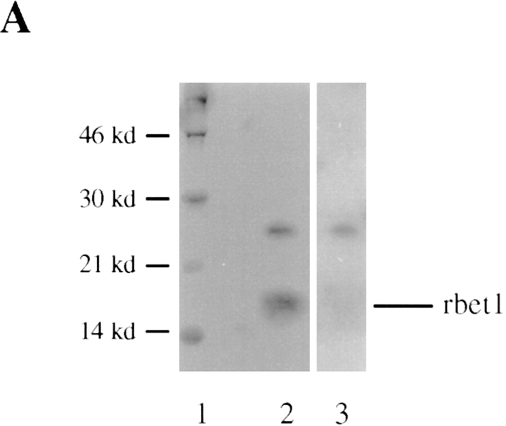
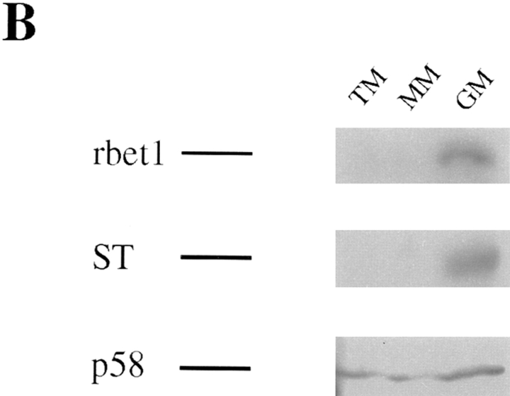
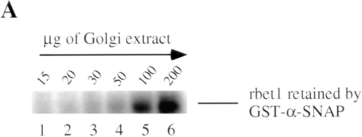
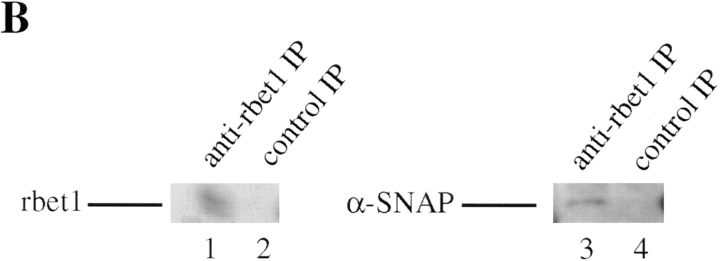
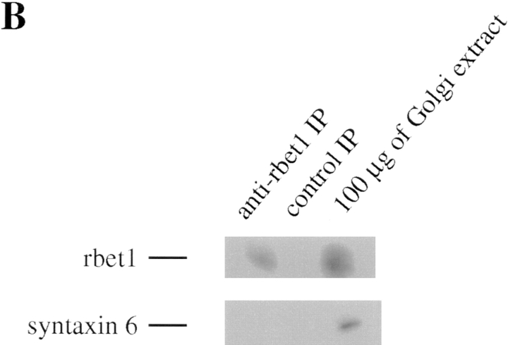
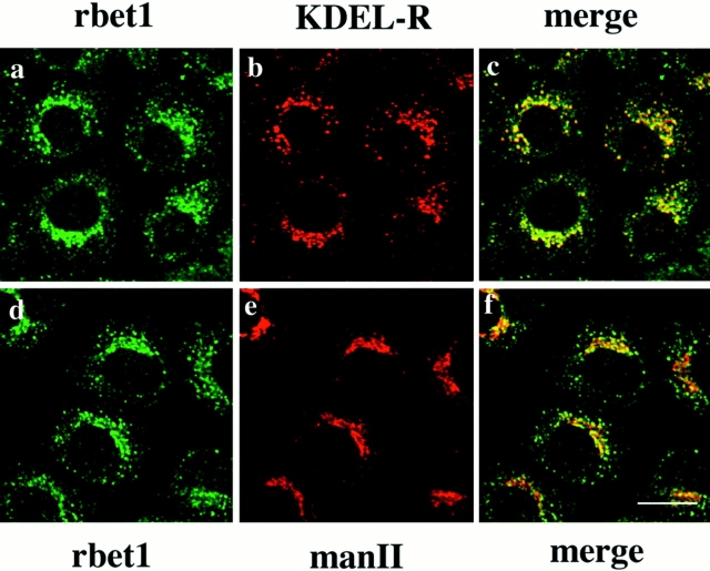
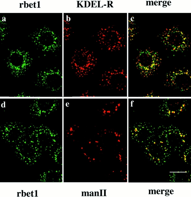
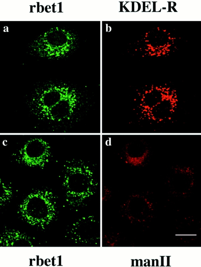
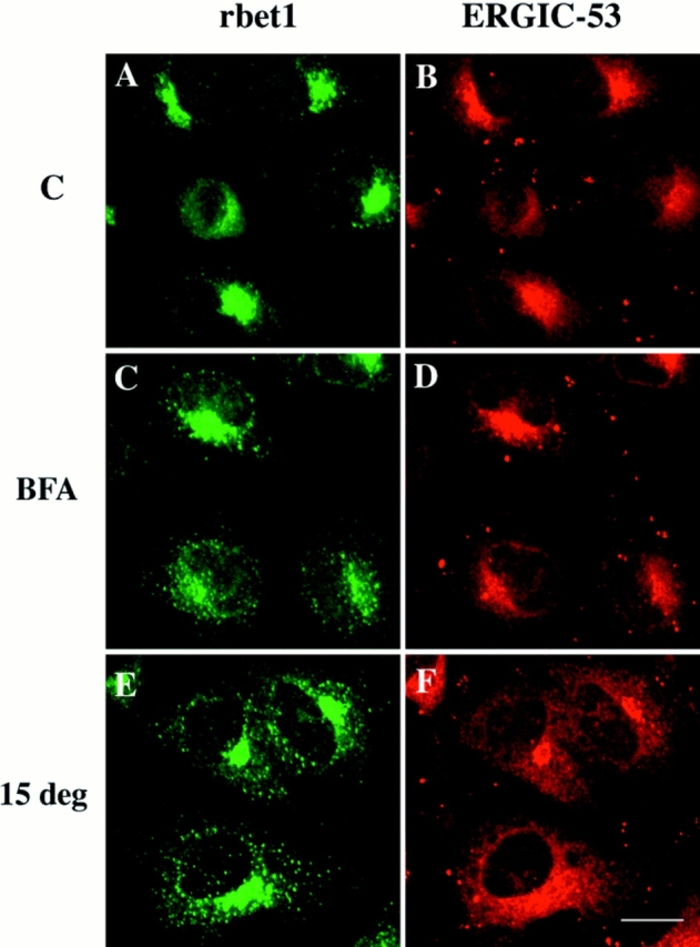
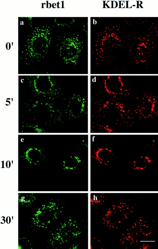
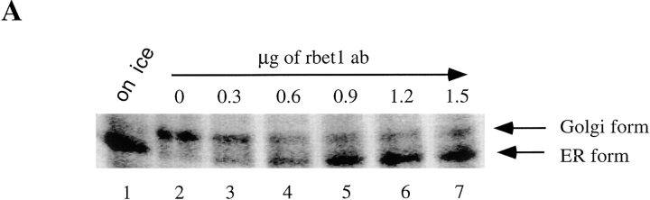
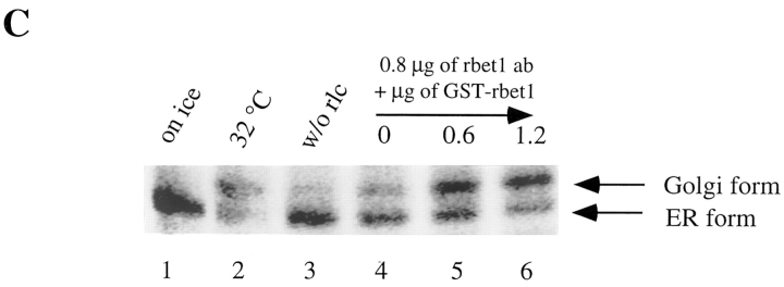
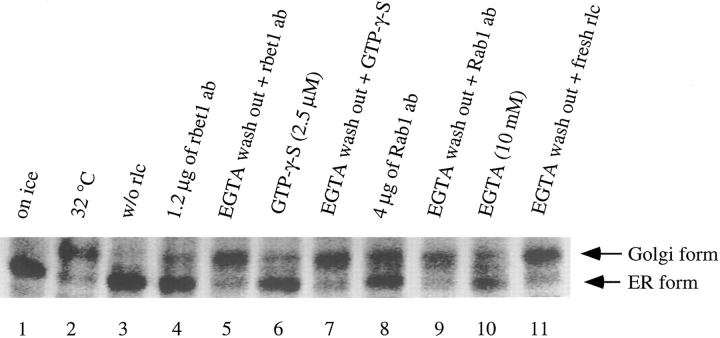
Yeast Bet1p participates in vesicular transport from the endoplasmic reticulum to the Golgi apparatus and functions as a soluble N-ethylmaleimide-sensitive factor attachment protein receptor (SNARE) associated with ER-derived vesicles. A mammalian protein (rbet1) homologous to Bet1p was recently identified, and it was concluded that rbet1 is associated with the Golgi apparatus based on the subcellular localization of transiently expressed epitope-tagged rbet1. In the present study using rabbit antibodies raised against the cytoplasmic domain of rbet1, we found that the majority of rbet1 is not associated with the Golgi apparatus as marked by the Golgi mannosidase II in normal rat kidney cells. Rather, rbet1 is predominantly associated with vesicular spotty structures that concentrate in the peri-Golgi region but are also present throughout the cytoplasm. These structures colocalize with the KDEL receptor and ERGIC-53, which are known to be enriched in the intermediate compartment. When the Golgi apparatus is fragmented by nocodazole treatment, a significant portion of rbet1 is not colocalized with structures marked by Golgi mannosidase II or the KDEL receptor. Association of rbet1 in cytoplasmic spotty structures is apparently not altered by preincubation of cells at 15 degrees C. However, upon warming up from 15 to 37 degrees C, rbet1 concentrates into the peri-Golgi region. Furthermore, rbet1 colocalizes with vesicular stomatitis virus G-protein en route from the ER to the Golgi. Antibodies against rbet1 inhibit in vitro transport of G-protein from the ER to the Golgi apparatus in a dose-dependent manner. This inhibition can be neutralized by preincubation of antibodies with recombinant rbet1. EGTA is known to inhibit ER-Golgi transport at a stage after vesicle docking but before the actual fusion event. Antibodies against rbet1 inhibit ER-Golgi transport only when they are added before the EGTA-sensitive stage. These results suggest that rbet1 may be involved in the docking process of ER-derived vesicles with the cis-Golgi membrane.


Figures

Figure 1
Figure 1
The mammalian bet1 proteins are highly conserved. The amino acid sequences of human, rat, and mouse bet1 are aligned and residues identical among them are shaded.
Figure 2
Figure 2
rbet1 is a 17-kD protein enriched in the membrane fraction of the Golgi and the IC. (A) Total membrane fraction of NRK cells was resolved by SDS-PAGE and transferred to a filter. The filter was incubated with rbet1 antibodies in the absence (lane 2) or presence of GST-rbet1 (lane 3). (B) Total membrane (TM), microsomal membrane (MM), and Golgi membrane (GM) fractions derived from rat liver were analyzed by immunoblot to detect rbet1, α2,6-sialyltransferase (ST), and the IC-enriched protein p58.
Figure 2
Figure 2
rbet1 is a 17-kD protein enriched in the membrane fraction of the Golgi and the IC. (A) Total membrane fraction of NRK cells was resolved by SDS-PAGE and transferred to a filter. The filter was incubated with rbet1 antibodies in the absence (lane 2) or presence of GST-rbet1 (lane 3). (B) Total membrane (TM), microsomal membrane (MM), and Golgi membrane (GM) fractions derived from rat liver were analyzed by immunoblot to detect rbet1, α2,6-sialyltransferase (ST), and the IC-enriched protein p58.
Figure 3
Figure 3
rbet1 is a SNARE. (A) The indicated amounts of Golgi extract were incubated with 2 μg of immobilized GST–α-SNAP. After extensive washing, the beads were analyzed by immunoblot to detect rbet1. (B) Golgi extract was immunoprecipitated with rbet1 antibodies or control antibodies and the immunoprecipitates were analyzed by immunoblot to detect rbet1 and α-SNAP.
Figure 3
Figure 3
rbet1 is a SNARE. (A) The indicated amounts of Golgi extract were incubated with 2 μg of immobilized GST–α-SNAP. After extensive washing, the beads were analyzed by immunoblot to detect rbet1. (B) Golgi extract was immunoprecipitated with rbet1 antibodies or control antibodies and the immunoprecipitates were analyzed by immunoblot to detect rbet1 and α-SNAP.
Figure 4
Figure 4
rbet1 exists in a protein complex that contains GS28 and syntaxin 5. (A) Golgi extract was immunoprecipitated with rbet1 antibodies or control antibodies. The immunoprecipitates were analyzed by immunoblot to detect rbet1, GS28, and syntaxin 5. (B) The immunoprecipitate of rbet1 and control antibodies, together with 100 μg of Golgi extract, were analyzed by immunoblot to detect rbet1 and syntaxin 6.
Figure 4
Figure 4
rbet1 exists in a protein complex that contains GS28 and syntaxin 5. (A) Golgi extract was immunoprecipitated with rbet1 antibodies or control antibodies. The immunoprecipitates were analyzed by immunoblot to detect rbet1, GS28, and syntaxin 5. (B) The immunoprecipitate of rbet1 and control antibodies, together with 100 μg of Golgi extract, were analyzed by immunoblot to detect rbet1 and syntaxin 6.
Figure 5
Figure 5
Double-labeling of rbet1 (a and d) with the KDEL receptor (KDEL-R) (b) or Golgi mannosidase II (man II) (e) in NRK cells. The merged pictures (c and f) are also presented. Bar, 10 μm.
Figure 6
Figure 6
NRK cells were treated with 10 μg/ml nocodazole for 1 h, fixed, and double-labeled for rbet1 (a and d) with KDEL-R (b) or man II (e). Also shown are the merged images (c and f). Bar, 10 μm.
Figure 7
Figure 7
NRK cells were treated with 10 μg/ml brefeldin A for 1 h, fixed, and double-labeled for rbet1 (a and c) with KDEL-R (b) or man II (d). Bar, 10 μm.
Figure 8
Figure 8
Double-labeling of rbet1 with ERGIC-53 in control cells (A and B), cells pretreated with 10 μg/ml brefeldin A for 1 h (C and D), and cells preincubated at 15°C for 3 h (E and F). Bar, 10 μm.
Figure 9
Figure 9
NRK cells were incubated at 15°C for 3 h and then incubated at 37°C for the indicated time. The cells were fixed and double-labeled for rbet1 (a, c, e, and g) with KDEL-R (b, d, f, and h). Bar, 10 μm.
Figure 10
Figure 10
(A) Vero cells were infected with VSV ts045 and incubated at 40°C for 4 h. Cells were then shifted to 32°C for the indicated time and fixed. Double-labeling of rbet1 (a, d, and g) with the envelope protein of VSV (VSVG) (b, e, and h) was performed. The merged images (c, f, and i) are also shown. (B) Control Vero and NRK cells were fixed and labeled with rbet1 antibodies. Bars, 10 μm.
Figure 11
Figure 11
rbet1 antibodies specifically inhibit in vitro ER-Golgi transport. (A) In vitro ER-Golgi transport was performed either on ice (lane 1) or at 32°C (lanes 2–7) supplemented with the indicated amounts of rbet1 antibodies. (B) In vitro ER-Golgi transport was performed either on ice (lane 1) or at 32°C (lanes 2–4) supplemented with 1.2 μg of rbet1 antibodies (lane 3) or the heat-inactivated antibodies (lane 4). (C) In vitro ER-Golgi transport was performed either on ice (lane 1) or at 32°C (lanes 2–6) in the absence (lane 3) or in the presence of rat liver cytosol (rlc) (lanes 1 and 2, and 4–6) supplemented with 0.8 μg of rbet1 antibodies and indicated amounts of GST-rbet1.
Figure 11
Figure 11
rbet1 antibodies specifically inhibit in vitro ER-Golgi transport. (A) In vitro ER-Golgi transport was performed either on ice (lane 1) or at 32°C (lanes 2–7) supplemented with the indicated amounts of rbet1 antibodies. (B) In vitro ER-Golgi transport was performed either on ice (lane 1) or at 32°C (lanes 2–4) supplemented with 1.2 μg of rbet1 antibodies (lane 3) or the heat-inactivated antibodies (lane 4). (C) In vitro ER-Golgi transport was performed either on ice (lane 1) or at 32°C (lanes 2–6) in the absence (lane 3) or in the presence of rat liver cytosol (rlc) (lanes 1 and 2, and 4–6) supplemented with 0.8 μg of rbet1 antibodies and indicated amounts of GST-rbet1.
Figure 12
Figure 12
rbet1 antibodies must be present before the EGTA-sensitive stage to exhibit the inhibitory effect on in vitro ER-Golgi transport. In vitro ER-Golgi transport was performed either on ice (lane 1) or at 32°C (lanes 2–11) in the absence (lane 3) or presence of rat liver cytosol (rlc) (lanes 1 and 2, and 4–11). The standard transport was performed for lanes 1–4. For lanes 5–11, transport assay was first performed in the presence of 10 mM EGTA to arrest the transport at the EGTA-sensitive stage followed by a washing step and second incubation at 32°C to continue the transport. Reagents were supplemented as indicated.

Similar articles

Cited by

References

    1. Aridor M, Bannykh SI, Rowe T, Balch WE. Sequential coupling between COPII and COPI vesicle coats in endoplasmic reticulum to Golgi transport. J Cell Biol. 1995;131:875–893. - PMC - PubMed
    1. Balch WE, McCaffery JM, Plunter H, Farquhar MG. Vesicular stomatitis virus glycoprotein is sorted and concentrated during export from the endoplasmic reticulum. Cell. 1994;77:841–852. - PubMed
    1. Banfield DK, Lewis MJ, Rabouille C, Warren G, Pelham HRB. Localization of Sed5, a putative vesicle targeting molecule, to the cis-Golgi network involves both its transmembrane and cytoplasmic domains. J Cell Biol. 1994;127:357–371. - PMC - PubMed
    1. Bannykh SI, Balch WE. Membrane dynamics at the endoplasmic reticulum–Golgi interface. J Cell Biol. 1997;138:1–4. - PMC - PubMed
    1. Bannykh SI, Rowe T, Balch WE. The organization of endoplasmic reticulum export complexes. J Cell Biol. 1996;135:19–35. - PMC - PubMed

Publication types

MeSH terms

Associated data