. 1997 Nov 17;139(4):929-40.
doi: 10.1083/jcb.139.4.929.

The O-glycosylated stalk domain is required for apical sorting of neurotrophin receptors in polarized MDCK cells

Affiliations

The O-glycosylated stalk domain is required for apical sorting of neurotrophin receptors in polarized MDCK cells

C Yeaman et al. J Cell Biol. .

Abstract

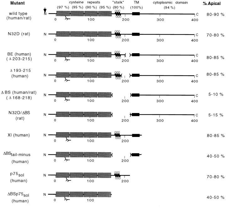
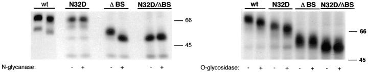
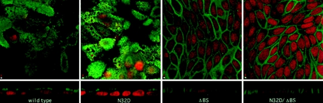
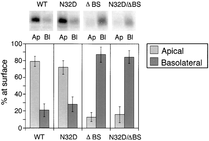
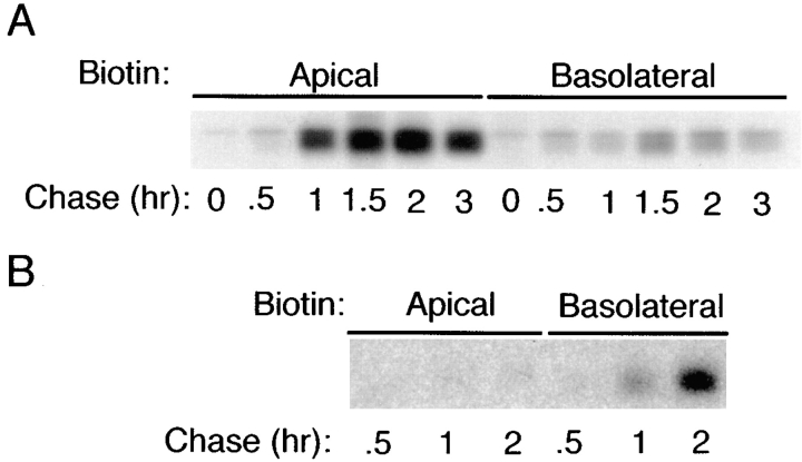
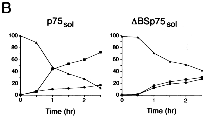
Delivery of newly synthesized membrane-spanning proteins to the apical plasma membrane domain of polarized MDCK epithelial cells is dependent on yet unidentified sorting signals present in the luminal domains of these proteins. In this report we show that structural information for apical sorting of transmembrane neurotrophin receptors (p75(NTR)) is localized to a juxtamembrane region of the extracellular domain that is rich in O-glycosylated serine/threonine residues. An internal deletion of 50 amino acids that removes this stalk domain from p75(NTR) causes the protein to be sorted exclusively of the basolateral plasma membrane. Basolateral sorting stalk-minus p75(NTR) does not occur by default, but requires sequences present in the cytoplasmic domain. The stalk domain is also required for apical secretion of a soluble form of p75(NTR), providing the first demonstration that the same domain can mediate apical sorting of both a membrane-anchored as well as secreted protein. However, the single N-glycan present on p75(NTR) is not required for apical sorting of either transmembrane or secreted forms.


Figures

Figure 1
Figure 1
Mutants constructed to characterize apical sorting information in p75NTR and their membrane distribution upon expression in MDCK cells. Degrees of amino acid conservation (percent identity) between human and rat p75NTR are shown in parentheses above each domain. Site of signal peptide cleavage is indicated by an arrow. Stippled boxes represent cysteine-rich repeat domains and black boxes represent the transmembrane (TM) domain. The N-linked glycosylation site in the first cysteine repeat is indicated. Lollipop structures represent O-linked carbohydrates in the juxtamembrane stalk domain. Internal deletions constructed within this domain are marked by parentheses. Amino acid residues are numbered, from amino to carboxy terminus, beneath schematic models. % Apical values are based on steady state biotinylation experiments discussed in the text.
Figure 2
Figure 2
Inhibition of N-glycosylation does not perturb apical sorting of p75NTR. (A) Tunicamycin inhibits N-glycosylation of p75NTR in a concentration-dependent manner. Adenovirus-mediated gene transfer into filter-grown MDCK with 50 pfu/cell AdCMVp75 was performed 48 h before experiment. Cells were pretreated 2.5 h with indicated concentrations of tunicamycin and then labeled with [35S]cysteine for 4 h in the continued presence of tunicamycin. p75NTR was immunoprecipitated from cell lysates and subjected to SDS-PAGE as described in Materials and Methods. (B) ER-to-Golgi transport and (C) polarized targeting of p75NTR in the absence or presence of tunicamycin. Adenovirus-mediated gene transfer into filter-grown MDCK with 50 pfu/cell AdCMVp75 was performed 48 h before experiment. Cells were pretreated 2 h with or without 10 μg/ml tunicamycin, starved in cysteine-free medium for 30 min and then pulse labeled with [35S]cysteine for 20 min in the continued presence of tunicamycin, where indicated. After the pulse, cultures were chased in the continued presence or absence of tunicamycin. At indicated chase times, filters were placed on ice and subjected to domain-selective biotinylation. p75NTR was immunoprecipitated from cell lysates and surface-labeled protein was recovered on streptavidin-agarose. Aliquots of immunoprecipitates from apically biotinylated cells are shown in B, and streptavidin-agarose precipitates from the same samples and from basolaterally biotinylated cells are shown in C. Position of albumin standard (66 kD) is indicated in B. Quantitation reflects the average of duplicate experiments, one of which is represented by gels shown in figure.
Figure 2
Figure 2
Inhibition of N-glycosylation does not perturb apical sorting of p75NTR. (A) Tunicamycin inhibits N-glycosylation of p75NTR in a concentration-dependent manner. Adenovirus-mediated gene transfer into filter-grown MDCK with 50 pfu/cell AdCMVp75 was performed 48 h before experiment. Cells were pretreated 2.5 h with indicated concentrations of tunicamycin and then labeled with [35S]cysteine for 4 h in the continued presence of tunicamycin. p75NTR was immunoprecipitated from cell lysates and subjected to SDS-PAGE as described in Materials and Methods. (B) ER-to-Golgi transport and (C) polarized targeting of p75NTR in the absence or presence of tunicamycin. Adenovirus-mediated gene transfer into filter-grown MDCK with 50 pfu/cell AdCMVp75 was performed 48 h before experiment. Cells were pretreated 2 h with or without 10 μg/ml tunicamycin, starved in cysteine-free medium for 30 min and then pulse labeled with [35S]cysteine for 20 min in the continued presence of tunicamycin, where indicated. After the pulse, cultures were chased in the continued presence or absence of tunicamycin. At indicated chase times, filters were placed on ice and subjected to domain-selective biotinylation. p75NTR was immunoprecipitated from cell lysates and surface-labeled protein was recovered on streptavidin-agarose. Aliquots of immunoprecipitates from apically biotinylated cells are shown in B, and streptavidin-agarose precipitates from the same samples and from basolaterally biotinylated cells are shown in C. Position of albumin standard (66 kD) is indicated in B. Quantitation reflects the average of duplicate experiments, one of which is represented by gels shown in figure.
Figure 3
Figure 3
Rat p75NTR mutants lack N- and/ or O-glycans. MDCK cells stably expressing native (wt) and mutant forms of rat p75NTR were labeled overnight with [35S]cysteine. p75NTR was immunoprecipitated from cell lysates with anti-cytoplasmic domain antibodies and deglycosylated with indicated enzymes as described in Materials and Methods. Note that N32D p75NTR lacks N-glycans but is O-glycosylated; ΔBS p75NTR lacks O-glycans but is N-glycosylated; and N32D/ΔBS p75NTR lacks both N- and O-glycans.
Figure 4
Figure 4
Immunolocalization of rat p75NTR glycosylation mutants. Stably transfected MDCK cultures expressing native and mutant forms of rat p75NTR were fixed and permeabilized as described in Materials and Methods. p75NTR was detected by indirect immunofluorescence using anti- cytoplasmic domain polyclonal antibodies. Each panel shows a single 1-μm optical section in the XY-plane (top) as well as a vertical section through the sample (bottom). Bar, 2 μm.
Figure 5
Figure 5
Steady state biotinylation of rat p75NTR glycosylation mutants. Stably transfected MDCK clones expressing native and mutant forms of rat p75NTR were grown on filters for 5 d and then subjected to domain-selective biotinylation with NHS-SS-biotin as described in Materials and Methods. Biotinylated proteins were precipitated from cell lysates with avidin-agarose and resolved by SDS-PAGE. Electrophoretic protein transfers were probed with rabbit polyclonal antibodies against the cytoplasmic domain of p75NTR and detected with 125I-anti–rabbit IgG. Blots were analyzed with a Molecular Dynamics phosphorimager. Bars represent the mean ± SD of multiple determinations from three separate experiments (n = 8).
Figure 6
Figure 6
ΔBS p75NTR is targeted directly to the basal-lateral domain of MDCK cells. Polarized MDCK cultures stably expressing (A) native rat p75NTR or (B) ΔBS rat p75NTR were starved in cysteine-free medium for 30 min and then pulse labeled with [35S]cysteine for 20 min. After the pulse, cultures were chased in medium containing an excess of unlabeled cysteine. At indicated chase times, filters were placed on ice and incubated with NHS-SS-biotin either in the apical or basolateral medium. p75NTR was immunoprecipitated from cell lysates and surface-labeled protein was recovered on streptavidin-agarose. Images shown were obtained after exposure of the gel to Kodak XAR-5 film (A) or Molecular Dynamics phosphorimager screen (B).
Figure 7
Figure 7
Amino acids 193–215 can be deleted from the juxtamembrane stalk domain without compromising apical sorting of p75NTR. (A) Stably transfected MDCK clones expressing Δ193–215 p75NTR were grown on filters for 5 d and then subjected to domain-selective biotinylation with NHS-SS-biotin as described in Materials and Methods. Biotinylated proteins were precipitated from cell lysates with avidin-agarose and resolved by SDS-PAGE. Electrophoretic protein transfers were probed with rabbit polyclonal antibodies against the cytoplasmic domain of p75NTR and detected with 125I-anti–rabbit IgG. Blots were analyzed with a Molecular Dynamics phosphorimager. Bars represent the mean ± SD of triplicate determinations from a representative experiment. (B) Stably transfected MDCK cultures expressing Δ193–215 p75NTR were starved in cysteine-free medium for 30 min and then pulse labeled with [35S]cysteine for 20 min. After the pulse, cultures were chased in medium containing an excess of unlabeled cysteine. At indicated chase times, filters were placed on ice and incubated with NHS-SS-biotin either in the apical or basolateral medium. Δ193–215 p75NTR was immunoprecipitated from cell lysates with an antibody against the cysteine-rich domain (ME 20.4) and surface-labeled protein was recovered on streptavidin-agarose.
Figure 7
Figure 7
Amino acids 193–215 can be deleted from the juxtamembrane stalk domain without compromising apical sorting of p75NTR. (A) Stably transfected MDCK clones expressing Δ193–215 p75NTR were grown on filters for 5 d and then subjected to domain-selective biotinylation with NHS-SS-biotin as described in Materials and Methods. Biotinylated proteins were precipitated from cell lysates with avidin-agarose and resolved by SDS-PAGE. Electrophoretic protein transfers were probed with rabbit polyclonal antibodies against the cytoplasmic domain of p75NTR and detected with 125I-anti–rabbit IgG. Blots were analyzed with a Molecular Dynamics phosphorimager. Bars represent the mean ± SD of triplicate determinations from a representative experiment. (B) Stably transfected MDCK cultures expressing Δ193–215 p75NTR were starved in cysteine-free medium for 30 min and then pulse labeled with [35S]cysteine for 20 min. After the pulse, cultures were chased in medium containing an excess of unlabeled cysteine. At indicated chase times, filters were placed on ice and incubated with NHS-SS-biotin either in the apical or basolateral medium. Δ193–215 p75NTR was immunoprecipitated from cell lysates with an antibody against the cysteine-rich domain (ME 20.4) and surface-labeled protein was recovered on streptavidin-agarose.
Figure 8
Figure 8
N-glycosylation is not required for apical secretion of soluble forms of p75NTR or N-CAM. Stably transfected MDCK clones expressing truncated ectodomains of either (A) p75NTR or (B) N-CAM were pretreated for 2.5 h with or without 10 μg/ml tunicamycin, starved for 30 min in medium lacking methionine and cysteine, and then pulse labeled with [35S]cysteine (for p75NTR labeling) or 35S-express (for N-CAM labeling) for 20 min. After the pulse, cultures were chased in the continued presence or absence of tunicamycin for 2 h. p75NTR and N-CAM were immunoprecipitated from apical and basolateral chase medium samples and resolved by SDS-PAGE. Fluorograms were analyzed by scanning densitometry. Bars represent mean ± SD for triplicate determinations from a representative experiment.
Figure 9
Figure 9
O-glycosylated domain is required for apical secretion of soluble p75NTR. (A) Clones of MDCK cells stably expressing human p75NTR truncated at amino acid 168 were pretreated for 2.5 h with or without 10 μg/ml tunicamycin. Cells were starved in cysteine-free medium for 30 min then pulse labeled with [35S]cysteine for 20 min. After the pulse, cultures were chased in the continued presence or absence of tunicamycin for 3 h. ΔBS p75NTR sol was immunoprecipitated from apical and basolateral chase medium and from cell lysates with a mAb against the cysteine-rich domain (ME 20.4). After SDS-PAGE, p75NTR was detected by fluorography. Results obtained with three independent clones are shown. (B) Time course of secretion of soluble p75NTR with and without O-glycosylated domain. Cells were starved in cysteine-free medium for 30 min, pulse labeled with [35S]cysteine for 20 min, and then chased in medium containing excess unlabeled cysteine for indicated times. p75NTR sol and ΔBS p75NTR sol were immunoprecipitated from apical and basolateral chase medium and from cell lysates with a mAb against the cysteine-rich domain (ME 20.4). Proteins were resolved by SDS-PAGE and gels were analyzed with a Molecular Dynamics phosphorimager. Apical, basolateral, and cells indicated by closed squares (▪), closed circles (•), and closed triangels (▴), respectively.
Figure 9
Figure 9
O-glycosylated domain is required for apical secretion of soluble p75NTR. (A) Clones of MDCK cells stably expressing human p75NTR truncated at amino acid 168 were pretreated for 2.5 h with or without 10 μg/ml tunicamycin. Cells were starved in cysteine-free medium for 30 min then pulse labeled with [35S]cysteine for 20 min. After the pulse, cultures were chased in the continued presence or absence of tunicamycin for 3 h. ΔBS p75NTR sol was immunoprecipitated from apical and basolateral chase medium and from cell lysates with a mAb against the cysteine-rich domain (ME 20.4). After SDS-PAGE, p75NTR was detected by fluorography. Results obtained with three independent clones are shown. (B) Time course of secretion of soluble p75NTR with and without O-glycosylated domain. Cells were starved in cysteine-free medium for 30 min, pulse labeled with [35S]cysteine for 20 min, and then chased in medium containing excess unlabeled cysteine for indicated times. p75NTR sol and ΔBS p75NTR sol were immunoprecipitated from apical and basolateral chase medium and from cell lysates with a mAb against the cysteine-rich domain (ME 20.4). Proteins were resolved by SDS-PAGE and gels were analyzed with a Molecular Dynamics phosphorimager. Apical, basolateral, and cells indicated by closed squares (▪), closed circles (•), and closed triangels (▴), respectively.
Figure 10
Figure 10
Basolateral expression of ΔBS p75 is dependent on the cytoplasmic domain. Clones of MDCK cells expressing human p75NTR with an internal deletion of the O-glycosylated membrane stalk domain (amino acids 168–218) and truncated at amino acid 248 were grown on filters for 5 d. (A) Cultures were subjected to domain-selective biotinylation with NHS-LC-biotin. ΔBS p75tail-minus was immunoprecipitated from cell lysates with a mAb against the cysteine-rich domain (ME 20.4) and resolved by SDS-PAGE. Electrophoretic protein transfers were probed with 125I-streptavidin. Blots were analyzed with a Molecular Dynamics phosphorimager. Bars (% apical, light grey, and % basolateral, dark grey, respectively) represent the mean ± SD of triplicate determinations in a representative experiment. Results obtained with two independent clones are shown. (B) Stably transfected MDCK cultures expressing ΔBS p75tail-minus were starved in cysteine-free medium for 30 min and then pulse labeled with [35S]cysteine for 20 min. After the pulse, cultures were chased in medium containing an excess of unlabeled cysteine. At indicated chase times, filters were placed on ice and incubated with NHS-SS-biotin either in the apical or basolateral medium. ΔBS p75tail-minus was immunoprecipitated from cell lysates and surface-labeled protein was recovered on streptavidin-agarose. % apical and % basolateral indicated by closed squares (▪) and closed circles (•), respectively.

Similar articles

Cited by

References

    1. Baldwin AN, Shooter EM. Zone mapping of the binding domain of the rat low affinity nerve growth factor receptor by the introduction of novel N-glycosylation sites. J Biol Chem. 1995;270:4594–4602. - PubMed
    1. Baldwin AN, Bitler CM, Welcher AA, Shooter EM. Studies on the structure and binding properties of the cysteine-rich domain of rat low affinity nerve growth factor receptor (p75NGFR) J Biol Chem. 1992;267:8352–8359. - PubMed
    1. Blochberger TC, Sabatine JM, Lee YC, Hughey RP. O-linked glycosylation of rat renal g-glutamylpeptidase adjacent to its membrane anchor domain. J Biol Chem. 1989;264:20718–20722. - PubMed
    1. Brown DA, Crise B, Rose JK. Mechanism of membrane anchoring affects polarized expression of two proteins in MDCK cells. Science. 1989;245:1499–1501. - PubMed
    1. Casanova JE, Apodaca G, Mostov KE. An autonomous signal for basolateral sorting in the cytoplasmic domain of the polymeric immunoglobulin receptor. Cell. 1991;66:65–75. - PubMed

Publication types