. 1997 Nov 1;11(21):2835-44.
doi: 10.1101/gad.11.21.2835.

Rescue of osteoclast function by transgenic expression of kinase-deficient Src in src-/- mutant mice

Affiliations

Rescue of osteoclast function by transgenic expression of kinase-deficient Src in src-/- mutant mice

P L Schwartzberg et al. Genes Dev. .

Abstract

The Src tyrosine kinase has been implicated in a wide variety of signal transduction pathways, yet despite the nearly ubiquitous expression of c-src, src-/- mice show only one major phenotype-osteopetrosis caused by an intrinsic defect in osteoclasts, the cells responsible for resorbing bone. To explore further the role of Src both in osteoclasts and other cell types, we have generated transgenic mice that express the wild-type and mutated versions of the chicken c-src proto-oncogene from the promoter of tartrate resistant acid phosphatase (TRAP), a gene that is expressed highly in osteoclasts. We demonstrate here that expression of a wild-type transgene in only a limited number of tissues can fully rescue the src-/- phenotype. Surprisingly, expression of kinase-defective alleles of c-src also reduces osteopetrosis in src-/- animals and partially rescues a defect in cytoskeletal organization observed in src-/- osteoclasts. These results suggest that there are essential kinase-independent functions for Src in vivo. Biochemical examination of osteoclasts from these mice suggest that Src may function in part by recruiting or activating other tyrosine kinases.


Figures

Figure 1
Figure 1
(A) Structure of the TRAPsrc transgene. The TRAP promoter, first exon, and intron were fused to the ATG of the chicken c-src cDNA. Encoded amino-acids important for activation of Src or mutated in transgenic constructs are indicated above the drawing. (U) Unique region; (SH2) Src homology 2 region; (SH3) Src homology 3. (B) Expression of the TRAPsrc transgene in cultured osteoclasts from transgenic mice. RNA samples isolated from fresh bone marrow and from bone marrow cultured in the presence of 10−9 m Vitamin D3 (Takahashi et al. 1988) were analyzed by RT-PCR, using primers that generate products extending from the TRAP first exon to both the chicken c-src and the endogenous TRAP-coding sequences. Products were hybridized with a P32-labeled probe that recognizes the TRAPsrc-specific band as well as unspliced TRAP RNA. (Lanes 1,2) Fresh bone marrow; (lanes 3,4) bone marrow cultured in the presense of 10−9 m Vitamin D3. (Lanes 1,3) Non-transgenic mice. (C) Northern analysis of RNA derived from tissues of trangenic line A and C mice. (Lanes 1–7) Line A (Lane 1) Testes; (lane 2) brain; (lane 3) heart; (lane 4) spleen; (lane 5) kidney; (lane 6) liver; (lane 7) small intestine; (Lane 8) line C small intestine. (top) The filter was hybridized with a probe consisting of the first 200 bp of the chicken c-src gene; (bottom) the hybridized to a probe for RNA encoding the L32 ribosomal protein.
Figure 1
Figure 1
(A) Structure of the TRAPsrc transgene. The TRAP promoter, first exon, and intron were fused to the ATG of the chicken c-src cDNA. Encoded amino-acids important for activation of Src or mutated in transgenic constructs are indicated above the drawing. (U) Unique region; (SH2) Src homology 2 region; (SH3) Src homology 3. (B) Expression of the TRAPsrc transgene in cultured osteoclasts from transgenic mice. RNA samples isolated from fresh bone marrow and from bone marrow cultured in the presence of 10−9 m Vitamin D3 (Takahashi et al. 1988) were analyzed by RT-PCR, using primers that generate products extending from the TRAP first exon to both the chicken c-src and the endogenous TRAP-coding sequences. Products were hybridized with a P32-labeled probe that recognizes the TRAPsrc-specific band as well as unspliced TRAP RNA. (Lanes 1,2) Fresh bone marrow; (lanes 3,4) bone marrow cultured in the presense of 10−9 m Vitamin D3. (Lanes 1,3) Non-transgenic mice. (C) Northern analysis of RNA derived from tissues of trangenic line A and C mice. (Lanes 1–7) Line A (Lane 1) Testes; (lane 2) brain; (lane 3) heart; (lane 4) spleen; (lane 5) kidney; (lane 6) liver; (lane 7) small intestine; (Lane 8) line C small intestine. (top) The filter was hybridized with a probe consisting of the first 200 bp of the chicken c-src gene; (bottom) the hybridized to a probe for RNA encoding the L32 ribosomal protein.
Figure 2
Figure 2
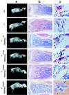
Radiographic and histological analysis of mice. (A) X-rays, lateral view. (B) Histological sections of tibia, low power (4× magnification). Sections were stained both with hematoxylin and eosin and for TRAP activity. (C) Histological sections, TRAP-stained osteoclasts in bone, high power (100×, note lacy ruffled border structures at the interface between osteoclasts and bone surface.) Genotypes of mice are indicated. Two examples of bones from src−/− mice rescued by the K295M transgene (TRAPsrcK295M) are shown to demonstrate the variability in phenotype. The bottom sample from a src−/−TRAPsrcK295M mouse is derived from the calvarium after treatment with IL-1 and is stained with hematoxylin and eosin only.
Figure 3
Figure 3
Histomorphometric analysis of rescued mice. Genotype of mice indicated on x axis. Percentage bone volume [amount of bone matrix as a percentage of the cancellous bone (Boyce et al. 1992)] is indicated on the y axis. A minimum of four samples were examined for each transgene. Histology was performed on samples from at least two transgenic lines for each construct, except for the TRAPsrcY416F rescued mice (histomorphometry was performed on line G only).
Figure 4
Figure 4
Rescue of abnormal morphology of src−/− osteoclasts by expression of TRAPsrc transgenes. (A) Morphology of src−/− osteoclasts. Osteoclasts were differentiated on plastic, partially purified, and stained for TRAP activity. (B) Actin organization of osteoclasts expressing TRAPsrc transgenes. Osteoclasts were stained with Rhodamine-phalloidin and examined by confocal microscopy. Genotypes of Osteoclasts are indicated. Note lack of peripheral actin ring in src−/− osteoclasts.
Figure 4
Figure 4
Rescue of abnormal morphology of src−/− osteoclasts by expression of TRAPsrc transgenes. (A) Morphology of src−/− osteoclasts. Osteoclasts were differentiated on plastic, partially purified, and stained for TRAP activity. (B) Actin organization of osteoclasts expressing TRAPsrc transgenes. Osteoclasts were stained with Rhodamine-phalloidin and examined by confocal microscopy. Genotypes of Osteoclasts are indicated. Note lack of peripheral actin ring in src−/− osteoclasts.
Figure 5
Figure 5
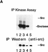
Osteoclasts from mice rescued with the TRAPsrcK295M transgene lack detectable Src kinase activity but have normal levels of tyrosine phosphorylation. (A) In vitro kinase assays. Osteoclasts derived from bone marrow were lysed in RIPA buffer, and extracts were normalized for both protein concentration (Biorad) and TRAP enzymatic activity (Sigma); after immunoprecipitation with anti-Src monoclonal 327 (Calbiochem), in vitro kinase assays with the exogenous substrate enolase were performed as described previously (Kaplan et al. 1995). One-half of each immunoprecipitate was analyzed by Western blotting probing with the Src2 antisera (Santa Cruz). (Lane 1) src−/−; (lane 2) src+/+; (lane 3) src−/−TRAPsrcWT line A; (lane 4) src−/−TRAPsrcWT line C; (lane 5) src−/−TRAPsrcK295M. (B) Tyrosine-phosphorylated proteins from cultured osteoclasts from wild-type src−/− and kinase-defective rescued mice. Protein extracts from cultured osteoclasts were normalized for both protein concentration (Bio-Rad) and TRAP enzymatic activity (Sigma), and analyzed with the 4G10, anti-phosphotyrosine antibody (UBI). Lanes are derived from the same exposure of a single gel.
Figure 5
Figure 5
Osteoclasts from mice rescued with the TRAPsrcK295M transgene lack detectable Src kinase activity but have normal levels of tyrosine phosphorylation. (A) In vitro kinase assays. Osteoclasts derived from bone marrow were lysed in RIPA buffer, and extracts were normalized for both protein concentration (Biorad) and TRAP enzymatic activity (Sigma); after immunoprecipitation with anti-Src monoclonal 327 (Calbiochem), in vitro kinase assays with the exogenous substrate enolase were performed as described previously (Kaplan et al. 1995). One-half of each immunoprecipitate was analyzed by Western blotting probing with the Src2 antisera (Santa Cruz). (Lane 1) src−/−; (lane 2) src+/+; (lane 3) src−/−TRAPsrcWT line A; (lane 4) src−/−TRAPsrcWT line C; (lane 5) src−/−TRAPsrcK295M. (B) Tyrosine-phosphorylated proteins from cultured osteoclasts from wild-type src−/− and kinase-defective rescued mice. Protein extracts from cultured osteoclasts were normalized for both protein concentration (Bio-Rad) and TRAP enzymatic activity (Sigma), and analyzed with the 4G10, anti-phosphotyrosine antibody (UBI). Lanes are derived from the same exposure of a single gel.

Similar articles

Cited by

References

    1. Baron R, Revesloot J-H, Neff L, Chakraborty M, Chatterjee D, Lomri A, Horne W. Cellular and molecular biology of bone. San Diego: Academic Press; 1993. Cellular and molecular biology of the osteoclast; pp. 445–495. California.
    1. Boyce B, Yoneda T, Lowe C, Soriano P, Mundy G. Requirement of pp60c-src expression for osteoclasts to form ruffled borders and to resorb bone in mice. J Clin Invest. 1992;90:1622–1627. - PMC - PubMed
    1. Boyce BF, Wright K, Reddy SV, Koop BA, Story B, Devlin R, Leach RJ, Roodman GD, Windle JJ. Targeting simian virus 40 T antigen to the osteoclast in transgenic mice causes osteoclast tumors and transformation and apoptosis of osteoclasts. Endocrinology. 1995;136:5751–5759. - PubMed
    1. Boyce BF, Aufdemorte TB, Garrett IR, Yates AJP, Mundy GR. Effects of interleukin-1 on bone turnover in normal mice. Endocrinology. 1989;125:1142–1150. - PubMed
    1. Broome, M. 1996. PhD thesis. University of California, San Diego, CA.

Publication types

MeSH terms

LinkOut - more resources