. 1997 Nov 3;139(3):639-49.
doi: 10.1083/jcb.139.3.639.

Major histocompatibility complex class II compartments in human and mouse B lymphoblasts represent conventional endocytic compartments

Affiliations

Major histocompatibility complex class II compartments in human and mouse B lymphoblasts represent conventional endocytic compartments

M J Kleijmeer et al. J Cell Biol. .

Abstract

In most human and mouse antigen-presenting cells, the majority of intracellular major histocompatibility complex (MHC) class II molecules resides in late endocytic MHC class II compartments (MIICs), thought to function in antigen processing and peptide loading. However, in mouse A20 B cells, early endocytic class II-containing vesicles (CIIVs) have been reported to contain most of the intracellular MHC class II molecules and have also been implicated in formation of MHC class II-peptide complexes. To address this discrepancy, we have studied in great detail the endocytic pathways of both a human (6H5.DM) and a mouse (A20.Ab) B cell line. Using quantitative immunoelectron microscopy on cryosections of cells that had been pulse-chased with transferrin-HRP or BSA-gold as endocytic tracers, we have identified up to six endocytic subcompartments including an early MIIC type enriched in invariant chain, suggesting that it serves as an important entrance to the endocytic pathway for newly synthesized MHC class II/invariant chain complexes. In addition, early MIICs represented the earliest endocytic compartment containing MHC class II- peptide complexes, as shown by using an antibody against an abundant endogenous class II-peptide complex. The early MIIC exhibited several though not all of the characteristics reported for the CIIV and was situated just downstream of early endosomes. We have not encountered any special class II-containing endocytic structures besides those normally present in nonantigen-presenting cells. Our results therefore suggest that B cells use conventional endocytic compartments rather than having developed a unique compartment to accomplish MHC class II presentation.


Figures
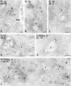
Figure 1
Figure 1
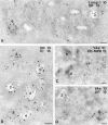
Endocytic compartments in 6H5.DM cells. Cells were incubated with Tf-HRP (Tf) for 3 or 10 min or with 5 nm BSA-gold (BSAg) for 3 or 10 min. Tf was detected with an HRP antibody (15- or 10-nm gold particles, A, B, C, and D, respectively). In addition, ultrathin cryosections were immunolabeled with antibodies specific for HLA-DR (A), I-chain NH2 terminus (C and D, IN) and I-chain (E, IC), or I-Ab (F). The distinct types of endocytic compartments are numbered 1 through 6. Tf is present in small vesicles and tubules (A and B), whereas 10-nm immunogold particles for DR are present only on the plasma membrane (A, PM). Type 2 compartments containing Tf are shown in C and D and characterized by an elongated tubular morphology with few internal vesicles. The typical electron lucent area can be seen in the curvature of the tubule (C). Type 3 compartments are shown in B, D, E, and F, and exhibit an irregular morphology and an electron lucent content with some internal vesicles. The type 3 compartment contains Tf labeling (C and D) and also 5 nm BSAg after 3 and 10 min of uptake (E and F, respectively). IN labeling is present on both the type 2 (C) and type 3 compartment (D). Immunolabeling for the luminal epitope of I-chain (IC) is abundant on the type 3 compartment (E). Multivesicular type 4 and multilaminar type 6 compartments (F) are negative for 5 nm BSAg after 10 min of uptake. Colocalization of I-Ab and BSAg is seen only in the type 3 compartment. Bar, 100 nm.
Figure 2
Figure 2
Characterization of MIICs in 6H5.DM cells. Ultrathin cryosections of 6H5.DM cells were immunolabeled with antibodies specific for LAMP-1 (A), I-chain COOH terminus, (B), CD-MPR (C), and peptide-loaded I-Ab (C and D, YAe). LAMP-1 (10-nm gold particles) is present on a multilaminar type 6 compartment (A), which also contains DR labeling (15-nm gold particles). In addition, both LAMP-1 and DR are found on a type 3 compartment, although to a smaller extent than on the type 6 compartment. I-chain COOH terminus labeling is restricted to type 2, 3, and 4 compartments (B), whereas DR is present in the multivesicular type 4 and more abundantly on the mixed type 5 compartment. Two typical type 6 compartments are shown in C; one of them shows labeling with YAe (10-nm gold particles). CD-MPR is absent from the type 6 compartments; labeling of CD-MPR (15 nm) is restricted to small, dense vesicles. YAe labels both on a type 3 and type 6 compartment (D). Bars, 100 nm.
Figure 3
Figure 3
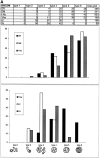
Quantitative analysis and graphical representation of Class II, DM, YAe, I-chain COOH terminus (IC), and I-chain NH2 terminus (IN) distribution in endocytic compartments of 6H5.DM (A) and A20.Ab (B) cells. Ultrathin cryosections of 6H5.DM cells were single immunolabeled for the indicated molecules using 10-nm gold particles. For each immunolabeling, 20 cell profiles were randomly selected and gold particles were counted directly in the microscope. (A) In 6H5.DM cells, six types of endocytic compartments could be distinguished, and only these were subjected to quantitation. These compartments were identified by their morphological characteristics, as shown in the bottom of the figure. The percentages of gold particles present in each compartment are summarized in the table. The total number of gold particles counted is shown in the last column of the table. Percentages of labeling for DR, DM, and I-Ab (top graph) and of YAe, I-chain COOH terminus, and I-chain NH2 terminus (bottom graph) are shown on the y axes. The graphs show strong enrichment of IC labeling in type 3 compartments, whereas labeling for DR, DM, and I-Ab is most abundant in types 5 and 6. IN labeling in types 3 and 4 is almost similar, while YAe is detected first in type 3 compartments and significantly enriched in the later types 4 to 6 (B). Quantitative analysis of A20.Ab cells was performed as described for 6H5.DM cells. I-chain NH2 terminus is strongly enriched in type 3 compartments, and both class II and DM are abundant in type 4 and 5/6 compartments.
Figure 3
Figure 3
Quantitative analysis and graphical representation of Class II, DM, YAe, I-chain COOH terminus (IC), and I-chain NH2 terminus (IN) distribution in endocytic compartments of 6H5.DM (A) and A20.Ab (B) cells. Ultrathin cryosections of 6H5.DM cells were single immunolabeled for the indicated molecules using 10-nm gold particles. For each immunolabeling, 20 cell profiles were randomly selected and gold particles were counted directly in the microscope. (A) In 6H5.DM cells, six types of endocytic compartments could be distinguished, and only these were subjected to quantitation. These compartments were identified by their morphological characteristics, as shown in the bottom of the figure. The percentages of gold particles present in each compartment are summarized in the table. The total number of gold particles counted is shown in the last column of the table. Percentages of labeling for DR, DM, and I-Ab (top graph) and of YAe, I-chain COOH terminus, and I-chain NH2 terminus (bottom graph) are shown on the y axes. The graphs show strong enrichment of IC labeling in type 3 compartments, whereas labeling for DR, DM, and I-Ab is most abundant in types 5 and 6. IN labeling in types 3 and 4 is almost similar, while YAe is detected first in type 3 compartments and significantly enriched in the later types 4 to 6 (B). Quantitative analysis of A20.Ab cells was performed as described for 6H5.DM cells. I-chain NH2 terminus is strongly enriched in type 3 compartments, and both class II and DM are abundant in type 4 and 5/6 compartments.
Figure 4
Figure 4
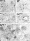
Endocytic compartments in A20.Ab cells. A20.Ab cells were incubated with 5-nm BSA-gold particles (BSAg) for 3 and 10 min (A, B, D, and E, respectively) or with Tf-HRP (Tf) for 10 min (C). After 3 min of BSA-gold uptake, particles are present in the type 1 compartment (A), which includes coated vesicles and some noncoated vesicles and tubules. BSA-gold is also present in the type 2 compartment at this time point (B) but is absent from the multivesicular type 4. In this picture, both types are immunolabeled for I-chain NH2 terminus. A type 3 compartment, shown in A and D, exhibits BSA-gold both after 3 min and 10 plus 50 min of uptake. Class II is present in type 3 compartments (A and D), which show the typical irregularly shaped morphology and electron lucent content with few internal vesicles (D). Type 5/6 compartments (E), with a dense content and few internal membranes, do not show BSA-gold after 10 min of uptake, while the type 2 compartment is positive for BSA-gold. Labeling for H-2M (DM, 10-nm gold particles) reveals little H-2M in the type 2, whereas type 5/6 compartments are strongly labeled for H-2M. Bars, 100 nm.
Figure 5
Figure 5
Characterization of MIICs in A20.Ab cells. Ultrathin cryosections of A20.Ab cells were double immunolabeled for class II and CD-MPR (A), cathepsin D (B), or LAMP-1 (C). Labeling for class II (5-nm gold particles) is present in type 4 and type 5/6 compartments (A), whereas CD-MPR labeling (10-nm gold particles) is detected only in small, dense vesicles (arrows). In B, a type 5/6 compartment positive for both class II (5-nm gold particles) and cathepsin D (10-nm gold particles) is shown. Labeling for LAMP-1 (10-nm gold particles) and class II (5-nm gold particles) is demonstrated in two multivesicular type 4 compartments (C). A type 3 compartment shown here contains some class II labeling, and only little LAMP-1 labeling. M, mitochondrion. Bars, 100 nm.
Figure 6
Figure 6
Immunogold labeling of I-chain in early MIICs of A20.Ab cells. A type 3/early MIIC with characteristic empty space is shown containing internalized BSA-gold particles and labeling for I-chain COOH terminus. Bar, 200 nm.
Figure 7
Figure 7
Comprehensive scheme of endocytic compartments and class II trafficking in B cells. The proposed relation between the different types of endocytic compartments (–6) including early and late MIICs as described in this study, and the classical terminology of endosomes and lysosomes are indicated. From our present morphological observations and the kinetic data in several recent fractionation studies (see Discussion), the following model of class II transport can be put forward. Class II/I-chain complexes derived from the TGN are transported to early MIICs and possibly to a minor extent to the plasma membrane and other endocytic compartments (see arrows with different thicknesses radiating from the TGN). After arrival in the endocytic pathway, class II/I-chain complexes migrate farther down the endocytic route (see arrows between types 1–6) together with endocytosed antigens, meanwhile being exposed to increasing proteolytic activity. This activity results in the removal of I-chain, a process probably already starting in type 3/early MIICs and progressing in later compartments. Once class II is freed from the I-chain peptide CLIP, antigenic peptides can bind to the class II molecules, a process catalyzed by HLA-DM. Peptide loading of class II commences in early MIICs and proceeds in later compartments, possibly depending on the type of class II and antigen. Escape of class II–peptide complexes from the degradative route for deposition at the cell surface has been shown to occur by exocytosis of MIICs by which the limiting membrane of MIICs is incorporated in the plasma membrane. During this process the internal vesicles of MIICs are externalized as exosomes (43). Other pathways of class II/peptide transport to the cell surface may very well exist but have not yet been identified.

Similar articles

Cited by

References

    1. Amigorena S, Drake JR, Webster P, Mellman I. Transient accumulation of new class II MHC molecules in a novel endocytic compartment in B lymphocytes. Nature (Lond) 1994;369:113–120. - PubMed
    1. Amigorena S, Webster P, Drake J, Newcomb J, Cresswell P, Mellman I. Invariant chain cleavage and peptide-loading in major histocompatibility complex class II vesicles. J Exp Med. 1995;181:1729–1741. - PMC - PubMed
    1. Bénaroche P, Yilla M, Raposo G, Ito K, Miwa K, Geuze HJ, Ploegh HL. How MHC class II molecules reach the endocytic pathway. EMBO (Eur Mol Biol Organ) J. 1995;14:37–49. - PMC - PubMed
    1. Bhattacharya A, Dorf ME, Springer TA. A shared alloantigenic determinant on Ia antigens encoded by the I-A and I-E subregions: evidence for I region gene duplication. J Immunol. 1981;127:2488–2495. - PubMed
    1. Bikoff EK, Germain RN, Robertson EJ. Allelic differences affecting invariant chain dependency of MHC class II subunit assembly. Immunity. 1995;2:301–310. - PubMed

Publication types

MeSH terms