. 1997 Oct 1;11(19):2557-68.
doi: 10.1101/gad.11.19.2557.

AU-rich elements target small nuclear RNAs as well as mRNAs for rapid degradation

Affiliations

AU-rich elements target small nuclear RNAs as well as mRNAs for rapid degradation

X C Fan et al. Genes Dev. .

Abstract

AU-rich elements (AREs, usually containing repeated copies of AUUUA), when present in the 3'-untranslated regions (UTRs) of many mammalian mRNAs, confer instability on their host RNA molecules. The viral small nuclear RNA (snRNA) Herpesvirus saimiri U RNA 1 (HSUR 1) also contains an AUUUA-rich sequence. Here, we report that this ARE induces rapid degradation of HSUR 1 itself and of other snRNAs including HSUR 2 and cellular U1. Mutational analyses of the viral ARE establish that sequence requirements for mRNA and snRNA decay are the same, suggesting a similar mechanism. Moreover, the in vivo degradation activity of mutant AREs correlates with their in vitro binding to the HuR protein, implicated previously as a component of the mRNA degradation machinery. Our results suggest that ARE-mediated instability can be uncoupled from both ongoing translation and deadenylation of the target RNA.


Figures

Figure 1
Figure 1
ARE-mediated HSUR 1 degradation in vivo. (A) HSUR RNA sequences and expression plasmid configuration. The HSUR 1 ARE and the nucleotides altered in Mut are underlined. The U1 promoter in the pUC–U1 vector is the 423-bp human genomic sequence upstream of U1 transcription initiation site, whereas the 3′ box is the 30-bp transcription termination signal downstream of the U1 coding sequence (Yuo and Weiner 1989). (B) T1 RNase protection analysis of wild-type and mutant HSUR 1 levels in transient transfection assays. The pUC–U1–HSUR 1 constructs were transiently cotransfected with a pUC–U1–HSUR 3 plasmid into mouse L929 cells (see Materials and Methods). Total RNA collected 48 hr after transfection was subjected to RNase T1 protection assays with wild-type and mutant HSUR 1 antisense RNA (lanes 2 and 3, respectively), together with antisense HSUR 3 RNA as an internal control. One-fiftieth of the amount of the anti-wild-type and anti-mutant HSUR 1 RNA probes used is shown in lanes 4 and 5. The data were quantitated with a Molecular Dynamics PhosphorImager and normalized to HSUR 3. Wild-type HSUR 1 levels were reproducibly one-eighth of those of mutant HSUR 1. (C) Whole-cell run-on assays of wild-type and mutant HSUR 1 transcription. The pUC–U1–HSUR 1 construct containing wild-type or mutant HSUR 1 sequences was cotransfected with pUC–U1–HSUR 3 into L cells, and whole-cell run-on assays were performed (see Materials and Methods). Total RNA was hybridized to nylon membranes that had been dot-blotted with wild-type (top) or mutant (bottom) HSUR 1 and HSUR 3 DNA fragments. Untransfected HSUR 4 DNA was also dotted as a negative control. The patterns of dots are illustrated at right; hybridizations with the run-on RNAs are at left. After quantitation and normalization against the cotransfected positive control HSUR 3 (also subtracting the untransfected negative control HSUR 4), the wild-type HSUR 1 (left dot, top) and the mutant (left dot, bottom) were found to have similar transcription rates (wild type:mutant = 0.95). (D) Immunoprecipitation of wild-type and mutant HSUR 1 from transfected mouse L929 cells. L cells were transiently transfected with the pUC–U1 constructs containing wild-type or mutant HSUR 1 genes, and whole-cell extracts were prepared by sonication. Equal amounts of extract were precipitated with anti-Sm monoclonal antibody Y12 or anti-U1 70K monoclonal antibody H111 as a control. RNA was harvested from the immunoprecipitation pellets (lanes 1,3,5,7) and supernatants (lanes 2,4,6,8), and wild-type (left) and mutant (right) HSUR 1 were assayed by T1 RNase protection. For both wild-type and mutant HSUR 1s, >90% of the RNA was in the anti-Sm precipitate (lanes 3,7), whereas >99% of the HSUR 1s remained in the supernatant with the anti-U1 70K antibody (lanes 2,6).
Figure 1
Figure 1
ARE-mediated HSUR 1 degradation in vivo. (A) HSUR RNA sequences and expression plasmid configuration. The HSUR 1 ARE and the nucleotides altered in Mut are underlined. The U1 promoter in the pUC–U1 vector is the 423-bp human genomic sequence upstream of U1 transcription initiation site, whereas the 3′ box is the 30-bp transcription termination signal downstream of the U1 coding sequence (Yuo and Weiner 1989). (B) T1 RNase protection analysis of wild-type and mutant HSUR 1 levels in transient transfection assays. The pUC–U1–HSUR 1 constructs were transiently cotransfected with a pUC–U1–HSUR 3 plasmid into mouse L929 cells (see Materials and Methods). Total RNA collected 48 hr after transfection was subjected to RNase T1 protection assays with wild-type and mutant HSUR 1 antisense RNA (lanes 2 and 3, respectively), together with antisense HSUR 3 RNA as an internal control. One-fiftieth of the amount of the anti-wild-type and anti-mutant HSUR 1 RNA probes used is shown in lanes 4 and 5. The data were quantitated with a Molecular Dynamics PhosphorImager and normalized to HSUR 3. Wild-type HSUR 1 levels were reproducibly one-eighth of those of mutant HSUR 1. (C) Whole-cell run-on assays of wild-type and mutant HSUR 1 transcription. The pUC–U1–HSUR 1 construct containing wild-type or mutant HSUR 1 sequences was cotransfected with pUC–U1–HSUR 3 into L cells, and whole-cell run-on assays were performed (see Materials and Methods). Total RNA was hybridized to nylon membranes that had been dot-blotted with wild-type (top) or mutant (bottom) HSUR 1 and HSUR 3 DNA fragments. Untransfected HSUR 4 DNA was also dotted as a negative control. The patterns of dots are illustrated at right; hybridizations with the run-on RNAs are at left. After quantitation and normalization against the cotransfected positive control HSUR 3 (also subtracting the untransfected negative control HSUR 4), the wild-type HSUR 1 (left dot, top) and the mutant (left dot, bottom) were found to have similar transcription rates (wild type:mutant = 0.95). (D) Immunoprecipitation of wild-type and mutant HSUR 1 from transfected mouse L929 cells. L cells were transiently transfected with the pUC–U1 constructs containing wild-type or mutant HSUR 1 genes, and whole-cell extracts were prepared by sonication. Equal amounts of extract were precipitated with anti-Sm monoclonal antibody Y12 or anti-U1 70K monoclonal antibody H111 as a control. RNA was harvested from the immunoprecipitation pellets (lanes 1,3,5,7) and supernatants (lanes 2,4,6,8), and wild-type (left) and mutant (right) HSUR 1 were assayed by T1 RNase protection. For both wild-type and mutant HSUR 1s, >90% of the RNA was in the anti-Sm precipitate (lanes 3,7), whereas >99% of the HSUR 1s remained in the supernatant with the anti-U1 70K antibody (lanes 2,6).
Figure 2
Figure 2
Mutational analysis of the HSUR 1 ARE. (A) Sequences of the six HSUR 1 point mutants, M1–M6, with the sites of U → G mutation underlined. (B) RNA levels of the HSUR 1 mutants. Wild-type HSUR 1 and mutants M1–M6, all driven by the same U1 promoter, were each transiently cotransfected with HSUR 3 into mouse L929 cells, and the RNA levels were assayed by T1 RNase protection (lanes 1–7, respectively). The antisense HSUR 1 probe covered a 120-nucleotide region at the 3′ end, which is common to wild-type HSUR 1 and all six mutants. When quantitated and normalized to HSUR 3, the mutant M1–M6 levels were found to be 7.0-, 10.1-, 1.8-, 5.8-, 6.4-, and 6.1-fold that of wild-type HSUR 1.
Figure 2
Figure 2
Mutational analysis of the HSUR 1 ARE. (A) Sequences of the six HSUR 1 point mutants, M1–M6, with the sites of U → G mutation underlined. (B) RNA levels of the HSUR 1 mutants. Wild-type HSUR 1 and mutants M1–M6, all driven by the same U1 promoter, were each transiently cotransfected with HSUR 3 into mouse L929 cells, and the RNA levels were assayed by T1 RNase protection (lanes 1–7, respectively). The antisense HSUR 1 probe covered a 120-nucleotide region at the 3′ end, which is common to wild-type HSUR 1 and all six mutants. When quantitated and normalized to HSUR 3, the mutant M1–M6 levels were found to be 7.0-, 10.1-, 1.8-, 5.8-, 6.4-, and 6.1-fold that of wild-type HSUR 1.
Figure 3
Figure 3
Degradation of β-globin mRNA containing AREs from HSUR 1 and its mutants. (A) Schematic diagram of the β-globin reporter construct and the sequences inserted into its 3′ UTR. Transcription of the rabbit β-globin construct was driven by the serum-inducible human c-fos promoter (Zubiaga et al. 1995). (B) Transient transfection analyses. The β-globin plasmids were transiently transfected into mouse L929 cells with cotransfection of the pEF–BOS–CAT plasmid, which expresses CAT mRNA constitutively (Zubiaga et al. 1995). After serum starvation, the transcription of β-globin was stimulated by serum addition. Cells were harvested when serum stimulation was initiated (T = 0) and at 1.0, 2.5, 4.0, and 5.5 hr thereafter. Total RNA was isolated for T1 RNase protection analyses using both a β-globin and a CAT antisense probe. (C) Time course of β-globin mRNA decay. The β-globin signals in Fig. 3B were quantitated on a Molecular Dynamics PhosphorImager, standardized to the CAT internal control (the lower band of the doublet was quantitated as the major T1 digestion product), and plotted. One hundred percent RNA was arbitrarily assigned to the time point with the highest signal. These results are the average of several experiments.
Figure 3
Figure 3
Degradation of β-globin mRNA containing AREs from HSUR 1 and its mutants. (A) Schematic diagram of the β-globin reporter construct and the sequences inserted into its 3′ UTR. Transcription of the rabbit β-globin construct was driven by the serum-inducible human c-fos promoter (Zubiaga et al. 1995). (B) Transient transfection analyses. The β-globin plasmids were transiently transfected into mouse L929 cells with cotransfection of the pEF–BOS–CAT plasmid, which expresses CAT mRNA constitutively (Zubiaga et al. 1995). After serum starvation, the transcription of β-globin was stimulated by serum addition. Cells were harvested when serum stimulation was initiated (T = 0) and at 1.0, 2.5, 4.0, and 5.5 hr thereafter. Total RNA was isolated for T1 RNase protection analyses using both a β-globin and a CAT antisense probe. (C) Time course of β-globin mRNA decay. The β-globin signals in Fig. 3B were quantitated on a Molecular Dynamics PhosphorImager, standardized to the CAT internal control (the lower band of the doublet was quantitated as the major T1 digestion product), and plotted. One hundred percent RNA was arbitrarily assigned to the time point with the highest signal. These results are the average of several experiments.
Figure 4
Figure 4
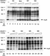
Binding of viral ARE sequences to HuR. The binding of the HuR protein to [α-32P]UTP-labeled wild-type HSUR 1 transcript in the presence of unlabeled wild-type or mutant HSUR 1 competitor RNA was assayed by UV cross-linking in HeLa cell nuclear extract. After RNase A treatment, label transfer was visualized by electrophoresis in a 12.5% SDS–polyacrylamide gel. The competitor RNAs, whose sequences are given in Figs. 1A and 2A, are indicated at top of the gel. (Lanes 1,14) Proteins cross-linked in the absence of specific competitor RNA. For each set of competitors, the first lane (lanes 2,5,8,11,15,18,21,24) contains a 10-fold molar excess, the second lane (lanes 3,6,12,16,19,22,25) a 20-fold molar excess, and the third lane (lanes 4,7,10,13,17,20,23,26) a 50-fold molar excess over the labeled HSUR 1 RNA. The cross-linked HuR protein is indicated with an arrow.
Figure 5
Figure 5
AUUUA repeats target other snRNAs for rapid decay. (A) The 5′ ARE of wild-type HSUR 2 and its mutants. The single guanosine that interrupts the AUUUA repeat sequence in HSUR 2 was mutated to UA, CC, and CA (underlined) in HSUR 2 M1, HSUR 2 M2, and HSUR 2 M3, respectively. (B) RNA levels of wild-type and mutant HSUR 2s. Wild-type HSUR 2 or mutant M1, M2, or M3, all controlled by the same U1 promoter, were each transiently cotransfected with HSUR 3 into mouse L929 cells and analyzed by T1 RNase protection. The level of mutant M1 (lane 2), which has four tandem copies of AUUU, is one-fifth that of wild-type HSUR 2 (lane 1), whereas controls M2 and M3 (lanes 3 and 4, respectively), which have been mutated at the same two positions as M1, have levels 1.3- and 1.1-fold of the wild-type HSUR 2, respectively. (C) The 5′-end sequences of wild-type U1 and two U1 mutants. The 5′ splice site recognition sequence of U1 and the sequences replacing it in the mutants are underlined. The AU3–U1 mutant has four tandem copies of AUUU, whereas in the AGU–U1 mutant, there are four AUUU repeats interrupted by 3 Gs (the same sequence as in HSUR 1 M2). (D) Levels of AU3–U1 and AGU–U1 in duplicate transfection experiments. Each U1 mutant was transiently cotransfected with HSUR 3 into mouse L929 cells. U1 RNA levels were analyzed by primer extension using an oligonucleotide complementary to the most 3′ 20 nucleotides of U1 (nucleotides 155–164), whereas HSUR 3 was assayed by T1 RNase protection assay as above. When normalized to HSUR 3 and averaged between the duplicate transfection experiments, the AU3–U1 level was found to be one-fourth that of AGU–U1.
Figure 5
Figure 5
AUUUA repeats target other snRNAs for rapid decay. (A) The 5′ ARE of wild-type HSUR 2 and its mutants. The single guanosine that interrupts the AUUUA repeat sequence in HSUR 2 was mutated to UA, CC, and CA (underlined) in HSUR 2 M1, HSUR 2 M2, and HSUR 2 M3, respectively. (B) RNA levels of wild-type and mutant HSUR 2s. Wild-type HSUR 2 or mutant M1, M2, or M3, all controlled by the same U1 promoter, were each transiently cotransfected with HSUR 3 into mouse L929 cells and analyzed by T1 RNase protection. The level of mutant M1 (lane 2), which has four tandem copies of AUUU, is one-fifth that of wild-type HSUR 2 (lane 1), whereas controls M2 and M3 (lanes 3 and 4, respectively), which have been mutated at the same two positions as M1, have levels 1.3- and 1.1-fold of the wild-type HSUR 2, respectively. (C) The 5′-end sequences of wild-type U1 and two U1 mutants. The 5′ splice site recognition sequence of U1 and the sequences replacing it in the mutants are underlined. The AU3–U1 mutant has four tandem copies of AUUU, whereas in the AGU–U1 mutant, there are four AUUU repeats interrupted by 3 Gs (the same sequence as in HSUR 1 M2). (D) Levels of AU3–U1 and AGU–U1 in duplicate transfection experiments. Each U1 mutant was transiently cotransfected with HSUR 3 into mouse L929 cells. U1 RNA levels were analyzed by primer extension using an oligonucleotide complementary to the most 3′ 20 nucleotides of U1 (nucleotides 155–164), whereas HSUR 3 was assayed by T1 RNase protection assay as above. When normalized to HSUR 3 and averaged between the duplicate transfection experiments, the AU3–U1 level was found to be one-fourth that of AGU–U1.

Similar articles

Cited by

References

    1. Aharon T, Schneider RJ. Selective degradation of short-lived mRNAs with the granulocyte-macrophage colony-stimulating factor AU-rich 3′ noncoding region is mediated by a cotranslational mechanism. Mol Cell Biol. 1993;13:1971–1980. - PMC - PubMed
    1. Albrecht J-C, Fleckenstein B. Nucleotide sequence of HSUR 6 and HSUR 7, two small RNAs of herpesvirus saimiri. Nucleic Acids Res. 1992;20:1810. - PMC - PubMed
    1. Belasco J, Brawerman G. Control of messenger RNA stability. San Diego, CA: Academic Press; 1993.
    1. Biesinger B, Trimble JJ, Desrosiers RC, Fleckenstein B. The divergence between two oncogenic herpesvirus saimiri strains in a genomic region related to the transforming phenotype. Virology. 1990;176:505–514. - PubMed
    1. Bohjanen PR, Petryniak B, June CH, Thompson CB, Lindsten T. An inducible cytoplasmic factor (AU-B) binds selectively to AUUUA multimers in the 3′ untranslated region of lymphokine mRNA. Mol Cell Biol. 1991;11:3288–3295. - PMC - PubMed

Publication types

MeSH terms

LinkOut - more resources