. 1997 Sep 15;11(18):2323-34.
doi: 10.1101/gad.11.18.2323.

Diabetes, defective pancreatic morphogenesis, and abnormal enteroendocrine differentiation in BETA2/neuroD-deficient mice

Affiliations

Diabetes, defective pancreatic morphogenesis, and abnormal enteroendocrine differentiation in BETA2/neuroD-deficient mice

F J Naya et al. Genes Dev. .

Abstract

Candidate transcription factors involved in pancreatic endocrine development have been isolated using insulin gene regulation as a paradigm. The cell-type restricted basic helix-loop-helix (bHLH) gene, BETA2/NeuroD, expressed in pancreatic endocrine cells, the intestine, and the brain, activates insulin gene transcription and can induce neurons to differentiate. To understand the importance of BETA2 in pancreatic endocrine cell differentiation, mice lacking a functional BETA2 gene were generated by gene targeting experiments. Mice carrying a targeted disruption of the BETA2 gene developed severe diabetes and died perinatally. Homozygous BETA2 null mice had a striking reduction in the number of insulin-producing beta cells and failed to develop mature islets. Islet morphogenesis appeared to be arrested between E14.5 and E17.5, a period characterized by major expansion of the beta cell population. The presence of severe diabetes in these mice suggests that proper islet structure plays an important role in blood glucose homeostasis. In addition, secretin- and cholecystokinin-producing enteroendocrine cells failed to develop in the absence of BETA2. The absence of these two pancreatic secretagogs may explain the abnormal cellular polarity and inability to secrete zymogen granules in pancreatic acinar exocrine cells. The nervous system appeared to develop normally, despite abundant expression of BETA2 in differentiating neurons. Thus, BETA2 is critical for the normal development of several specialized cell types arising from the gut endoderm.


Figures

Figure 1
Figure 1
Disruption of BETA2 gene by homologous recombination and genotype analysis of heterozygote intercrosses. (A) Maps of wild-type BETA2 locus, targeting vector, and disrupted BETA2 allele. I and II designate exons in the BETA2 gene. The 5′ external probe used for Southern blotting is shown (▪). The diagnostic restriction fragments with their expected sizes are indicated as lines. The bacterial lacZ gene was inserted in the same orientation as the BETA2 genomic locus. The neomycin resistance (neoR) gene was inserted in the opposite transcriptional orientation. The introduction of the lacZ–neoR cassette disrupted all [including the bHLH and transactivation (R. Stein, unpubl.) domains] but the first 66 amino acids of BETA2, resulting in a BETA2–LacZ fusion protein with a novel SacI site. (B) Southern analysis of SacI-digested DNA from the litter of a BETA2 +/− intercross. Sizes of wild-type and targeted alleles are indicated by 13 and 4 kb, respectively. Subsequent genotype analysis was performed by PCR on tail DNA using the primers B2.1, B2.2, and B2.lacZ. Arrows indicate the primers and hatched box designates the bHLH domain.
Figure 1
Figure 1
Disruption of BETA2 gene by homologous recombination and genotype analysis of heterozygote intercrosses. (A) Maps of wild-type BETA2 locus, targeting vector, and disrupted BETA2 allele. I and II designate exons in the BETA2 gene. The 5′ external probe used for Southern blotting is shown (▪). The diagnostic restriction fragments with their expected sizes are indicated as lines. The bacterial lacZ gene was inserted in the same orientation as the BETA2 genomic locus. The neomycin resistance (neoR) gene was inserted in the opposite transcriptional orientation. The introduction of the lacZ–neoR cassette disrupted all [including the bHLH and transactivation (R. Stein, unpubl.) domains] but the first 66 amino acids of BETA2, resulting in a BETA2–LacZ fusion protein with a novel SacI site. (B) Southern analysis of SacI-digested DNA from the litter of a BETA2 +/− intercross. Sizes of wild-type and targeted alleles are indicated by 13 and 4 kb, respectively. Subsequent genotype analysis was performed by PCR on tail DNA using the primers B2.1, B2.2, and B2.lacZ. Arrows indicate the primers and hatched box designates the bHLH domain.
Figure 2
Figure 2
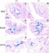
Abnormal pancreatic islet morphogenesis in BETA2 −/− mice. (A–D) Sagittal sections of BETA2 +/− (left panels) and BETA2 −/− (right panels) embryos at E9.5 (A,B) and pancreas at E14.5 (C,D) stained with X-gal. There were no obvious differences in morphology of the early developing pancreas or in the number of β-gal-positive cells. β-Gal-positive cells were similarly distributed in both +/− and −/− tissue. β-Gal expression was also detected in both the dorsal and ventral pancreas. (E,F) Pancreata of +/− and −/− mice at E17.5 stained with X-gal. At E17.5 (E) there was evidence of islet formation in the +/− pancreas, as indicated by β-gal-positive cells, which formed a sphere-shaped structure with a distinct border surrounded by acinar cells (arrows). In contrast, distinct islets were not present in −/− pancreas at E17.5 (F), although β-gal-positive cells were seen without distinctive shape or border. Sections were counterstained in nuclear fast red. (drg) Dorsal root ganglion; (sc) spinal cord. Original magnification, 500× (A,B); 250× (C–F).
Figure 3
Figure 3
Multiple islet cell types coexpress BETA2. Localization of pancreatic hormones in BETA2-expressing cells of +/− (A–H) and −/− (I–P) by multiple labeling. Single sections of newborn mice were examined for β-galactosidase activity by X-gal-staining and for hormones by immunofluorescence. Well defined islets, heterogeneous in size were seen with X-gal-stained cells in +/− mice (B,D,F,H). X-Gal-stained cells within these islets showed immunofluorescent staining for insulin (A), glucagon (C), somatostatin (E), and peptide YY (G) visualized by Cy3-conjugated (A) and FITC-conjugated (C,E,G) secondary antibodies. Endocrine cell types in the +/− pancreas were distributed normally with the β cells (Ins) in the core of the islet and the other cell types in the periphery of the islet. (I–P) Localization of hormones and β-gal in the pancreatic endocrine cells of −/− mice by immunofluorescence (I,K,M,O) and X-gal staining (J,L,N,P) as in A–H. Endocrine cells in the −/− pancreas coexpressed β-gal but did not appear as organized islets. Each cell type appeared in clusters in contrast to their distribution in +/− mice. Original magnification, 1000×.
Figure 4
Figure 4
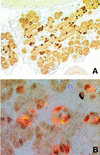
PDX-1 expression in BETA2 −/− pancreas. (A) At E16.0, PDX-1 was expressed throughout the pancreatic epithelium (original magnification, 250×); (B) coimmunolocalization of insulin (imunofluorescence, red, cytoplasmic) and PDX-1 (imunoperoxidase staining, brownish, nuclear) in −/− E16.0 pancreas (original magnification, 1000×). Most insulin-producing cells also coexpressed PDX-1.
Figure 5
Figure 5
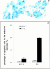
Increase in the number of apoptotic cells in the BETA2 −/− endocrine pancreas. X-Gal-stained E17.5 and P0 pancreatic sections were subjected to TUNEL assay. For each individual animal, the numbers of cells positive for apoptosis and/or X-gal were counted from five slide sets of four serial 7-μm sections, which included an average of five islet clusters per section. Results are the average of numbers from three different animals and are shown as percent of the number of endocrine cells; error bars represent standard deviation. At P0 there were ∼14-fold more (P < 0.00001, n = 3) apoptotic cells in the −/− endocrine pancreas (B,C) than in its +/− counterpart (A,C). However, at E17.5 (C), the −/− endocrine pancreas already showed a tendency of having fivefold more (P = 0.0204, n = 3) apoptotic cells than its +/− counterpart. Two-sided Student’s t-test was used to determine the significance of the differences.
Figure 6
Figure 6
Acinar cell abnormalities in BETA2 −/− mice. (A–F) Hematoxylin and eosin stain of pancreas of E17.5, P0, and P2 BETA2 +/− and −/− mice (original magnification, 400×). Acinar cell nuclei were randomly distributed within the cellular compartment of −/− mice (F) in contrast to +/− mice (C). Acinar cells in the −/− mice (F) lacked the distinct cellular polarity as well as the intense basophilic staining in the basal portion of acinar cells and acidophilic staining at the apical surface (arrows) evident at this developmental stage in +/− pancreas (C). (G–L) Ultrastructural analysis of exocrine pancreas. Polarization was evident in +/− pancreas at birth (P0) and at 2 at days of age (H,I) (original magnification, 2400×) but not in −/− pancreas at comparable stages (K,L) (original magnification, 3000×). Note the abundance of secretory granules and vacuoles in mutant pancreas at 2 days of age (L). (v) Secretory vesicles; (n) nuclei.
Figure 7
Figure 7
Western blot analysis of α-amylase in the P2-pancreas. A representative result is shown. Equal amounts of total cellular extracts (40 μg) from each pancreas examined were subjected to SDS-PAGE; BETA2 −/− (lane 1), +/− (lane 2), and +/+ (lane 3). Western blot analysis was performed by using anti-α-amylase antibody.
Figure 8
Figure 8
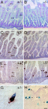
Absence of enteroendocrine cells expressing secretin and cholecystokinin in BETA2 −/− mice. Immunostaining for secretin (A,B), CCK (C,D), and serotonin (E,F) (arrows) in the small intestine of 2 day old BETA2 +/− (left panels) and BETA2 −/− (right panels) mice. Note the absence of secretin- and CCK-staining cells in the −/− mice (B,D) compared to +/− mice (A,C). Nuclear β-galactosidase staining in +/− (G) and −/− (H). G shows a single cell with brown cytoplasmic staining for secretin and blue nuclear X-gal staining from an adult BETA2 +/− mouse. Several isolated mucosal epithelial cells (arrows) in the small intestine of a −/− mouse showing nuclear β-galactosidase activity. Bar, 100 μm for A–D; 50 μm for E,F; 5 μm for G; and 20 μm for H. Sections were stained using the ABC method with True Blue peroxidase substrate counterstained with contrast red (A,B) or DAB substrate counterstained with hematoxylin (C–H).

Similar articles

Cited by

References

    1. Ahlgren U, Pfaff SL, Jessell TM, Edlund T, Edlund H. Independent requirement for ISL1 in formation of pancreatic mesenchyme and islet cells. Nature. 1997;385:257–260. - PubMed
    1. Alpert S, Hanahan D, Teitelman G. Hybrid insulin genes reveal a developmental lineage for pancreatic endocrine cell and imply a relationship with neurons. Cell. 1988;53:295–308. - PubMed
    1. Bain G, Maandag ECR, Izon DJ, Amsen D, Kruisbeek AM, Weintraub BC, Krop I, Schlissel MS, Feenyey AJ, Roon MV. E2A proteins are required for proper B cell development and initiation of immunoglobulin gene rearrangements. Cell. 1994;79:885–892. - PubMed
    1. Bartholoma A, Nave K A. NEX-1: A novel brain specific helix loop helix protein with autoregulation and sustained expression in mature cortical neurons. Mech Dev. 1994;48:217–228. - PubMed
    1. Bonner-Weir S, Trent D, Weir GC. Partial pancreatectomy in the rat and subsequent defect in glucose-induced insulin secretion. J Clin Invest. 1983;71:1544–1553. - PMC - PubMed

Publication types

MeSH terms